Introduction
Squamous cell carcinoma (SCC) is the most common cancer of the oral cavity and oropharynx. These tumours represent the sixth most common cancer subsite in the world, with 710 000 cases diagnosed annually and over 350 000 deaths per year worldwide.Reference Rahman, Iocca, Kufta and Shanti1
Standard treatment for oropharyngeal SCC includes concurrent chemoradiotherapy for advanced tumours, or radiotherapy alone, with or without initial surgical resection (by way of transoral robotic surgery or transoral laser surgery), for less advanced disease.Reference Pignon, le Maitre, Maillard and Bourhis J;2–Reference Glenny, Furness, Worthington, Conway, Oliver and Clarkson4
Major oropharyngeal haemorrhage is a rare but life-threatening complication after treatment of these cancers. Over the past seven years, our state-wide head and neck cancer service has managed multiple cases of major oropharyngeal haemorrhage following non-surgical treatment of oropharyngeal SCCs. These patients were all treated by our Neurological Intervention and Imaging Service of Western Australia (‘NIISwa’) team with endovascular embolisation of the offending tumour vessels. Whilst major post-treatment haemorrhage after chemoradiotherapy has been reported in some small case series, no previously published studies have, to our knowledge, investigated risk factors for haemorrhage in those receiving chemoradiotherapy or radiotherapy alone.Reference Self, Bumpous, Ziegler, Wilson and Potts5,Reference Chou, Rath, Johnson and Goyal6
We present here a case series of our experience in Western Australia, with the aim of describing the characteristics that may pre-dispose patients to bleeding in this context, including radiological predictors of haemorrhage.
Materials and methods
Patient selection
This retrospective case series received approval from the North and South Metropolitan Health Service review committees in Perth, Western Australia.
A state-wide database of interventional neuroradiology procedures performed in Western Australia was used to extract relevant cases between 13 September 2013 and 8 January 2021. The study included patients with oropharyngeal SCCs who had suffered a major haemorrhage event requiring endovascular embolisation, either during or after treatment with chemoradiotherapy or radiotherapy alone. We excluded those patients who had not yet undergone treatment, those who were surgically managed and those who had a histological diagnosis other than SCC. A multidisciplinary head and neck cancer database was used to extract a tumour stage matched control group of patients who did not experience oropharyngeal bleeding.
Data extraction
A retrospective chart review was conducted of the electronic and hard-copy medical records. Patient records were reviewed for basic demographic details (age, sex, immunosuppression, smoking history), tumour characteristics (histology, location, tumour stage, primary vs recurrence) and treatment details (chemoradiotherapy vs radiotherapy alone, radiotherapy dose, chemotherapy agent, length of therapy).
Tumours were staged using the American Joint Committee on Cancer AJCC Cancer Staging Manual, eighth edition.Reference Amin, Edge, Greene, Byrd, Brookland and Washington7 For those with no documented American Joint Committee on Cancer stage, we used the available clinical information, imaging and pathology to retrospectively stage these patients.
Immunodeficiency was defined as meeting one of the following criteria (which were chosen because of their incorporation in the Charlson Comorbidity IndexReference Charlson, Pompei, Ales and MacKenzie8): known diagnosis of primary immunodeficiency, secondary immunodeficiency (human immunodeficiency virus, poorly controlled type 2 diabetes mellitus, organ transplant), immunomodulator medications or long-term high dose steroid therapy, and presence of concurrent haematological malignancy.
Imaging review
A review of pre- and post-treatment computed tomography (CT) images was conducted by a specialist head and neck radiologist with 14 years’ experience, who was blinded to the patient's treatment regimen (i.e. chemoradiotherapy vs radiotherapy alone).
Pre-treatment CT imaging was reviewed and the index lesion was assessed in terms of several parameters. These included: tumour primary site and contiguous secondarily involved subsites (tonsil, base of tongue, glosso-tonsillar sulcus, soft palate, pharyngeal wall, oral tongue), primary tumour morphology (endophytic or exophytic, dependent on whether the tumour bulk was primarily deep to or growing externally to the pharyngeal mucosa), maximum axial tumour size in antero-posterior and transverse dimensions, maximum anterior extent of the tumour from vallecula or base of tongue mucosa, and the presence or absence of effacement of parapharyngeal fat.
Pre-treatment CT images were also used to assess the proximity of the culprit bleeding vessel to the tumour mass (distant (more than 5 mm) or near (less than 5 mm), abutting, or encased (more than 180 degrees’ circumferential encasement of vessel)), and to determine which vessel was involved and if the vessel was ipsilateral or contralateral to the tumour mass.
Pre- and post-treatment CT imaging was reviewed to assess for treatment-related primary tumour deep cavitating or ulcerative changes, which was defined as non-anatomical gas deep to the native mucosal plane within the treated tumour mass.
Statistical analysis
Analysis was conducted on the patient group as a whole, then on the treatment-related subgroups of haemorrhage and non-haemorrhage groups. Data were collated, and analysed using simple descriptive statistics. Categorical variables were compared using the Fisher exact test. Results with p-values of less than 0.05 were considered statistically significant.
Results
Seven patients with oropharyngeal SCC, treated with chemoradiotherapy (n = 5) or radiotherapy alone (n = 2), who suffered a major haemorrhage event were identified from the interventional neuroradiology procedure database. Seven tumour stage matched patients, who underwent chemoradiotherapy (n = 5) or radiotherapy alone (n = 2), who did not bleed were included as a control group.
The endovascular embolisation procedures were all performed by board-certified interventional neuroradiologists using biplanar neuroangiography equipment (Axiom™; Siemens). Embolic material was delivered, via radial or femoral artery access, through a 0.0165 inch (0.4191 mm) inner-diameter microcatheter (Headway® Duo; Microvention) into the relevant artery. The embolic materials were selected on a case-by-case basis, and included n-butyl cyanoacrylate (Histoacryl®; Braun), polyvinyl alcohol particles (Contour™; Boston Scientific) and platinum coils (Axium™; Medtronic).
Of the 14-patient cohort, 13 were male. The median patient age was 65 years (range, 56–69 years) in the haemorrhage group and 70 years (range, 65–74 years) in the non-haemorrhage group. Two patients were immunocompromised because of a history of type 2 diabetes mellitus. Four patients in the non-haemorrhage group were taking antiplatelet medications. No patients in the haemorrhage group were taking antiplatelet or anticoagulant medications. No patients in either group suffered from a medical coagulopathy during treatment or at the time of oropharyngeal haemorrhage.
Both patients who received radiotherapy alone in the haemorrhage group were treated with non-curative intent; both of those who received radiotherapy alone in the non-haemorrhage group were treated curatively. All patients in both treatment groups (chemoradiotherapy or radiotherapy alone) received a radiotherapy dose of 70 Gy in 35 fractions. All patients who had chemotherapy were treated with cisplatin monotherapy (n = 10), with an average treatment duration of 44.3 days. Patient demographics, tumour staging and treatment details are presented in Table 1.
Patient and treatment characteristics

*n = 5; †n = 2; ‡n = 5; **n = 2. CRT = chemoradiotherapy; RT = radiotherapy; HPV = human papilloma virus; N/A = not applicable; CT = computed tomography
Four patients from the cohort died (three from the haemorrhage group and one from non-haemorrhage group). Two of the haemorrhage group died from large recurrent oropharyngeal bleeds. The other two patients both died from aspiration pneumonia. The 12-month overall survival rate for the cohort was 79 per cent; there was no statistically significant difference in survival between the haemorrhage and non-haemorrhage groups (p = 0.51). The median follow-up duration for the cohort was 13 months (range, 8–44 months).
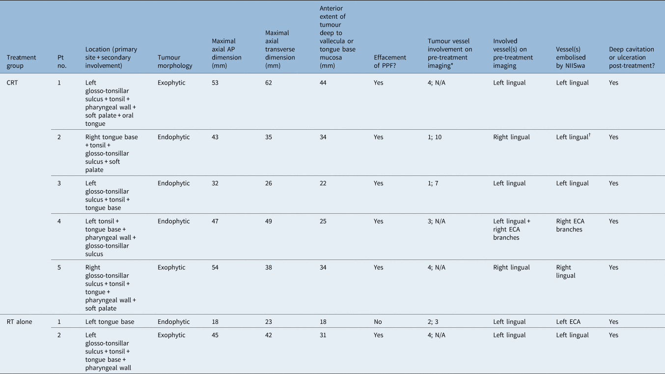
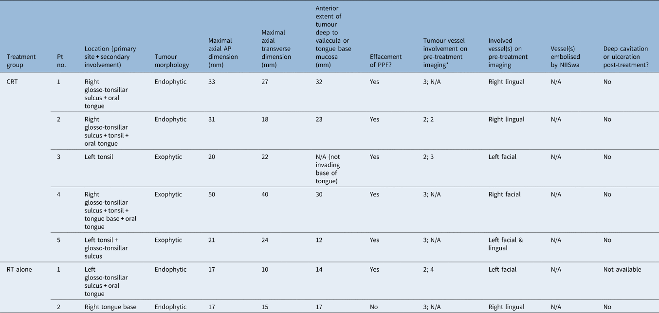
The results of the CT imaging review are detailed in Tables 2 and 3. Because of the small sample size, statistically significant differences could not be determined between the radiotherapy-only haemorrhage and non-haemorrhage groups for any variable. As such, the analysis presented here reports haemorrhage versus non-haemorrhage groups as a whole.
Haemorrhage group: tumour characteristics on CT imaging

*Data represent the proximity of the culprit bleeding vessel to the tumour mass, in terms of: assigned category (i.e. 1 = distant, 2 = near, 3 = abutting or 4 = encased) and distance from tumour in millimetres. †Tumour crossed midline, post-treatment ulceration with left lingual artery pseudoaneurysm. CT = computed tomography; pt. no. = patient number; AP = antero-posterior; PPF = parapharyngeal fat; NIISwa = Neurological Intervention and Imaging Service of Western Australia; CRT = chemoradiotherapy; N/A = not applicable; ECA = external carotid artery; RT = radiotherapy
Non-haemorrhage group: tumour characteristics on CT imaging

*Data represent the proximity of the culprit bleeding vessel to the tumour mass, in terms of: assigned category (i.e. 1 = distant, 2 = near, 3 = abutting or 4 = encased) and distance from tumour in millimetres. CT = computed tomography; pt. no. = patient number; AP = antero-posterior; PPF = parapharyngeal fat; NIISwa = Neurological Intervention and Imaging Service of Western Australia; CRT = chemoradiotherapy; N/A = not applicable; RT = radiotherapy
There was a statistically significant difference in the maximal axial transverse tumour dimension, with a median of 38 mm (range, 26–62 mm) in the haemorrhage group and 22 mm (range, 10–40 mm) in the non-haemorrhage group (p = 0.02). There was no statistically significant difference in the maximum antero-posterior axial dimension between the haemorrhage and non-haemorrhage groups (p = 0.06).
There were no statistically significant associations between bleeding and tumour stage, morphology (exophytic vs endophytic growth pattern) or effacement of parapharyngeal fat on pre-treatment CT. There were also no significant differences between bleeding and the particular vessel involved (as seen on pre-treatment CT) or the laterality of the bleeder vessel comparative to the tumour mass.
Although there was no difference in tumour distance from the closest or culprit vessels on pre-treatment CT between the haemorrhage and non-haemorrhage groups (p = 0.67), all haemorrhage group tumours involved the proximal trunks of vessels, whilst all in the non-haemorrhage group involved the distal aspects of the culprit vessels. Additionally, the haemorrhage group had a median anterior tumour invasion from the base of tongue or vallecula mucosa of 31 mm (range, 18–44 mm), compared to 20 mm (range, 12–32 mm) in the non-haemorrhage group (p = 0.08).
All patients in the haemorrhage group (n = 7) developed deep cavitating or ulcerative changes at the tumour site between pre- and post-treatment CT imaging, whilst no patients in the non-haemorrhage group had cavitating or ulcerative changes. This difference was statistically significant (p < 0.0001).
Discussion
Only two previous publications in the searchable literature have investigated haemorrhage after chemoradiation for oropharyngeal cancers. Self et al. reviewed a single tertiary centre database of 139 patients managed between 2005 and 2010, of whom 10 had a large-volume oropharyngeal haemorrhage.Reference Self, Bumpous, Ziegler, Wilson and Potts5 They identified advanced tumour (T) stage as the most important factor in patients developing haemorrhage after chemoradiation.Reference Self, Bumpous, Ziegler, Wilson and Potts5 Building on those findings, Chou et al. published a case series of 10 patients in 2021, investigating clinical and radiological factors that might lead to haemorrhage in this patient subset.Reference Chou, Rath, Johnson and Goyal6 They found that all five patients who bled had exophytic tumours on pre-treatment CT imaging; of those five, four had significant parapharyngeal fat effacement and all had deeply ulcerated tumour beds on post-treatment CT.Reference Chou, Rath, Johnson and Goyal6 These authors also identified the facial artery as the main vessel involved by these tumours.Reference Chou, Rath, Johnson and Goyal6
The current study further contributes to this body of literature. We found that the median maximal axial transverse dimension of tumours was much larger in the haemorrhage cohort (38 mm vs 22 mm), which correlated with a larger median maximum anterior tumour invasion from the base of tongue or vallecula mucosa in the haemorrhage group (31 mm vs 20 mm). Interestingly, we found the primary vessel involved with bleeding tumours was the lingual not facial artery, and all bleeding tumours involved the proximal trunk of this vessel. This could explain the catastrophic high-volume nature of these bleeds and the need for urgent interventional neuroradiology input in these cases.
Unlike Chou et al., we did not identify any significant difference in effacement of parapharyngeal fat between haemorrhage or non-haemorrhage groups. Congruous with the findings of Chou et al.,Reference Chou, Rath, Johnson and Goyal6 however, we found that all patients in the haemorrhage group developed deep cavitating or ulcerative changes at the tumour site between pre- and post-treatment CT imaging, whilst no patients in the non-haemorrhage group developed these changes. Post-chemoradiotherapy clinic-based nasendoscopy, performed to identify deeply cavitating tumour beds, could prove a useful adjunct in determining the risk of bleeding within this patient group and could be useful for early interventional neuroradiology opinion.
The present study has the same limitations as those of any retrospective review of clinical data: treatment selection bias is hard to trace, and information is reliant on the accuracy and completeness of medical records. The use of the interventional neuroradiology database means that patients with post-surgical haemorrhage who bled and were treated with palliative intent, or those who died prior to reaching hospital, are not captured. The authors’ initial intention was to assess haemorrhage versus non-haemorrhage groups, and to include an analysis of chemoradiation versus radiation alone. Despite using a large state-wide Western Australian database, our subset of radiotherapy-only patients was too small to draw statistically significant conclusions.
• Squamous cell carcinoma (SCC) is the most common cancer of the oropharynx
• Major oropharyngeal haemorrhage is a rare and potentially life-threatening complication after chemoradiotherapy for these tumours
• Such haemorrhage is managed with endovascular embolisation of the bleeding vessel by interventional neuroradiologists
• A retrospective case series was conducted of oropharyngeal SCC patients who suffered haemorrhage requiring embolisation during or after chemoradiotherapy or radiotherapy
• The study investigated radiological and clinical predictors of haemorrhage
• Tumour size, tumour bed ulceration or cavitation, and involvement of proximal aspect of external carotid branches were predictive of haemorrhage
This study's findings build on the small volume of literature currently available on this topic. Tumour bed ulceration and cavitation appears to be highly predictive of bleeding, and a direct product of the erosive tissue damage that occurs with combined chemoradiotherapy to mucosal surfaces and vascular structures.Reference Self, Bumpous, Ziegler, Wilson and Potts5 Future research could be directed at identifying means of preventing this tissue destruction, such as optimising nutritional intake during treatment, preventing treatment-related weight loss, or altering treatment regimens to prevent collateral destruction of healthy oropharyngeal tissue.
Acknowledgement
We would like to acknowledge Dr Felipe Cardemil for conducting the statistical analysis for this paper.
Competing interests
None declared