




No CrossRef data available.
Article contents
Specific sites in the Beta Interaction Domain of a schistosome Ca2+ channel β subunit are key to its role in sensitivity to the anti-schistosomal drug praziquantel
Published online by Cambridge University Press: 17 October 2003
Abstract
Praziquantel, the drug of choice against schistosomiasis, disrupts calcium (Ca2+) homeostasis in schistosomes via an unknown mechanism. Voltage-gated Ca2+ channels are heteromultimeric transmembrane protein complexes that contribute to impulse propagation and also regulate intracellular Ca2+ levels. β subunits modulate the properties of the pore-forming α1 subunit of high voltage-activated Ca2+ channels. Unlike other Ca2+ channel β subunits, which have current stimulatory effects, a β subunit subtype found in S. mansoni (SmβA) and S. japonicum (Sjβ) dramatically reduces current levels when co-expressed with Ca2+ channel α1 subunits in Xenopus oocytes. It also confers praziquantel sensitivity to the mammalian Cav2.3 α1 subunit. The Beta Interaction Domains (BIDs) of SmβA and Sjβ lack 2 conserved serines that each constitute a consensus site for protein kinase C (PKC) phosphorylation. Here, we use site-directed mutagenesis of schistosome β subunits to show that these unique functional properties are correlated with the absence of these consensus PKC sites in the BID. Furthermore, a second schistosome β subunit subtype contains both serines in the BID, enhances currents through α1 subunits, and does not confer praziquantel sensitivity. Thus, phosphorylation sites in the BID may play important roles in defining the modulatory properties and pharmacological sensitivities of schistosome Ca2+ channel β subunits.
Information
- Type
- Research Article
- Information
- Copyright
- 2003 Cambridge University Press
INTRODUCTION
Praziquantel (PZQ) is the drug of choice against schistosomiasis. It causes a disruption of calcium (Ca2+) homeostasis in adult worms via an unknown mechanism (reviewed by Andrews, 1985; Day, Bennett & Pax, 1992; Redman et al. 1996). We are investigating the interaction of PZQ with voltage-gated Ca2+ channels, which have been proposed as a possible target of this drug.
Voltage-gated Ca2+ channels are important regulators of Ca2+ homeostasis in excitable cells, and are involved in coupling cellular excitation to a wide variety of intracellular functions (reviewed by Catterall, 2000; Hofmann, Lacinová & Klugbauer, 1999). Ca2+ channels can be divided broadly into high voltage-activated (HVA; Cav1 and Cav2), and low voltage-activated (LVA; Cav3) classes. Associated with the main, pore-forming α1 subunit of HVA Ca2+ channels are auxiliary β and α2/δ subunits, which modulate channel properties and facilitate plasma membrane expression (reviewed by Walker & DeWaard, 1998). In particular, co-expression of Ca2+ channel α1 subunits with β subunits results in increases in current amplitude, changes in the kinetics and voltage dependence of channel activation and inactivation, increases in ligand binding (reviewed by Walker & DeWaard, 1998; Birnbaumer et al. 1998; Hanlon & Wallace, 2002), and changes in the rate of recovery from inactivation (Jeziorski, Greenberg & Anderson, 2000).
Recently, we characterized two Ca2+ channel β subunits from schistosomes (SjCavβ from Schistosoma japonicum, henceforth called Sjβ within this report; SmCavβA from S. mansoni, henceforth called SmβA within this report) that have unusual structural features and modulatory properties (Kohn et al. 2001 a). Two highly conserved serine residues that constitute consensus protein kinase C (PKC) phosphorylation sites in the Beta Interaction Domain (BID), the primary site of interaction with the α1 subunit (DeWaard, Pragnell & Campbell, 1994), are not conserved in these schistosome β subunits. In contrast with other known β subunits, co-expression of Sjβ or SmβA in Xenopus oocytes with either a jellyfish (CyCav1) or human (Cav2.3) α1 subunit results in a dramatic decrease in current levels. Furthermore, both of these β subunits confer PZQ sensitivity to the otherwise PZQ-insensitive Cav2.3 α1 subunit, implicating these schistosome Ca2+ channel subunits in PZQ action.
One hypothesis to explain the unusual modulatory properties and pharmacological sensitivities of Sjβ and SmβA is that they result from the absence of the conserved consensus PKC phosphorylation sites found in the BIDs of other β subunits. In this report, we use site-directed mutagenesis and expression in Xenopus oocytes to test that hypothesis. We assess the effects of these mutations by co-expressing wild-type and mutant β subunits with the mammalian Cav2.3 α1 subunit. This α1 subtype presumably codes for R-type Ca2+ channels, which have been implicated in important neuronal functions (see for example, Wu, Borst & Sakmann, 1998). It expresses very robustly in Xenopus oocytes and has been used by others to monitor the effects of auxiliary subunits on Ca2+ channel properties (Jones, Wei & Yue, 1998; Olcese et al. 1994; Jeziorski, Greenberg & Anderson, 1999).
We find that creation of a consensus PKC motif at either or both of the two conserved sites in the BID of SmβA results in a β subunit that, in contrast with wild-type SmβA, enhances current levels through Cav2.3 and no longer confers PZQ sensitivity to Cav2.3. Also in contrast with wild-type SmβA, a second subunit we have cloned from S. mansoni (SmCavβB, henceforth called SmβB) contains both consensus PKC sites in the BID, has current stimulatory effects similar to those found for typical subunits, and does not confer PZQ sensitivity to Cav2.3. Our results indicate that phosphorylation in the BID by PKC likely plays an important role in regulating the types of modulatory effects of these schistosome β subunits on associated α1 subunits. It also indicates that lack of phosphorylation in the BID is associated with the ability of β subunits to confer PZQ sensitivity to Ca2+ channels.
MATERIALS AND METHODS
Cloning of a Ca2+ channel subunit cDNA from S. mansoni
The preparation of RNA from adult S. mansoni has been previously described for cloning of SmβA and other schistosome Ca2+ channel subunits (Kohn et al. 2001 a,b). Amplification of the initial fragment of a second S. mansoni β subunit, SmβB, employed the same strategy and primers described for cloning of SmβA (Kohn et al. 2001 a). Degenerate primers were based on highly conserved regions of β subunits. PCR using these primers against either a cDNA library (from P. LoVerde) or amplified, adapter-ligated cDNA pools (Matz, 2002) generated a 390 bp band, which was cloned into the pCR-4 TOPO TA cloning vector (Invitrogen). Both the 5′ and 3′ ends of the cDNA were obtained by PCR from the same sources, using a combination of vector- or adapter-specific primers and gene-specific primers. A consensus sequence (agreement of at least 2 out of 3 sequences derived from independent sources) was determined for the entire cDNA. PCR was used to amplify a full-length copy of the coding sequence, which was cloned into pCR-4 TOPO (Invitrogen) and sequenced.
Construction of mutants
All mutations of cloned β subunit sequences (see Fig. 1) were created using the Quick-Change mutagenesis kit (Stratagene) with appropriate oligonucleotide primers. Mutations in the SmβA subunit were at positions 247, converting the wild-type cysteine to serine (C247S), 257, converting alanine to serine (A257S), and 259, converting lysine to threonine (K259T). C247S, A257S and A257S, K259T double mutants were also constructed. The sequence of each mutated β subunit was confirmed by sequencing of the entire coding region.

Fig. 1. Subunits and mutations. (A) Alignment of the BID regions from schistosome Sjβ, SmβA, SmβB and rat β2a. The sites corresponding to the conserved serine residues are shaded. (B) SmβA mutants. A portion of the BID from SmβA is shown with mutated residues. (C) Phylogenetic tree of β subunits. Amino acid sequences were aligned using ClustalW (Thompson, Higgins & Gibson, 1994), and a neighbour-joining tree constructed as implemented in Mega v.2.1 (Kumar et al. 2001). Trees derived using other algorithms gave similar results. Sequences (and NCBI accession numbers) are: Human β1 (NM_000723); Rabbit β1 (M25817); Human β2 (NM_000724); Rabbit β2 (X64298); Human β3 (NM_000725); Rabbit β3 (CAA45578); Xenopus laevis (toad) β3 (U33217); Human β4 (NM_000726); Rat β4 (L02315); Caenorhabditis elegans (nematode) βA (AF000198); C. elegans βB (AF043703); Musca domestica (housefly) β (X78561); Drosophila melanogaster (fruitfly) β (U11074); Cyanea capillata (jellyfish) β (U93076); S. japonicum β (AY033597); S. mansoni βA (AY033598); S. mansoni βB (AY033599). The schistosome β subunits are shaded.
Expression in Xenopus oocytes
The α1 and β subunit constructs were linearized with appropriate enzymes (Pme I for SmβA and SmβB; Hind III for Cav2.3), purified, and transcribed using the T7 version of the mMessage mMachine kit (Ambion). The amounts of transcribed RNA were estimated on glyoxal denaturing gels. The subunit RNAs were combined with the Cav2.3 α1 subunit RNA at a 3[ratio ]1 molar ratio in order to ensure saturating levels of the subunits.
As SmβA reduces, rather than enhances, currents through Cav2.3, we were concerned that the endogenous β subunit found in Xenopus oocytes (Tareilus et al. 1997) might complicate interpretation of our data. For this reason, in most experiments, we co-injected an antisense oligonucleotide (B24; 5′ACTTGGGTGAAGACTCAGAAGTTT3′) complementary to the endogenous β subunit mRNA (see Tareilus et al. 1997) to minimize effects of that subunit on Cav2.3. As was described by Tareilus et al. (1997), the amount of B24 oligonucleotide we injected (12 μM), blocks the majority (but not all) of the expression of the endogenous β subunit. Higher concentrations of the B24 oligonucleotide completely blocked expression of Cav2.3 in our hands, as was also previously reported by Tareilus et al. (1997). Water and the complementary sense oligonucleotide were used as controls.
Experiments including the antisense oligonucleotide were more reproducible, presumably because the current-enhancing effects of the endogenous β subunit had been minimized. However, as we have previously shown (Kohn et al. 2001 a), SmβA also reduces currents in the absence of the B24 oligonucleotide. Furthermore, in our previous experiments, as well as in those reported here, the types of shifts we record in current-voltage relationships and steady-state inactivation were dependent entirely on the particular type of exogenous β subunit being expressed.
Preparation of oocytes was as described (Kohn et al. 2001 a). Stage V and VI oocytes were injected with a total of 10–20 ng transcribed RNA (46 nl total volume) and incubated at 17 °C in ND96 medium (96 mM NaCl, 2 mM KCl, 1 mM MgCl2, 1·8 mM CaCl2, and 5 mM HEPES, pH 7·4) supplemented with 2·5 mM sodium pyruvate, 100 units/ml penicillin (Sigma), 100 μg/ml streptomycin (Sigma), and 5% horse serum (Sigma). Oocytes were injected with 100 mM BAPTA dissolved in 10 mM HEPES at least 1 h prior to recording to eliminate potential artifacts from the endogenous Ca2+-activated chloride current. The standard bath solution consisted of 40 mM Sr(OH)2, 40 mM N-methlyglucamine, 10 mM glucose, and 10 mM HEPES, adjusted to pH 7·4 with methanesulfonic acid. Sr2+ was used as the charge carrier because Cav2.3 is most permeable to that ion (Bourinet et al. 1996).
Currents were recorded under two-electrode voltage clamp at room temperature. Borosilicated microelectrodes were filled with 3 M KCl and had impedances of 0·1–1 MΩ. Oocytes were clamped at −90 mV or (in experiments studying steady-state inactivation) −120 mV. Data were recorded and analysed with pClamp 8.2 software (Axon Instruments), and leakage currents were subtracted on-line. Other analyses of data are described in the figure legends.
Praziquantel (Sigma) was prepared as a 10−2M stock solution in dimethyl sulfoxide (DMSO) and diluted in bath solution to the final concentration. DMSO had no effects on expressed currents at the dilutions used.
Statistics
Error bars in all figures are S.E.M. Analysis of Variance followed by Dunnett's post-test was used to determine statistical differences between means, except where noted. P values are shown in the figure legends.
Accession numbers
The SmβB nucleotide sequence reported in this article has been deposited in the NCBI Nucleotide Database under accession number AY033599. Other sequences used include human Cav2.3 (Acc. no. L27745) and S. mansoni SmβA (Acc. no. AY033598).
RESULTS
Mutations in the BID of SmβA dramatically alter its effects on current levels through Cav2.3
The BID of SmβA lacks 2 conserved serine residues that constitute consensus PKC sites in other β subunits (Fig. 1A). In both SmβA and Sjβ, the residues at these sites are cysteine (residue 247 in SmβA) and alanine (residue 257 in SmβA). We tested the significance of these residues to SmβA function by mutating either or both of them to serine residues (Fig. 1B), and then co-expressing the mutated subunits with Cav2.3 in Xenopus oocytes. As shown in Fig. 2A and B, conversion of either C247 or A257 or both residues to serine results in increased current levels, in contrast to the dramatic decrease found with wild-type SmβA. Indeed, co-expression of Cav2.3 with any of these 3 SmβA mutants results in an approximate doubling of current amplitude, indicating that a serine residue at either of these sites is sufficient for producing a schistosome subunit with modulatory properties more typical of other known β subunits.

Fig. 2. Modulatory effects of SmβA mutants on Cav2.3. (A) Mutations in the BID alter the modulatory effects of SmβA on Cav2.3. Typical current traces recorded from Xenopus oocytes injected with Cav2.3 RNA alone, with Cav2.3 plus SmβA RNA, or Cav2.3 plus the SmβA C247S, A257S RNA. Wild-type SmβA reduces current amplitude through the Cav2.3 α1 subunit, while the mutated SmβA subunit enhances currents. (B) Mutation to serine of cysteine 247, alanine 257, or both eliminates the reduction of Cav2.3 peak current found with wild-type SmβA. Shown are average peak currents recorded from Xenopus oocytes for Cav2.3 expressed alone [Alone; number of independent experiments (n)=10]; Cav2.3 plus wild-type SmβA (Wild-type; n=9); Cav2.3 plus the SmβA C247S, A257S double mutant (C247S, A257S; n=11); Cav2.3 plus the SmβA C247S mutant (C247S; n=11); Cav2.3 plus the SmβA A257S mutant (A257S; n=10); and Cav2.3 plus the SmβA A257S, K259T double mutant (A257S, K259T; n=9). Asterisks indicate significant differences compared to wild-type SmβA (P<0·01). (C) Wild-type and mutated SmβA shift the current/voltage relationship of Cav2.3 in a hyperpolarizing direction to differing degrees. Current-voltage relationships were recorded in oocytes expressing Cav2.3 alone ([squf ]), Cav2.3 plus wild-type SmβA (□; data from Kohn et al. 2001 a), Cav2.3 plus the SmβA C247S mutant ([utri ]), Cav2.3 plus the SmβA A257S mutant ([dtri ]), Cav2.3 plus the SmβA C247S, A257S mutant (○), and Cav2.3 plus the SmβA A257S, K259T mutant ([loz ]), and normalized to peak current values. Peak current measurements were determined by 250 ms voltage steps from a holding potential of −90 mV. The curves represent fits of the Boltzmann function Inorm=gnorm(V−Vrev)/(1+exp[−(V−V1/2)]/k), where the normalized conductance gnorm=0·021, 0·020, 0·018, 0·022, 0·016, and 0·022; the reversal potential Vrev=69·7, 55·4, 64·0, 62·1, 67·1, and 66 mV; the slope factor k=5·5, 5·2, 5·5, 5·0, 4·1, and 4·0; and the half-activation potential V1/2=+4·6, −9·0, −6·4, +2·0, −9·3, and +6·2 mV for Cav2.3 expressed alone (n=7), or with wild-type (n=8), C247S (n=4), A257S (n=6), C247S, A257S (n=10), or A257S, K259T (n=7) SmβA subunits, respectively. (D) Effects of wild-type and mutant SmβA on the voltage dependence of Cav2.3 steady-state inactivation. Steady-state inactivation in oocytes expressing the different subunit combinations was determined by a 5 s prepulse from a holding potential of −120 mV to the voltages shown, followed by a 50 ms pulse to +20 mV. Symbols are as in (B). Lines represent fits to the Boltzmann function I=Imin+Imax/[1+exp(V−V1/2/k)], where the slope factor k=−8·4, −10·2, −11·3, −11·5, −11·9, and −11·3, and half-inactivation potential V1/2=−23·7, −32·6, −36·8, −34·8, −24·0, and −24·3 mV for Cav2.3 expressed alone (n=5), or with wild-type (n=6), C247S (n=4), A257S (n=4), C247S, A257S (n=6), or A257S, K259T (n=5) SmβA, respectively. (E) SmβA subunits containing serines at residue 247, 257, or both no longer confer PZQ sensitivity to Cav2.3. The ratio of the peak current in the presence and absence of 100 nM PZQ is shown for Cav2.3 expressed with different SmβA mutants (X-axis). If PZQ has no effect, then the ratio will equal one. Results are shown for Cav2.3 alone (Alone; n=5); Cav2.3 plus wild-type SmβA (Wild-type; n=5); Cav2.3 plus the SmβA C247S, A257S mutant (C247S, A257S; n=5); Cav2.3 plus the SmβA C247S mutant (C247S; n=8); Cav2.3 plus the SmβA A257S mutant (A257S; n=6); and Cav2.3 plus the SmβA A257S, K259T mutant (A257S, K259T; n=5). Asterisks indicate significant differences from the wild-type SmβA (P<0·01).
In order to test the potential role of phosphorylation, we constructed and expressed a SmβA mutant containing both the A257S mutation and a K259T mutation (see Fig. 1B). This mutated BID contains a serine, but the associated consensus PKC site has been eliminated. Thus, this mutated BID resembles that of wild-type SmβA in that it contains no consensus PKC phosphorylation sites. As with wild-type SmβA, this A257S, K259T mutant dramatically reduces current levels when co-expressed with Cav2.3 (Fig. 2B). Another mutation of SmβA constructed to test the possible role of phosphorylation substituted a negatively charged aspartic acid (D) at residue 257 to potentially mimic the effects of phosphorylation at that site. However, this construct appeared to be non-functional, and was not pursued further.
Other modulatory properties of the SmβA subunit are also affected by mutations at these sites. As with wild-type SmβA, the mutated SmβA subunits shift the current/voltage (I/V) relationship of expressed Cav2.3 in a hyperpolarizing direction, though to varying degrees (Fig. 2C). The least dramatic shifts are seen with the A257S and the A257S, K259T mutants. The leftward shift in the voltage-dependence of steady-state inactivation seen with wild-type SmβA is also apparent in the mutated subunits (Fig. 2D). However, the two subunits with single mutations exhibit a more dramatic shift than wild-type SmβA, while the 2 SmβA double mutants (C247S, A257S and A257S, K259T) show a hyperpolarizing shift that is less pronounced than wild-type.
Presence of consensus PKC phosphorylation sites in the SmβA BID eliminates the capability of SmβA to confer PZQ sensitivity to Cav2.3
The SmβA mutants described above were co-expressed with Cav2.3 to test for sensitivity to 100 nM PZQ. The presence of a serine at either residue 247 or 257, or at both residues 247 and 257, resulted in the loss of the ability of SmβA to confer PZQ sensitivity to Cav2.3 (Fig. 2E). In contrast, the A257S, K259T mutant, which contains the serine residue but not the consensus PKC site, is sensitive to PZQ, producing a 1·5-fold increase in current in the presence of the drug.
A second subunit from S. mansoni contains consensus PKC sites in the BID, shows more conventional modulation of Cav2.3, and does not confer PZQ sensitivity
We have cloned a second β subunit from S. mansoni (SmβB). This β subunit is more closely related phylogenetically to other β subunits (see Fig. 1C) and, like other schistosome coding regions, has a high A-T content (62%). The coding region of SmβB is much shorter than SmβA, with a predicted MW of 67519 (SmβA predicts a MW of 87478). Even though it is smaller, SmβB contains more consensus PKC (15 vs 11) and PKA (3 vs 1) sites than SmβA. Interestingly, the isoelectric points of these proteins are also quite different. The isoelectric point of SmβA is 8·96, which is similar to that of most β subunits, while that of SmβB is 5·80, which is similar to that of the β3 class of vertebrate β subunits.
Most strikingly, unlike SmβA, SmβB contains both conserved serines in the BID, each of which constitutes a consensus PKC phosphorylation site (Fig. 1A). We have co-expressed SmβB in Xenopus oocytes with Cav2.3. In contrast to wild-type SmβA, SmβB increases current levels through Cav2.3 (Fig. 3A). SmβB also causes a hyperpolarizing shift in the current/voltage relationship (data not shown) and a dramatic hyperpolarizing shift in steady-state inactivation (Fig. 3B).

Fig. 3. Modulatory effects and pharmacological sensitivities of SmβB, a schistosome β subunit that contains both consensus PKC sites in the BID. (A) SmβB increases current levels through Cav2.3. Shown are average peak currents recorded from Xenopus oocytes for Cav2.3 expressed alone (Alone; n=6); Cav2.3 expressed with SmβA (SmβA; n=4); and Cav2.3 expressed with SmβB (SmβB; n=5). Asterisks indicate significant differences from Cav2.3 expressed alone (P<0·01). (B) Effects of SmβB on the voltage-dependence of steady-state inactivation of Cav2.3. The experimental protocol is as described in Fig. 2D. Fits of the Boltzmann function described in Fig. 2D produced values of k=−8·4, −10·2, and −8·6, and V1/2=−23·7, −32·6, and −53·2 mV for Cav2.3 expressed alone ([squf ]; n=5; data from Fig. 2D, for comparison), or with SmβA ([utrif ]; n=6; data from Fig. 2D, for comparison) or SmβB ([bull ]; n=6) subunits, respectively. (C) SmβB does not confer PZQ sensitivity to Cav2.3. The ratio of the peak current in the presence and absence of 100 nM PZQ is shown for the different α1/β combinations (X-axis). If PZQ has no effect, the ratio will equal one. Results are shown for Cav2.3 alone (Alone; n=5; data from Fig. 2E, shown for comparison); Cav2.3 plus SmβA (SmβA; n=5; data from Fig. 2E, shown for comparison); and Cav2.3 plus SmβB (SmβB; n=4). Asterisk indicates that the ratio differs significantly from that found with Cav2.3 alone (P<0·01).
A dramatic difference between the two S. mansoni subunits is their respective abilities to confer PZQ sensitivity to Cav2.3. Unlike SmβA, Cav2.3 co-expressed with SmβB is not affected by 100 nM PZQ (Fig. 3C).
DISCUSSION
The SmβA and Sjβ Ca2+ channel β subunits from schistosomes have at least 2 very unusual properties. First, instead of enhancing currents through α1 subunits, as do all other known β subunits, they dramatically reduce currents. Second, when Cav2.3 is co-expressed in Xenopus oocytes with these β subunits, currents increase significantly in the presence of 100 nM PZQ, an anti-schistosomal drug that disrupts Ca2+ homeostasis in adult worms. The experiments described in this report demonstrate that these unusual properties of these schistosome β subunits are dependent upon the absence of 2 highly conserved consensus PKC phosphorylation sites found in the BIDs of other known β subunits.
The BID is a 30 amino acid region that is the primary site of β subunit interaction with the α1 subunit. The BID binds to the Alpha Interaction Domain of the α1 subunit and has been shown to be capable of modulating current amplitude, voltage-dependence of activation, and kinetics and voltage-dependence of inactivation (DeWaard, Pragnell & Campbell, 1994). Phosphorylation of this region may thus play an important role in regulating modulatory effects of the β subunit.
Creation of one or both consensus PKC sites in the BID of SmβA results in a β subunit that more closely resembles other β subunits in its modulatory effects. Thus, when mutated SmβA is co-expressed with Cav2.3, current levels increase instead of decreasing, and sensitivity to PZQ is essentially eliminated. The presence of a serine residue at either of the two conserved sites is sufficient to reverse the unique effects found for the wild-type SmβA subunit. One explanation for these results is that phosphorylation at these sites plays an important role in regulating the modulatory effects of subunits on α1 subunits. Wild-type SmβA, which cannot be phosphorylated at these sites in the BID, does not enhance current levels, and in fact acts instead to reduce current levels; it also confers PZQ sensitivity to Cav2.3. This hypothesis is supported by the results found with the SmβA mutant (A257S, K259T) that was designed to contain a serine residue that does not constitute a consensus PKC phosphorylation site. Co-expression of this β subunit with Cav2.3 results in dramatic current reduction, as well as the ability to confer PZQ sensitivity.
Interestingly, S. mansoni also expresses a second β subunit, SmβB, which does contain the PKC sites in the BID. This β subunit is more closely related phylogenetically to other β subunits than either SmβA or Sjβ. SmβB enhances current through Cav2.3, and it does not confer PZQ sensitivity to Cav2.3. Mutational analysis of the consensus PKC sites in the BID of SmβB will help determine whether the functional distinctions between these 2 S. mansoni β subunits map primarily to these sites. Furthermore, the presence of this second, more conventional β subunit raises questions about the respective roles of each schistosome subtype, how they are differentially expressed, and whether other flatworms or other organisms contain both SmβA- and SmβB-like β subunits.
Phosphorylation, including phosphorylation by PKC, plays an important role in regulating the properties of voltage-gated Ca2+ channel subunits (reviewed by Rossie, 1999; Kamp & Hell, 2000; Keef, Hume & Zhong, 2001). A recombinant β2a subunit can be phosphorylated in vitro by PKC, with a reported stoichiometry of 1–2 moles of phosphate per mole of β2a subunit (Puri et al. 1997). There are several consensus PKC sites in the schistosome and other β subunits, and, other than in schistosomes and other flatworms (manuscript in preparation), 2 of them are found consistently in the BID. The experiments in this report indicate that phosphorylation of the BID by PKC likely plays an important role in the modulatory properties of these channel subunits. They also indicate that the lack of phosphorylation sites in the BIDs of one subtype of schistosome β subunits may be a key factor in the mechanism of PZQ action and the susceptibility of schistosomes to PZQ.
We thank Lynn Milstead for help with manuscript preparation. Schistosome-infected mice were supplied by Dr Fred Lewis, Biomedical Research Institute, through NIH-NIAID contract N01-AI-55270. Dr Edward Perez-Reyes provided the human Cav2.3 clone, and Dr Philip T. LoVerde provided schistosome cDNA libraries. N. K. was supported by NSF Research Experiences for Undergraduates grant no. DBI-9732151 to the Whitney Laboratory. This work was supported by NSF grant no. IBN-0110550 to P. A. A. and NIH grant no. AI 40522 to R. M. G.
References
REFERENCES
ANDREWS, P.
(1985).
Praziquantel: mechanisms of anti-schistosomal activity. Pharmacology and Therapeutics 29, 129–156.CrossRefGoogle Scholar
BIRNBAUMER, L.,
QIN, N.,
OLCESE, R.,
TAREILUS, E.,
PLATANO, D.,
COSTANTIN, J. &
STEFANI, E.
(1998).
Structures and functions of calcium channel subunits.
Journal of Bioenergetics and Biomembranes 30, 357–375.CrossRefGoogle Scholar
BOURINET, E.,
ZAMPONI, G. W.,
STEA, A.,
SOONG, T. W.,
LEWIS, B. A.,
JONES, L. P.,
YUE, D. T. &
SNUTCH, T. P.
(1996).
The α1E calcium channel exhibits permeation properties similar to low-voltage-activated calcium channels.
Journal of Neuroscience 16, 4983–4993.Google Scholar
CATTERALL, W. A.
(2000).
Structure and regulation of voltage-gated Ca2+ channels.
Annual Review of Cell and Developmental Biology 16, 521–555.CrossRefGoogle Scholar
DAY, T. A.,
BENNETT, J. L. &
PAX, R. A.
(1992).
Praziquantel: the enigmatic antiparasitic.
Parasitology Today 8, 342–344.CrossRefGoogle Scholar
DEWAARD, M. D.,
PRAGNELL, M. &
CAMPBELL, K. P.
(1994).
Ca2+ channel regulation by a conserved β subunit domain.
Neuron 13, 495–503.CrossRefGoogle Scholar
HANLON, M. R. &
WALLACE, B. A.
(2002).
Structure and function of voltage-dependent ion channel regulatory β subunits.
Biochemistry 41, 2886–2894.CrossRefGoogle Scholar
HOFMANN, F.,
LACINOVA, L. &
KLUGBAUER, N.
(1999).
Voltage-dependent calcium channels: from structure to function.
Reviews of Physiology, Biochemistry, and Pharmacology 139, 33–87.CrossRefGoogle Scholar
JEZIORSKI, M. C.,
GREENBERG, R. M. &
ANDERSON, P. A. V.
(1999).
Cloning and expression of a jellyfish calcium channel β subunit reveal functional conservation of the α1-β interaction.
Receptors and Channels 6, 375–386.Google Scholar
JEZIORSKI, M. C.,
GREENBERG, R. M. &
ANDERSON, P. A. V.
(2000).
Calcium channel β subunits differentially modulate recovery of the channel from inactivation.
FEBS Letters 483, 125–130.CrossRefGoogle Scholar
JONES, L. P.,
WEI, S.-K. &
YUE, D. T.
(1998).
Mechanism of auxiliary subunit modulation of neuronal α1E calcium channels.
Journal of General Physiology 112, 125–143.CrossRefGoogle Scholar
KAMP, T. J. &
HELL, J. W.
(2000).
Regulation of cardiac L-type calcium channels by protein kinase A and protein kinase C.
Circulation Research 87, 1094–1102.CrossRefGoogle Scholar
KEEF, K. D.,
HUME, J. R. &
ZHONG, J.
(2001).
Regulation of cardiac and smooth muscle Ca2+ channels (Cav1.2a,b) by protein kinases.
American Journal of Physiology: Cell Physiology 281, C1743–C1755.Google Scholar
KOHN, A. B.,
ANDERSON, P. A. V.,
ROBERTS-MISTERLY, J. M. &
GREENBERG, R. M.
(2001 a).
Schistosome calcium channel β subunits: unusual modulatory effects and potential role in the action of the antischistosomal drug praziquantel.
Journal of Biological Chemistry 276, 36873–36876.Google Scholar
KOHN, A. B.,
LEA, J. M.,
ROBERTS-MISTERLY, J. M.,
ANDERSON, P. A. V. &
GREENBERG, R. M.
(2001 b).
Structure of three high voltage-activated Ca2+ channel α1 subunits from Schistosoma mansoni.
Parasitology 123, 489–497.Google Scholar
KUMAR, S.,
TAMURA, K.,
JAKOBSEN, I. B. &
NEI, M.
(2001).
MEGA2: molecular evolutionary genetics analysis software.
Bioinformatics 17, 1244–1245.CrossRefGoogle Scholar
MATZ, M. V.
(2002).
Amplification of representative cDNA samples from microscopic amounts of invertebrate tissue to search for new genes. In
GFP: Methods in Molecular Biology (ed. Hicks, U. B. W.), pp. 3–18.
Humana Press,
Totowa.CrossRef
OLCESE, R.,
QIN, N.,
SCHNEIDER, T.,
NEELY, A.,
WEI, X.,
STEFANI, E. &
BIRNBAUMER, L.
(1994).
The amino terminus of a calcium channel β subunit sets rates of channel inactivation independently of the subunit's effect on activation.
Neuron 13, 1433–1438.CrossRefGoogle Scholar
PURI, T. S.,
GERHARDSTEIN, B. L.,
ZHAO, X.-L.,
LADNER, M. B. &
HOSEY, M. M.
(1997).
Differential effects of subunit interactions on protein kinase A- and C-mediated phosphorylation of L-type calcium channels.
Biochemistry 36, 9605–9615.CrossRefGoogle Scholar
REDMAN, C. A.,
ROBERTSON, A.,
FALLON, P. G.,
MODHA, J.,
KUSEL, J. R.,
DOENHOFF, M. J. &
MARTIN, R. J.
(1996).
Praziquantel: an urgent and exciting challenge.
Parasitology Today 12, 14–20.CrossRefGoogle Scholar
ROSSIE, S.
(1999).
Regulation of voltage-sensitive sodium and calcium channels by phosphorylation.
Advances in Second Messenger and Phosphorylation Research 33, 23–48.CrossRefGoogle Scholar
TAREILUS, E.,
ROUX, M.,
QIN, N.,
OLCESE, R.,
ZHOU, J.,
STEFANI, E. &
BIRNBAUMER, L.
(1997).
A Xenopus oocyte β subunit: evidence for a role in the assembly/expression of voltage-gated calcium channels that is separate from its role as a regulatory subunit.
Proceedings of the National Academy of Sciences, USA 94, 1703–1708.CrossRefGoogle Scholar
THOMPSON, J. D.,
HIGGINS, D. G. &
GIBSON, T. J.
(1994).
CLUSTAL W: improving the sensitivity of progressive multiple sequence alignment through sequence weighting, position-specific gap penalties and weight matrix choice.
Nucleic Acids Research 22, 4673–4680.CrossRefGoogle Scholar
WALKER, D. &
DEWAARD, M.
(1998).
Subunit interaction sites in voltage-dependent Ca2+ channels: role in channel function.
Trends in Neuroscience 21, 148–154.CrossRefGoogle Scholar
WU, L. G.,
BORST, J. G. &
SAKMANN, B.
(1998).
R-type Ca2+ currents evoke transmitter release at a rat central synapse.
Proceedings of the National Academy of Sciences, USA 95, 4720–4725.CrossRefGoogle Scholar
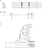
Fig. 1. Subunits and mutations. (A) Alignment of the BID regions from schistosome Sjβ, SmβA, SmβB and rat β2a. The sites corresponding to the conserved serine residues are shaded. (B) SmβA mutants. A portion of the BID from SmβA is shown with mutated residues. (C) Phylogenetic tree of β subunits. Amino acid sequences were aligned using ClustalW (Thompson, Higgins & Gibson, 1994), and a neighbour-joining tree constructed as implemented in Mega v.2.1 (Kumar et al. 2001). Trees derived using other algorithms gave similar results. Sequences (and NCBI accession numbers) are: Human β1 (NM_000723); Rabbit β1 (M25817); Human β2 (NM_000724); Rabbit β2 (X64298); Human β3 (NM_000725); Rabbit β3 (CAA45578); Xenopus laevis (toad) β3 (U33217); Human β4 (NM_000726); Rat β4 (L02315); Caenorhabditis elegans (nematode) βA (AF000198); C. elegans βB (AF043703); Musca domestica (housefly) β (X78561); Drosophila melanogaster (fruitfly) β (U11074); Cyanea capillata (jellyfish) β (U93076); S. japonicum β (AY033597); S. mansoni βA (AY033598); S. mansoni βB (AY033599). The schistosome β subunits are shaded.
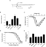
Fig. 2. Modulatory effects of SmβA mutants on Cav2.3. (A) Mutations in the BID alter the modulatory effects of SmβA on Cav2.3. Typical current traces recorded from Xenopus oocytes injected with Cav2.3 RNA alone, with Cav2.3 plus SmβA RNA, or Cav2.3 plus the SmβA C247S, A257S RNA. Wild-type SmβA reduces current amplitude through the Cav2.3 α1 subunit, while the mutated SmβA subunit enhances currents. (B) Mutation to serine of cysteine 247, alanine 257, or both eliminates the reduction of Cav2.3 peak current found with wild-type SmβA. Shown are average peak currents recorded from Xenopus oocytes for Cav2.3 expressed alone [Alone; number of independent experiments (n)=10]; Cav2.3 plus wild-type SmβA (Wild-type; n=9); Cav2.3 plus the SmβA C247S, A257S double mutant (C247S, A257S; n=11); Cav2.3 plus the SmβA C247S mutant (C247S; n=11); Cav2.3 plus the SmβA A257S mutant (A257S; n=10); and Cav2.3 plus the SmβA A257S, K259T double mutant (A257S, K259T; n=9). Asterisks indicate significant differences compared to wild-type SmβA (P<0·01). (C) Wild-type and mutated SmβA shift the current/voltage relationship of Cav2.3 in a hyperpolarizing direction to differing degrees. Current-voltage relationships were recorded in oocytes expressing Cav2.3 alone ([squf ]), Cav2.3 plus wild-type SmβA (□; data from Kohn et al. 2001 a), Cav2.3 plus the SmβA C247S mutant ([utri ]), Cav2.3 plus the SmβA A257S mutant ([dtri ]), Cav2.3 plus the SmβA C247S, A257S mutant (○), and Cav2.3 plus the SmβA A257S, K259T mutant ([loz ]), and normalized to peak current values. Peak current measurements were determined by 250 ms voltage steps from a holding potential of −90 mV. The curves represent fits of the Boltzmann function Inorm=gnorm(V−Vrev)/(1+exp[−(V−V1/2)]/k), where the normalized conductance gnorm=0·021, 0·020, 0·018, 0·022, 0·016, and 0·022; the reversal potential Vrev=69·7, 55·4, 64·0, 62·1, 67·1, and 66 mV; the slope factor k=5·5, 5·2, 5·5, 5·0, 4·1, and 4·0; and the half-activation potential V1/2=+4·6, −9·0, −6·4, +2·0, −9·3, and +6·2 mV for Cav2.3 expressed alone (n=7), or with wild-type (n=8), C247S (n=4), A257S (n=6), C247S, A257S (n=10), or A257S, K259T (n=7) SmβA subunits, respectively. (D) Effects of wild-type and mutant SmβA on the voltage dependence of Cav2.3 steady-state inactivation. Steady-state inactivation in oocytes expressing the different subunit combinations was determined by a 5 s prepulse from a holding potential of −120 mV to the voltages shown, followed by a 50 ms pulse to +20 mV. Symbols are as in (B). Lines represent fits to the Boltzmann function I=Imin+Imax/[1+exp(V−V1/2/k)], where the slope factor k=−8·4, −10·2, −11·3, −11·5, −11·9, and −11·3, and half-inactivation potential V1/2=−23·7, −32·6, −36·8, −34·8, −24·0, and −24·3 mV for Cav2.3 expressed alone (n=5), or with wild-type (n=6), C247S (n=4), A257S (n=4), C247S, A257S (n=6), or A257S, K259T (n=5) SmβA, respectively. (E) SmβA subunits containing serines at residue 247, 257, or both no longer confer PZQ sensitivity to Cav2.3. The ratio of the peak current in the presence and absence of 100 nM PZQ is shown for Cav2.3 expressed with different SmβA mutants (X-axis). If PZQ has no effect, then the ratio will equal one. Results are shown for Cav2.3 alone (Alone; n=5); Cav2.3 plus wild-type SmβA (Wild-type; n=5); Cav2.3 plus the SmβA C247S, A257S mutant (C247S, A257S; n=5); Cav2.3 plus the SmβA C247S mutant (C247S; n=8); Cav2.3 plus the SmβA A257S mutant (A257S; n=6); and Cav2.3 plus the SmβA A257S, K259T mutant (A257S, K259T; n=5). Asterisks indicate significant differences from the wild-type SmβA (P<0·01).
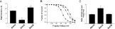
Fig. 3. Modulatory effects and pharmacological sensitivities of SmβB, a schistosome β subunit that contains both consensus PKC sites in the BID. (A) SmβB increases current levels through Cav2.3. Shown are average peak currents recorded from Xenopus oocytes for Cav2.3 expressed alone (Alone; n=6); Cav2.3 expressed with SmβA (SmβA; n=4); and Cav2.3 expressed with SmβB (SmβB; n=5). Asterisks indicate significant differences from Cav2.3 expressed alone (P<0·01). (B) Effects of SmβB on the voltage-dependence of steady-state inactivation of Cav2.3. The experimental protocol is as described in Fig. 2D. Fits of the Boltzmann function described in Fig. 2D produced values of k=−8·4, −10·2, and −8·6, and V1/2=−23·7, −32·6, and −53·2 mV for Cav2.3 expressed alone ([squf ]; n=5; data from Fig. 2D, for comparison), or with SmβA ([utrif ]; n=6; data from Fig. 2D, for comparison) or SmβB ([bull ]; n=6) subunits, respectively. (C) SmβB does not confer PZQ sensitivity to Cav2.3. The ratio of the peak current in the presence and absence of 100 nM PZQ is shown for the different α1/β combinations (X-axis). If PZQ has no effect, the ratio will equal one. Results are shown for Cav2.3 alone (Alone; n=5; data from Fig. 2E, shown for comparison); Cav2.3 plus SmβA (SmβA; n=5; data from Fig. 2E, shown for comparison); and Cav2.3 plus SmβB (SmβB; n=4). Asterisk indicates that the ratio differs significantly from that found with Cav2.3 alone (P<0·01).
 You have
Access
- 48
- Cited by