Introduction
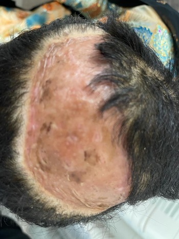
A young patient with Down’s syndrome, with dermatofibrosarcoma protuberans (DFSP), was intended for adjuvant radiotherapy. The lesion area was on the parietal region of the patient’s head (Figure 1). The standard for radiotherapy for DFSP with positive margins is 50–66 Gy given in 25–33 fractions through external beam radiation therapy (EBRT). Reference Williams, Morris, Kirwan, Dagan and Mendenhall1 However, there are several potential complications with treating this patient with EBRT.
The location of the lesion on the parietal region of the patient head where the skin was replaced with a flap. Given the proximity to brain tissue and curvature of the lesion mould, brachytherapy was chosen to treat this case.

Patients with Down’s syndrome typically require treatment under anaesthesia. Complications of anaesthesia for patients with Down’s syndrome are well documented. Reference Kobel, Creighton and Steward2 Patients with Down’s syndrome usually have a larger tongue and smaller subglottic area, which requires a smaller tube for intubation. Literature also suggests that patients with Down’s syndrome are more prone to airway infections. Reference Kobel, Creighton and Steward2 A major risk in intubating a patient with Down’s syndrome is atlanto-occipital dislocation. Reference Kobel, Creighton and Steward2 Another potential complication is bradycardia, where the heart rate drops during the induction of anaesthesia or sedation. Reference Borland, Colligan and Brandom3 Since anaesthesia-induced ventilator dysfunction can occur, close observation is necessary until full recovery from anaesthesia.
Considering the young age of the patient, it was necessary to reduce radiation dose to the brain. Only brachytherapy and electron beam could deliver the high-dose gradient necessary to minimise the dose outside the lesion. The curvature and location of the lesion would make electron beam dose distribution non-uniform.
Brachytherapy was chosen as the treatment for this patient as it offers several advantages: rapid dose fall-off, fewer number of fractions and a mould applicator can be made around an irregular/curved surface. There have been many cases where brachytherapy was administered to treat DFSP in patients with an irregular anatomy. Reference Jamora, Cereno, Inocencio and Hizon4,Reference Bandyopadhyay, Basu, Biswas and Ghosh5
In this case report, we present a custom surface applicator that was prepared on a 3D-printed model of the anatomy of a patient with Down’s syndrome in order to reduce the potential risks from anaesthesia. Though 3D printing is a popular technology often used in various medical fields, 3D printing in radiotherapy is considered as a novel modality which has various applications (boluses, phantoms for quality assurance, compensator blocks, proton range compensators and brachytherapy custom applicators). Reference Bandyopadhyay, Basu, Biswas and Ghosh5–Reference Arenas, Sabater and Sintas10 In brachytherapy, 3D printing is used to create custom applicators. Reference Arenas, Sabater and Sintas10,Reference Casey, Awotwi-Pratt and Bahl11 However, a major uncertainty with custom moulding is the difference in attenuation of the 3D-printed material in comparison to water, which is the medium for brachytherapy dose calculation algorithm TG-43. Reference Nath, Anderson, Luxton, Weaver, Williamson and Meigooni12 TG-43 calculates radiation dose in an infinite medium of water without accounting for true geometry. Any presence of air gaps and denser materials are ignored. There is little literature available validating TG-43 for 3D-printed applicators. One material has been shown to be water equivalent. Reference Bassi, Langan and Malone13 It is recommended to verify TG-43 when using a custom-printed applicator, or alternatively use TG186. Reference Fowler, Buyyounouski, Jenkins and Fahimian14
Method
Treatment setup
For surface brachytherapy, a surface mould applicator is prepared on the patient. The applicator is designed by embedding catheter tubes on dental wax placed on a thermoplastic mask. The expected time to prepare such an applicator neatly on a cooperative patient would be around three quarters to one hour. Since the patient in this study was uncooperative, a computed tomography (CT) scan of the patient was performed under sedation. Since full general anaesthesia was considered risky, the patient was sedated intravenously using propofol and ketamine and their airway was maintained with oropharyngeal airway. The boundaries of the lesion were marked with wires by the radiation oncologist before the CT scan.
3D Printing
The lesion and the associated clinical target volume (CTV) were delineated by the radiation oncologist. The body contour of the patient was generated automatically and was verified. 3D printing the whole head of the patient would require a large amount of 3D-printing material. Instead, the surface of the head was printed with a 5 mm thickness. The surface extended from the frontal region through to the occipital region, covering the temporal region as well. This provided enough area to prepare the thermoplastic mask for custom applicator. Along with this, the CTV was fused on the concave side of the contour at an additional depth of 5 mm. This visualised the CTV when the catheters and wax were placed on the 3D-printed model. Once the contour was ready, it was exported as a dicom file from ECLIPSE planning station.
3D-printing software reads the Standard Triangle Language (STL) format. The Dicom-to-STL conversion was performed using 3D Slicer. 3D Slicer is a platform-independent open source application for medical image computing. Reference Fedorov, Beichel and Kalpathy-Cramer15 It is capable of dicom visualisation, image fusion, image segmentation and various other tasks. Reference Fedorov, Beichel and Kalpathy-Cramer15 The segmentation module from 3D Slicer allows dicom structures to be converted to other formats such as STL, NIFTI, OBJ and NRRD. Please note that 3D Slicer is not an FDA approved product, and the user is responsible for testing 3D Slicer before clinical use. Reference Fedorov, Beichel and Kalpathy-Cramer15
The 3D printer used was the ProJet MJP 3600 Max from 3D Systems. The ProJet MJP 3600 Max is a MultiJet printer with UV light for resin material curing. The printer is interfaced by 3D Systems’ 3D Sprint Software. 3D Sprint allows one to check for any inconsistencies in design, to split large designs and to align designs in order to reduce print time.
The time for printing is proportional to the height of the design, hence the head design was split into three parts with male–female connectors to save time. The print time for this design was 22 h in ultra-high definition (18 um thickness layer). The MJP 3600 uses the VisiJet M3 crystal as resin material and wax as support material.
The cured M3 crystal was CT scanned to analyse the CT number, which was found to be 300 HU. The material was too rigid to 3D print a surface applicator, and the attenuation properties are unknown. Once printing was completed, the support material was removed from the 3D print by post-processing.
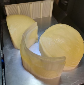
There are many possible methods for post-processing. We used vegetable oil to melt away the wax, washed the 3D-print with soap and water and dried it. After post-processing, the three parts were assembled by male–female plugs and fixed with super glue (Figure 2).
The figure above shows a 3D-printed model of the patient’s head, divided into three parts. These must be post-processed to remove support material (wax) by dissolving it in hot vegetable oil. After post-processing, they can be assembled by auto-generated male–female plugs and super glue.

Brachytherapy mould applicator
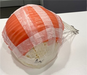
A thermoplastic mask was prepared on the 3D-printed model (Figure 3). Since the 3D-printed model incorporated the CTV, catheter placement was very convenient. The area above the CTV was cut out from the thermoplastic mask and replaced by dental wax sheets of 2 mm to provide material for attenuation. Eleven applicator mould probes (catheters) from a Varian mould applicator set, 10 mm apart from each other, were sandwiched between wax sheets of 2 mm. This sandwiched catheter set was placed above the dental wax on the thermoplastic mask. TG-43 can overestimate dose to surface up to 15%; however, adding a few mm of bolus material reduces the overestimation to 3%. Reference Boman, Satherley, Schleich, Paterson, Greig and Louwe16 Metal markers were placed at the distal end of the mould probe for easy channel reconstruction. Each probe was marked from 1 to 11 from the near end. The length of each probe was 32 cm, and the length of a whole channel is 132 cm.
Varian mould applicator that was prepared on the 3D-printed model. As seen from the image, 11 probes were placed equidistant at 1cm.

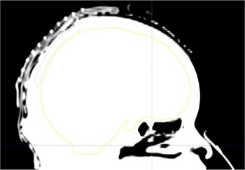
The prepared mould applicator was tested on the patient. Upon satisfactory fitting, a second CT scan was performed on the patient under sedation. The second scan of the patient was performed for brachytherapy planning. Ultrasound gel with cotton gauze was placed inside the applicator to avoid any air gaps (Figure 4). Presence of air gaps can cause over estimation of dose by TG-43 at the surface Reference Boman, Satherley, Schleich, Paterson, Greig and Louwe16 ; however, there is no clinically significant difference at 1 cm depth due to small air cavities. Reference Granero, Perez-Calatayud, Vijande, Ballester and Rivard17
The fitting of the mould applicator on the patient’s head is shown. Air gaps in the scan are minimum. The largest observed was 3 mm. The fitting of the mould applicator on the patient was satisfactory.

Brachytherapy planning
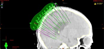
Prescription for brachytherapy was 32·5 Gy in 5 fractions, delivering 6·5 Gy in each fraction. This would be an equivalent dose (EQD2) of 44·69 Gy. Each channel was reconstructed manually from the metal marker on the distal end in the CT scan. TG-43 volumetric optimisation was done on ECLIPSE planning station to deliver 95% of the prescribed dose to 95% of the target volume and not exceeding 120% of the prescribed dose to the patient skin (Figure 5). The CTV was 5 mm into the patient skin, 7·5 cm long and 10 cm wide. Volume of the CTV was 32·34 cm3. In the final plan, 2970 cGy covered 95% of CTV volume and maximum dose in the CTV was kept below 120%. Treatment time on the initial day was 16 min with an Ir-192 source of 7·474 Ci strength. A QA plan was generated to check for clearance for all channels prior to treatment delivery on each day.
Image shows the isodose for 90% of the prescription dose. The coverage for 90% isodose line was 95% of the CTV volume.

Discussion
Around 1 in 800 babies are born with Down’s syndrome. Reference Kobel, Creighton and Steward2 The risks associated with using general anaesthesia on patients with Down’s syndrome are well documented. Reference Kobel, Creighton and Steward2,Reference Borland, Colligan and Brandom3 Patients with Down’s syndrome are prone to respiratory infection, airway blockage and atlanto-occipital dislocation. Reference Kobel, Creighton and Steward2,Reference Borland, Colligan and Brandom3 In this case report, we were able to avoid using general anaesthesia by using brachytherapy and 3D printing.
General anaesthesia would be administered to a non-cooperative patient during mould applicator preparation and radiotherapy simulation. The applicator preparation would normally take about 45 min. Since general anaesthesia is risky for patients with Down’s syndrome, only a limited amount of time can be spent under sedation (∼20 min).
Instead, we used the patient body contour from a normal CT scan to create a 3D-printed model, on which the applicator preparation was performed. This allowed unrushed and neat placement of the catheter probes. Having the CTV on the 3D-printed model enabled better placement of catheters, placed evenly and well beyond the CTV region to provide excellent coverage. A lot of pressure could be applied to the thermoplastic mask and the dental wax, relative to what could have been applied directly to the patient.
The fitting of the mould applicator on the patient prior to the treatment planning CT scan was satisfactory. Ultrasound gel was used to minimise air gaps. Brachytherapy-enabled delivery of a similar equivalent dose as EBRT (44·69 Gy EQD2) with a lower dose to the brain (1cc less than 80% of the prescription dose) in fewer fractions. The patient was adapting to the sedation and the dosage of sedation needed to be increased each treatment day. On the third treatment session, the patient woke up during the treatment. If an external beam treatment of 20–25 fractions was intended, the dosage of the sedation would have been very high by the end of the treatment.
A drawback of 3D printing is the amount of time that it takes to print a model. The time taken for printing this patient head model was about 22 h. Additional time was needed to prepare the anatomy to be printed and post-processing the 3D-printed model.
Conclusion
There are two rare conditions in this case: DFSP and Down’s syndrome. Since there are several complications with general anaesthesia for a patient with Down’s syndrome, it is best to avoid general anaesthesia and provide sedation with minimal dosage and minimal sessions. Brachytherapy is an excellent option to reduce the number of fractions and hence the number of anaesthesia sessions. We used a 3D-printed model to prepare the mould applicator, significantly reducing the sedation during radiotherapy simulation. 3D printing a patient’s anatomy is an excellent option to prepare mould applicators for surface brachytherapy. It can greatly reduce potential risks from anaesthesia, improve patient comfort and allow relaxed environment for the preparation of the applicator.
Acknowledgement
We would like to thank Dr. Salim Chaib Rassou, radiation oncology consultant, for this patient. We would like to thank Dr. Rajini Kausalya, anaesthesia consultant, for this case and also guiding us manuscript preparation for anaesthesia complications for this patient.
Declarations
Competing interest
The authors have no identified conflicts of interest.
Ethical approval
The study of this paper has been performed using 3D printing. The patient was subject to procedures that are part of brachytherapy with anaesthesia. No extra involvement from the patient was required.
Consent to Publish
Consent to use the images and data of the patient has been obtained from the patient.