As long ago as the early 1980s, Capelli et alReference Capelli, Andrade and Somerville 1 commented “it is not surprising that the nomenclature of [ventricular septal defects] has been debated, confused, defined didactically, and re-defined and has not resulted in a satisfactory simple classification acceptable to all the different disciplines involved in the management of patients or the heart with this common anomaly”. Little has changed since then. The current 10th iteration of the International Classification of Disease has long been recognised as inadequate for satisfactory coding of congenital cardiac malformations. Fortunately, the codes for the various lesions to be included in the 11th iteration will be determined by the International Society for Nomenclature of Paediatric and Congenital Heart Disease. However, Capelli et al would probably not be surprised that previous meetings of this committee, seeking to reach consensus on the best definitions for coding of congenital cardiac malformations, have not yet been conclusive. This is especially true for the anatomic substrates, or holes, providing the potential for interventricular shunting. These defects are usually described as “ventricular septal defects” or equivalent in Germanic languages, and as “interventricular communications” by those using Romance languages.Reference Anderson, Spicer, Giroud and Mohun 2 The two terms, however, differ significantly in their meaning, as an interventricular communication is not necessarily caused by a defect in the ventricular septum,Reference Anderson, Spicer, Giroud and Mohun 2 with the Romance languages being more pure in their description. Complicating the debate is the evolution of several systems of nomenclature that continues to create problems in communication, even between those using the same language. As of yet, there is no perfect correlation between the systems, despite the efforts of “cross-mapping” of the different approaches to codification. If the 11th iteration of the International Classification is to be accepted, and used widely by those diagnosing and treating patients with congenitally malformed hearts, it is essential that the definitions used are based on universally accepted, and anatomically precise, principles. This is particularly the case for the holes that permit interventricular shunting, as they are the most common congenital cardiac malformations.
In hopes of charting a potential pathway towards consensus, we review here the evolution and history of the various systems of nomenclature, highlighting some of the consistencies and inconsistencies that, in our opinion, remain challenging. We will present images demonstrating the embryological development of the ventricular septum, in this way providing a structure by which the normal septum can be described, and defects accurately categorised. It is our belief that understanding the reasoning behind the various current systems of nomenclature in the context of the knowledge provided by embryological development will engender productive discussions, hopefully permitting a final resolution to the naming of the channels that permit interventricular shunting.
How, and why, did the different classifications evolve?
In the early years, those diagnosing the defects that permitted shunting between the ventricles would most frequently describe them as the “maladie du Roger”, named after Henry-Louis Roger, who was born in 1809, and died in 1891. He described the hearts with “openings in the interventricular septum”. His approach was physiological rather than morphologic, being based on the small nature of the defects he observed. It was in the late 1800s that RokitanskyReference Von Rokitansky 3 first provided a morphologic approach to the ventricular septum, dividing it into a posterior muscular septum, a membranous portion, and an anterior muscular septum. He divided the latter muscular component itself into front and hind portions. Despite his appreciation of the anatomy of the ventricular septum, Rokitansky did not describe in his extensive analysisReference Von Rokitansky 3 defects of the “pars membranacea”, which PeacockReference Peacock 4 had illustrated years ago as the cause of almost all defects found in the upper ventricular septum.
In the 1930s, Abbott, in her groundbreaking classification, introduced the concept of descriptions of defects based on their borders.Reference Abbott 5 She distinguished between defects, described by her, to the open anterior to the membranous part of the septum, and into the side of the right ventricle behind the septal leaflet of the tricuspid valve, and those “less common” defects lying more anteriorly, and opening into the outlet of the right ventricle, which she called bulbar defects. Taussig,Reference Taussig 6 in the 1940s, chose a simple approach, and merely differentiated between “high” and “low” defects. WoodReference Wood 7 criticised this simplified system on the basis that it failed to provide a manner of describing what he, in his experience, considered to be the most common defects, namely those “located in the anterior part of the membranous septum”. Coincident with the appearance of Wood’s second edition of Diseases of the Heart and Circulation,Reference Wood 7 the investigation of Becu et alReference Becu, Fontana, Dushane, Kirklin, Burchell and Edwards 8 in 1956 at the Mayo Clinic provided the first detailed anatomical categorisation of channels permitting interventricular shunting. Their categorisation was based on the division of the ventricular septum into the outlet, inlet, and muscular regions, with defects categorised as “related to the right ventricular outflow tract” or “not related to the right ventricular outflow tract”. Defects related to the outflow tracts were further subdivided into those that were located posterior to the supraventricular crest, then called the crista supraventricularis, and therefore related to the membranous septum, or those located anterior to the crest. The investigators further commented that “most of the defects [were] related to the membranous septum”, and that the rest of the defects were anterior to the crest and therefore adjacent to the leaflets of the pulmonary valve, or else not related to the right ventricular outflow tract, and therefore encased within the muscular septum. Becu et alReference Becu, Fontana, Dushane, Kirklin, Burchell and Edwards 8 also emphasised that the lesions adjacent to the membranous septum, and bordering upon it, existed not because the membranous septum itself was perforate, although it could be, but rather because of deficiencies of the muscular septum surrounding the persisting membranous component. This crucial feature of a defect related to the membranous septum was then reinforced by Sherman,Reference Sherman 9 who analysed the specimens stored in the museum of Pittsburgh Children’s Hospital. Like Becu et al,Reference Becu, Fontana, Dushane, Kirklin, Burchell and Edwards 8 Sherman emphasised that, in those defects abutting on the membranous septum, “the size of the defect is largely determined by the degree of myocardial deficiency”.Reference Sherman 9 Sherman further noted that the defects differed in position “according to the direction that they extend from the membranous septum at the expense of adjacent myocardium”.Reference Sherman 9 It is this observation, supported by Becu et al,Reference Becu, Fontana, Dushane, Kirklin, Burchell and Edwards 8 that underscores the justification for defining defects based on their relations, and their extension from the membranous septum.
It was the description by Becu et alReference Becu, Fontana, Dushane, Kirklin, Burchell and Edwards 8 of the channels relative to the supraventricular crest, moreover, that likely promoted the subsequently popular distinction of defects as being “infracristal” and “supracristal”, and then to some as being “intracristal”, as suggested by Rosenquist et al.Reference Rosenquist, Sweeney, Stemple, Christianson and Rowe 10 The feature of the “intracristal” channels was the exclusive nature of their muscular borders, but the term has not been widely accepted. Therefore, at this stage of play, the substrates for interventricular shunting were generally distinguished as to whether they were infracristal and membranous, supracristal, or muscular.
We presume it was the differences noted by the Mayo groupReference Becu, Fontana, Dushane, Kirklin, Burchell and Edwards 8 that provided the basis for the subsequent numerical system of categorisation.Reference Wells and Lindesmith 11 In this system, defects adjacent to the membranous septum were assigned to a Type 2 category, those encased within the muscular septum to Type 4, and those adjacent to the leaflets of the pulmonary valve to Type 1. Within the system, however, there was also a Type 3, which was an inlet, or “atrioventricular canal” type of defect.Reference Wells and Lindesmith 11 Sherman,Reference Sherman 9 much earlier, had identified these lesions as a subset of the membranous variant, stating that “the criterion for this subclassification is fusion of the anterior mitral leaflet with the septal leaflet of the tricuspid valve through the defect so that the posterior wall of the defect is bordered by the smooth continuous undersurface of these two leaflets”. This is an excellent description of the defects that border the membranous septum, yet extend inferiorly towards the crux of the heart (Fig 2). It is unclear to us as to why this particular variant should then comprise a discrete entity, as it can appropriately be assigned to the Type 2 category. It is the separate designation of Type 3 defects, or defects of “atrioventricular canal type”, which continues to create one of the greatest obstacles in achieving consensus in classification.
Subsequent problems in description
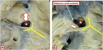
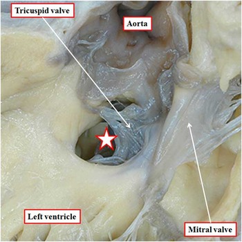
An early essay in this field by one of the current authorsReference Soto, Becker, Moulaert, Lie and Anderson 12 introduced a term that has subsequently remained highly contentious, namely, the adjective perimembranous. In this essay, Soto et al,Reference Soto, Becker, Moulaert, Lie and Anderson 12 like Becu et al,Reference Becu, Fontana, Dushane, Kirklin, Burchell and Edwards 8 and Sherman,Reference Sherman 9 noted that the primary feature of defects adjacent to the membranous septum was the degree of deficiency in the muscular edges of the hole.Reference Soto, Becker, Moulaert, Lie and Anderson 12 Therefore, they proposed that these types of defects could be called perimembranous, emphasising as their diagnostic feature the presence of fibrous continuity between the leaflets of the tricuspid and aortic valves in the roof of the channels (Fig 2). Soto et alReference Soto, Becker, Moulaert, Lie and Anderson 12 further suggested that these defects, grouped together because of aortic-to-tricuspid continuity, could differ depending on whether they excavated the inlet, outlet, or trabecular components of the muscular ventricular septum. Although Soto et alReference Soto, Becker, Moulaert, Lie and Anderson 12 supported the approach of the Mayo groupReference Becu, Fontana, Dushane, Kirklin, Burchell and Edwards 8 by classifying all defects into one of three groups, they differed when they chose not to define defects based on their relationship to the supraventricular crest, or crista supraventricularis. Instead, they defined defects based on distinctive phenotypic features. The first was the presence of aortic-to-tricuspid valvar continuity, which was their chosen criterion for the perimembranous lesions (Figs 1a, 2). The second was the presence of exclusively muscular borders, and the third was the finding of fibrous continuity between the leaflets of the pulmonary and aortic valves, the latter feature making some channels directly subarterial (Fig 1b).
The illustrations show the types of defects that Becu et alReference Wood 7 from the Mayo Clinic categorised as being “infracristal” ( a ) or “supracristal ( b ). They failed to note, however, an important inconsistency in this logic for distinction. The hole shown in ( a ) (star) opens to the right ventricle between the limbs of the septomarginal trabeculation, or septal band, the muscular strap that reinforces the septal surface of the morphologically right ventricle (yellow Y). The structure defined as the “crista” (double headed arrow) is the muscular outflow septum, supporting the septal aspect of the subpulmonary infundibulum. In the hole considered to be “supracristal” [star in ( b )], the subpulmonary infundibulum has failed to form, and the outlet septum is no more than a fibrous ridge between the arterial valves. The hole remains cradled within the limbs of the septomarginal trabeculation, or septal band, with muscular continuity postero-caudally between the trabeculation and the inner heart curvature (white arrow). It is the structure that has been defined as the “crista” that has changed in this system, rather than the location of the hole relative to the apical muscular septum.

The image shows the defect (star) considered by ShermanReference Becu, Fontana, Dushane, Kirklin, Burchell and Edwards 8 to represent an “inlet” defect, and forming one of the lesions grouped as “Type 3” of the numerical classification.Reference Wells and Lindesmith 11 It is viewed from the left ventricle. Its postero-caudal border is formed by an extensive area of fibrous continuity between the leaflets of the tricuspid and mitral valves. Note also the tricuspid–aortic valvar continuity.

In 1983, Capelli et alReference Capelli, Andrade and Somerville 1 offered a different approach, preferring to describe the location of the channels permitting ventricular shunting according to their proximity to the cardiac valves. This approach had already been introduced by Lev et alReference Lev, Bharati, Meng, Liberthson, Paul and Idriss 13 for the hearts with double outlet from the right ventricle. Perhaps because the classification suggested by Capelli et alReference Anderson, Spicer, Giroud and Mohun 2 included eight categories of defects, with four of the types being uncommon, it lacked practicality and was not widely accepted.
By the late 1980s, therefore, two different approaches were emerging for classification of the defects. The classification proposed by Soto and his European colleaguesReference Soto, Becker, Moulaert, Lie and Anderson 12 had been modified to make it applicable for surgeons. It continued to emphasise phenotypic variability on the basis of the “border-forming structures around the defect because in closing the defect, sutures must be placed around it”.Reference Soto, Ceballo and Kirklin 14 In the modification proposed for surgical use, Soto and his colleagues from Birmingham, Alabama,Reference Soto, Ceballo and Kirklin 14 combined the previously proposed definitions based on borders with geographical terms. They willingly used the term perimembranous as a synonym for the defect considered by others to be conoventricular when it was also juxtatricuspid and juxtaaortic.
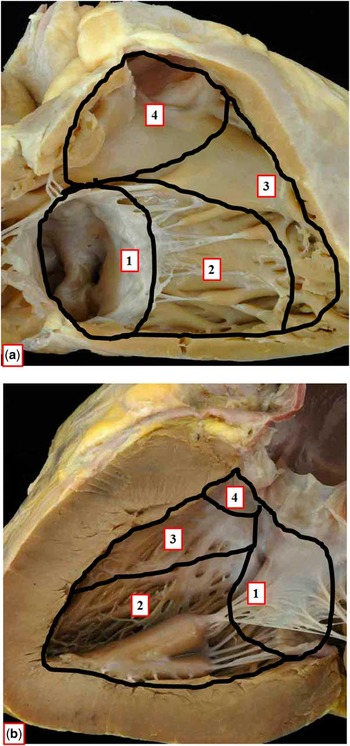
The alternative popular approach, summarised by Van Praagh et alReference Van Praagh, Geva and Kreutzer 15 in a commentary on the proposed surgical classification,Reference Soto, Ceballo and Kirklin 14 segregated the substrates for interventricular shunting solely on the basis of their location relative to their perceived components of the ventricular septum. Although based on a developmental concept, they acknowledged that “from the embryologic standpoint, there [were] many more components” to the ventricular septum than their selected four components.Reference Van Praagh, Geva and Kreutzer 15 Van Praagh et alReference Van Praagh, Geva and Kreutzer 15 applied this abridged developmental approach to define morphological components of the normal ventricular septum (Fig 3). They then distinguished defects based on their geographical location within these components, categorising the channels as being conoventricular, conoseptal, atrioventricular canal type, and muscular.Reference Van Praagh, Geva and Kreutzer 15
The photographs of the normal heart have been marked to show the suggested separation of the muscular ventricular septum into four components as proposed by Van Praagh et al.Reference Van Praagh, Geva and Kreutzer 15 The panels show the septum as viewed from the aspects of the right ( a ) and left ( b ) ventricles. The component designated as #1 was considered to be the septum of the atrioventricular canal. Component #2 was the muscular ventricular septum, or sinus septum. Component #3 was the septal band, or proximal conal septum, whereas component #4 was the parietal band, or distal conal septum.

The differences in approaches between these two classification systems have resulted in controversial and ongoing debates regarding both the widely accepted perimembranous defects and inlet defects. Van Praagh et alReference Van Praagh, Geva and Kreutzer 15 criticised nomenclature utilising the concept of “perimembranous” defects, arguing that only when defects were small, and limited to the area of the membranous septum, was such a description justified. Despite these suggested caveats, the term remains widely utilised internationally, partly owing to its historical description dating to the findings of Becu et al,Reference Becu, Fontana, Dushane, Kirklin, Burchell and Edwards 8 Sherman,Reference Sherman 9 and even the earliest description by Peacock.Reference Peacock 4 More recently, Soto et alReference Soto, Ceballo and Kirklin 14 commented that the “perimembranous” descriptor was “useful and well understood”, as it emphasised the surgically relevant exposure of the penetrating atrioventricular bundle in conoventricular defects adjacent to the membranous septum.
With regard to the “inlet”, or “AV canal-type defect”, Soto et al,Reference Soto, Ceballo and Kirklin 14 in contrast with Van Praagh et al,Reference Van Praagh, Geva and Kreutzer 15 specifically avoided using the term “AV canal type”. They emphasised the difference in the nature of the borders of the “inlet defect” when compared with the “AV canal-type defect”, despite their similar geographical locations. Specifically, Soto et alReference Soto, Ceballo and Kirklin 14 argued that the term “AV canal type” to describe a defect that was juxtatricuspid and perimembranous could confusingly imply the presence of a defect with common atrioventricular junction, but which permitted only ventricular shunting. Soto and his surgical colleaguesReference Soto, Ceballo and Kirklin 14 commented that this is an example of why defining the borders of a defect, rather than relying solely on its geographical location in the ventricular septum, is clinically relevant.
Our review of the evolution of the various methodologies of describing holes between the ventricles suggests that ongoing discrepancies in defining normal ventricular septal anatomy, which lack substantiation on the basis of precise knowledge of its developmental heritage, continue to create most of the impediments now preventing the achievement of consensus in the description of holes between the ventricles.
What, then, is or is not the structure of the normal ventricular septum?
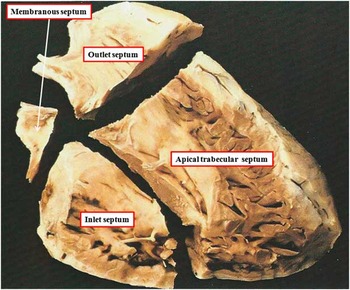
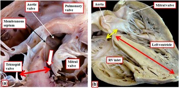
When Soto and his European colleaguesReference Soto, Becker, Moulaert, Lie and Anderson 12 produced their initial concept for phenotypic distinction of the defects that permit interventricular shunting, they suggested that the differences could be attributed to deficiencies of either the inlet or outlet components of the muscular ventricular septum. To show these features, they created arbitrary lines radiating from the membranous septum across the right ventricular aspect of the septum (Fig 4). This approach permitted differentiation of the defects in the membranous region according to the way in which they opened to the right ventricle, a characteristic that had been recognised from the outset of the emergence of systems of classification.Reference Becu, Fontana, Dushane, Kirklin, Burchell and Edwards 8 , Reference Sherman 9 We now recognise, however, that this approach (Fig 4), which focuses on the septum as seen from the right ventricle, as with the concept proposed by Van Praagh et al (Fig 3),Reference Van Praagh, Geva and Kreutzer 15 oversimplifies its complex geometry. This is partly because the subaortic outlet component of the normal left ventricle is deeply wedged between its inlet component, guarded by the mitral valve, and the septum. Thus, the muscular ventricular septum separates the inlet of the right ventricle, guarded by the tricuspid valve, from the outlet, rather than the inlet, of the left ventricle (Fig 5a). Therefore, it is incorrect to consider the infero-posterior part of the muscular ventricular septum as representing an “inlet septum”. There is no muscular septal entity in the normal heart that interposes between the ventricular inlets. The greater part of the muscular septum interposes between the apical ventricular components, with the fibrous or membranous septum being relatively insignificant when the ventricles are normally septated (Fig 5b).
The dissection of the normal ventricular mass, seen from the right side, reveals the concept initially used by Soto and his European colleaguesReference Soto, Becker, Moulaert, Lie and Anderson 12 when they sought to distinguish the components of the normal ventricular septum. They argued that by taking cuts as shown through the muscular septum, having separated it from the membranous septum, it could be divided into inlet, outlet, and apical trabecular components. We now recognise that the relationships of the components of the two normal ventricles are too complex to substantiate this simplistic division of the septum (see Figs 5 and 6). By the same token, our findings also cast doubt on the division suggested by Van Praagh et alReference Van Praagh, Geva and Kreutzer 15 (Fig 3).

Cuts taken across the normal ventricular septum demonstrate the deficiencies in the concept initially espoused by Soto and his European colleagues,Reference Soto, Becker, Moulaert, Lie and Anderson 12 and similarly the approach taken by Van Praagh et al.Reference Van Praagh, Geva and Kreutzer 15 The cut shown in ( a ), taken in short axis, shows how the right ventricular inlet, guarded by the tricuspid valve, is separated by the muscular ventricular septum (double-headed red arrow) from the inferior extension of the subaortic outlet of the left ventricle (white arrow). ( b ) Cutting the normal ventricular mass in its long axis so as to simulate the five-chamber echocardiographic section that incorporates the aortic root. It shows again how the muscular septum (red double-headed arrow) separates, at the base, the inlet of the right from the outlet of the left ventricle. The membranous septum is insignificant in terms of size. In this section, it interposes between the outlet of the left ventricle and the cavity of the right atrium (yellow double-headed arrow).

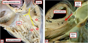
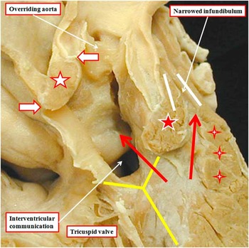
Problems also exist with regard to the “outlet septum” as described initially by Soto et al.Reference Soto, Becker, Moulaert, Lie and Anderson 12 It is the differentiation of the anatomical components of the right ventricular outflow tract that is key to the understanding of the make-up of the supraventricular crest, or “crista supraventricularis”.Reference Anderson, Becker and Van Mierop 16 As we have discussed, it was this structure that was used by Becu et alReference Becu, Fontana, Dushane, Kirklin, Burchell and Edwards 8 when they distinguished between the so-called “Type 1” and “Type 4” defects. In the normally septated heart, the crest is made up largely of the inner heart curvature, or ventriculo-infundibular fold (Fig 6a). This fold continues cranially as the free-standing muscular subpulmonary infundibulum (Fig 6b).Reference Anderson, Spicer, Giroud and Mohun 2 Only a small part of the crest, at the point where it inserts into the septal surface of the right ventricle, can be removed to create a channel into the left ventricular outlet.Reference Anderson, Spicer, Giroud and Mohun 2 As can be seen in Figure 6a, there is no anatomic boundary in the normal heart to show where the small part of the crest interposed between the ventricular outlets becomes continuous with the free-standing inner curve and the infundibular sleeve. As is also shown in Figure 6, the crest itself inserts to the septal surface of the right ventricle between the limbs of an obvious strap-like muscular entity. This prominent right ventricular landmark is a reinforcement of the apical muscular ventricular septum. Described as the septomarginal trabeculation, or the septal band, it was often considered to be part of the “crista”. It does not, however, occupy a “supraventricular position”. These building blocks of the outlet of the right ventricle can better be identified as discrete entities in the setting of deficient ventricular septation (Fig 7).Reference Capuani, Uemura, Ho and Anderson 17 , Reference Hosseinpour, Jones, Barron, Brawn and Anderson 18 The ventriculo-infundibular fold can then be identified as the structure interposed between the hinges of the atrioventricular and arterial valves. The true outlet septum that separates the ventricular outflow tracts,Reference Anderson, Becker and Van Mierop 16 in contrast, is discrete from the infundibular muscular sleeve, which itself can be deficient without transgressing on the left ventricular cavity (Fig 6b). Finally, complexities exist with the septoparietal trabeculations, which can be distinguished from the septomarginal trabeculations, and are muscular bundles that arise from the anterior aspect of the septal band (Fig 6a). The septoparietal trabeculations, including the moderator band, have also been shown to exhibit marked individual variation in appearance, differing in both number and prominence in normal hearts.
The view of the right ventricle in ( a ) is obtained by removing its parietal wall. It shows how it is accurate to describe inlet, apical, and outlet ventricular components. Note how the supraventricular crest, or crista supraventricularis, inserts on the septal surface between the limbs of the septomarginal trabeculation, or septal band (yellow Y). However, it is impossible to distinguish different parts of the crest itself. Also note the multiple septoparietal trabeculations (stars) that arise from the anterior aspect of the septomarginal trabeculation. The cut made in ( b ), replicating the parasternal long-axis echocardiographic section, shows how the larger part of the outlet of the right ventricle is made up of the free-standing subpulmonary muscular infundibulum, with an extracardiac tissue plane (red arrow) interposed between the infundibulum and the aortic root.

The image is from a heart obtained from a patient with tetralogy of Fallot, but with the muscular tissues of the inner heart curve interposed between the leaflets of the tricuspid and aortic valves, and also between the leaflets of the aortic and mitral valves. Because of the deficient ventricular septation, the components of the right ventricular outflow tract can be recognised in their own right. The muscular outlet septum (red star with white borders) is exclusively a right ventricular structure, and is recognised because it is interposed between the ventricular outflow tracts (long red arrows). Note the free-standing subpulmonary muscular infundibular sleeve (parallel white lines). The ventriculo-infundibular fold (white star with red borders) is defined because it is interposed between the leaflets of the atrioventricular and arterial valves (white arrows with red borders). The septomarginal trabeculation, or septal band (yellow Y) is the strap-like configuration that reinforces the septal surface of the right ventricle.

All of these findings regarding the differences between the right ventricular structures in the heart with normal as opposed to deficient septation point to the difficulties in relying solely on geographical terms when seeking to define the subtrates for interventricular shunting. But can additional evidence be adduced if, as suggested by Van Praagh et al,Reference Van Praagh, Geva and Kreutzer 15 we take note of the mechanisms of development?
How does the ventricular septum develop?
It has long been argued that knowledge of cardiac development would facilitate the understanding and the categorisation of the congenitally malformed heart.Reference Abbott 19 Until recently, however, the evidence relating to cardiac embryology has not always been sufficiently robust to permit accurate correlations. All this has now changed with the development of high-resolution episcopic microscopy.Reference Mohun and Weninger 20 This technique has now permitted us to examine large numbers of developing mouse hearts. The ability to cut the hearts in any desired plane, combined with accurate three-dimensional reconstructions, show in exquisite detail the morphologic changes that occur during cardiac development. Such analysis now provides evidence on the formation of the ventricular septum that permits inferences to be made to contribute to the optimal categorisation of the anatomic substrates for interventricular shunting.
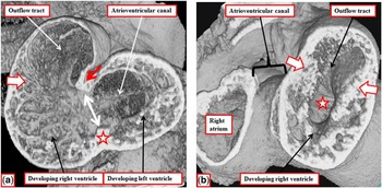
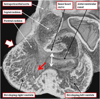
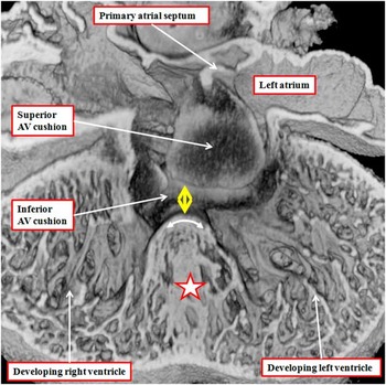
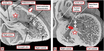
The right and left ventricles develop by the process of ballooning of their apical components from the inlet and outlet parts of the ventricular loop.Reference Moorman and Christoffels 21 In the earliest stages of ballooning, the atrioventricular canal is supported exclusively by the developing left ventricle (Fig 8a), whereas the outflow tract arises exclusively from the developing right ventricle. The right wall of the atrioventricular canal, nonetheless, is in direct continuity with the parietal wall of the ballooning right ventricle from the outset of development (Fig 8b). Rightward expansion of the atrioventricular canal (Fig 9) brings the cavity of the right atrium into direct communication with that of the right ventricle, thus providing the inlet part of the right ventricle, although initially this is very small relative to the extensive apical right ventricular component. The trabeculations in the developing right ventricle extend cranially to the origins of the outlet cushions. The outlet cushions are extensive collections of endothelial cells that proliferate, and eventually join together, so as to divide the outflow tract into the aortic and pulmonary channels. When first seen, the two outflow cushions are separate but continuous entities that spiral as they extend towards the aortic sac.Reference Kramer 22 At their proximal origins within the right ventricle, they are located in septal and parietal positions.Reference Anderson, Chaudhry and Mohun 23 When the aortic component of the outflow tract is produced by eventual fusion of the cushions, it is located dorsally and rightward at its proximal origin. At the end of the 11th day after conception (embryonic day 11.5 or E11.5), it still arises from the developing right ventricle (Fig 9). As the entirety of the outflow tract continues to arise from the developing right ventricle, the blood entering the left ventricle must traverse the embryonic interventricular communication so as to reach the developing aortic outflow tract. The communication, at this stage, has the inner heart curvature as its roof, and the crest of the developing muscular ventricular septum as its floor (Fig 9). As the atrioventricular canal continues to expand to give the right ventricle its own inlet, thus the inferior atrioventricular cushion fuses with the crest of the dorsal part of the apical muscular interventricular septum. Therefore, by E12.5, it is possible to recognise the developing tricuspid valve at the base of the right ventricle. The dorsal border of the interventricular communication is now formed by the fused atrioventricular cushions, which have divided the atrioventricular canal into the developing mitral and tricuspid valvar orifices (Fig 10). The location of the embryonic interventricular communication is now inferior to the inner heart curvature, occupying the area that, in the normally formed heart, will eventually be closed by the supraventricular crest and the membranous septum (Fig 11a). The outflow cushions have themselves fused by E12.5, with their proximal margins now forming an arch above the cavity of the developing right ventricle (Fig 11a). The aortic root, nonetheless, still remains supported exclusively by the right ventricle (Fig 11b). Left ventricular blood, therefore, must still traverse the embryonic interventricular communication, roofed by the inner heart curvature, to flow into the systemic outflow channel.
The images show the developing mouse heart at embryonic day 11.5 (E11.5), or mid way through the 11th embryonic day after conception. ( a ) The ventricular apical components begin to balloon from the heart tube, the atrioventricular canal is in communication only with the developing cavity of the left ventricle. The interventricular communication (double-headed white arrow) is between the crest of the developing apical septum (star) and the inner heart curvature (red arrow). The right ventricular trabeculations extend cranially to the origin of the outflow cushions (white arrow), which extend throughout the extent of the developing outflow tract. At this stage, the outflow tract arises exclusively from the developing right ventricle. ( b ) The oblique cut through the right atrium and the ballooning apical component of the right ventricle. The right wall of the atrioventricular canal already provides muscular continuity between the walls of the right atrium and the incomplete right ventricle. The star shows the embryonic interventricular communication. The trabeculations of the developing right ventricle extend to the origin of the outflow cushions in the outflow tract (white arrows).

The image shows a frontal section through the developing ventricles late on E11.5. Expansion of the atrioventricular canal has brought the rightward margin of the cavity of the atrioventricular canal into direct communication with the developing right ventricle (red arrow). The outflow tract continues to be supported exclusively by the right ventricle, with the trabeculations extending distally to the origins of the outflow cushions. At this stage, the cushions have yet to fuse, and are positioned septally and parietally at their proximal origins. The embryonic interventricular communication (double-headed white arrow) has the apical muscular septum as its floor, and the inner heart curvature forming its roof.

The image shows a “four chamber” cut through the atrioventricular canal region at the beginning of E12.5. The atrioventricular cushions have now fused with each other, and the primary atrial septum, with its mesenchymal cap, has fused with the cushions to close the primary atrial foramen. The inferior atrioventricular cushion has also fused with the dorsal part of the muscular ventricular septum so that the developing tricuspid valvar orifice is now committed to the right ventricle. The interventricular communication is now bounded caudally by the crest of the apical muscular septum (star), which carries the developing atrioventricular conduction axis (double-headed white arrow) on its crest. The dorsal margin of the communication (double-headed yellow arrow) is formed by the fused atrioventricular cushions.

The images show different section through a developing mouse heart at E12.5. ( a ) The rightward expansion of the atrioventricular canal, coupled with fusion of the inferior atrioventricular cushion with the dorsal part of the apical ventricular septum, has provided the scaffold for formation of the tricuspid valve. The embryonic interventricular communication now empties centrally into the cavity of the right ventricle. By this stage, the outflow cushions have fused (star), with the proximal parts separating the entrances into the developing arterial roots. As shown in ( b ), however, the aortic root remains supported exclusively by the right ventricle, with the embryonic interventricular communication (double-headed white arrow) still floored by the muscular septum (star) and roofed by the inner heart curvature. The proximal ends of the outflow cushions now form an arch in the roof of the right ventricle (double-headed red arrow).

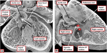
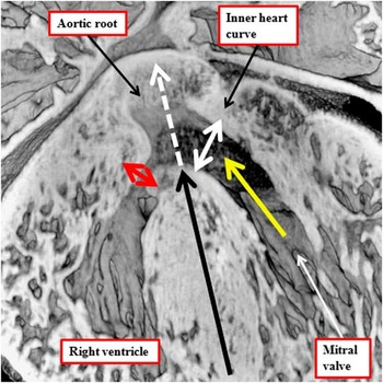
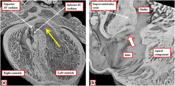
With continuing development during E12.5, there is gradual leftward shift of the subaortic component of the outflow tract. This brings the arch formed by the fused proximal outflow cushions into better alignment with the apical muscular septum (Fig 12a). These changes mean that, by E13.5, the rightward tubercles of the fused atrioventricular cushions are able to begin the closure of the persisting interventricular communication between the developing ventricles (Fig 12b). The aortic root continues its transfer leftward towards the cavity of the left ventricle during E13.5. Concomitant with this transfer, the plane roofed by the inner heart curvature itself becomes realigned leftward so as to form the boundary between the apical part of the left ventricle and its newly acquired subaortic outflow tract (Fig 13). By E14.5, the rightward part of the subaortic outflow tract, which during the process of transfer on E13.5 provided a communication with the cavity of the right ventricle (Fig 13), has closed. This process completes ventricular septation, with the hole being closed by fusion of the tubercles of the atrioventricular cushions with each other (Fig 14a), and with the base of the septal outflow cushion. This produces the membranous, or fibrous, part of the ventricular septum. The site of closure is positioned directly inferior to the inner curvature of the right ventricle, now recognisable as the newly formed supraventricular crest (Fig 14b). By this stage of development, the subaortic outflow tract, subsequent to its transfer to the left ventricle, has become deeply wedged between the mitral valve and the muscular ventricular septum (Fig 15), showing why there is no part of the muscular ventricular septum that is interposed postnatally between the ventricular inlets.
The images show different sections through a developing mouse heart at E13.5. As shown in ( a ), the blood from the left ventricle now flows into the aortic roof dorsally and cranially relative to the arch formed by the fused proximal outflow cushions, but the aortic root remains positioned above the developing right ventricle. ( b ) The oblique long-axis section, revealing how the anterior tubercles of the atrioventricular cushions (white stars with red borders), together with the base of the septal outflow cushion, are narrowing the persisting embryonic atrioventricular communication.

The image shows how the aortic root, during E13.5, is being transferred leftward so as to acquire direct origin from the developing left ventricle (yellow arrow). During this process, the cranial continuation of the long axis of the ventricular septum (black arrow) traverses the roof of the aortic root (dotted white arrow). This plane, at this stage of development, represents the geometric interventricular communication. The previous interventricular communication, as shown in Figure 11b, is now the outflow tract of the left ventricle (double-headed white arrow), and is roofed by the inner heart curvature. The red double-headed arrow shows the closing persisting embryonic interventricular communication. This is the plane of eventual ventricular septation.

The images are from a developing mouse heart at E14.5. ( a ) The tubercles of the atrioventricular (AV) cushions have fused together, and with the crest of the muscular septum, so as to commit the aortic root to the left ventricle (yellow arrow). With ongoing development, this part of the fused cushions will become the membranous septum. ( b ) The location of the dimple at the site of fusion of the cushions (white arrow) as seen from the right ventricle. At this stage, there has been no delamination of the septal leaflet of the tricuspid valve. Note how, with the completion of ventricular septation, the right ventricle has smooth walled inlet and outlet components, and an extensive apical trabecular component.

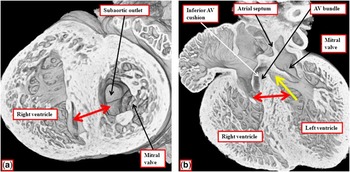
The short axis cut across the septated mouse heart at E14.5 ( a ) shows how the commitment of the subaortic outflow to the left ventricle moves the orifice of the mitral valve away from the ventricular septum. Because of this, the caudal part of the muscular ventricular septum (double-headed red arrow) separates the inlet of the right from the outlet of the left ventricle (Compare with Fig 5a). The four-chamber section through the same heart ( b ) shows how the location of the atrioventricular bundle confirms that the initial apical ventricular septum forms the entirety of the definitive muscular septum. There is no separate “atrioventricular canal septum”. The caudal part of the septum (double-headed red arrow) separates the inlet of the right ventricle from the outlet of the left ventricle (yellow arrow).

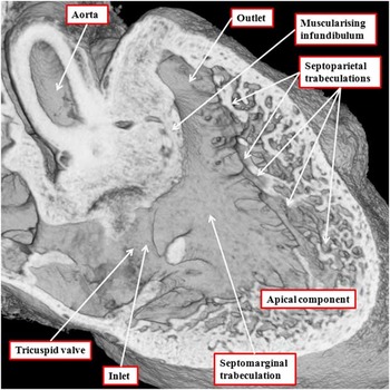
The location of the atrioventricular bundle, as shown in Figure 15b, confirms that the entirety of the definitive muscular septum is derived from the original apical muscular septum, itself formed contemporaneously with ballooning of the ventricular apical components (see Figs 8 and 10). An earlier investigation of the developing human heart has shown that a ring of cardiomyocytes surrounds the initial embryonic interventricular communication.Reference Lamers, Wessels and Verbeek 24 Part of this ring, with ongoing development, is converted into the atrioventricular conduction axis. The location of this axis, sandwiched between the crest of the muscular ventricular septum and the inferior atrioventricular cushion, indicates also that there is no separate formation of an “atrioventricular canal septum”. The extensive nature of the trabeculations within the fully formed right ventricle (Fig 16), along with recognition that the free-standing subpulmonary infundibulum is formed by muscularisation of the surface of the fused proximal outflow cushions,Reference Sizarov, Anderson, Mohun, Brown, Lamers and Moorman 25 shows in turn that there is very little muscular outlet septum to be found in the normally septated heart.
The cut of the developing right ventricle at E14.5, replicating the oblique subcostal echocardiographic section, shows how, with closure of the embryonic interventricular communication, it is now possible to recognise the inlet, apical trabecular, and outlet components of the right ventricle. The section shows how the surface of the outflow cushions has muscularised to form the supraventricular crest, with a tissue plane now forming between this structure and the aortic root. The trabecular layer of the ventricular wall is compacting to form the septoparietal trabeculations.

The description of the anatomic substrates for interventricular shunting
The changes observed during cardiac development in the orientation and margins of the embryonic interventricular communication are paralleled by the anatomy of the various channels that permit interventricular shunting in congenitally malformed hearts. As we have shown, at the early stage of development, subsequent to formation of the ventricular loop, the atrioventricular canal is committed exclusively to the developing left ventricle, whereas the outflow tract is supported exclusively above the developing right ventricle (Fig 8). This stage is comparable to congenitally malformed hearts in which the atrial chambers are connected exclusively to the dominant left ventricle, but both arterial trunks arise from the incomplete right ventricle. In this setting, the hole between the ventricles has exclusively muscular borders, and provides the outlet from the left ventricle. It is also serves as the only inlet to the incomplete right ventricle. The hole is best described as an interventricular communication. In the strictest terms, however, it is not yet a “ventricular septal defect”. This is because it is roofed by the inner heart curvature, rather than being bounded by two ventricular septal components. At the stage of cardiac development shown in Figure 8, the outlet part of the septum has yet to be formed. The only ventricular septal component of this communication is the apical muscular septum. The structure of the heart at this stage also provides strong evidence that the small outflow chamber found when the atrial chambers are connected to a dominant left ventricle is much more than an infundibulum. It is in fact an incomplete right ventricle, incomplete because it lacks its inlet component.Reference Anderson, Mohun and Moorman 26
At the end of E11.5 in the mouse, by the process of rightward expansion of the atrioventricular canal, the right ventricle has acquired its inlet component. This process in itself effectively shifts the right atrial vestibule across the dorsal margin of the initial apical muscular septum. During this process, there is no separate formation of an “atrioventricular canal septum”. Subsequent to the acquisition by the right ventricle of its inlet component, the channel existing between the ventricles can be compared with the arrangement seen in congenitally malformed hearts with double outlet right ventricle.Reference Lev, Bharati, Meng, Liberthson, Paul and Idriss 13 The communication is unequivocally interventricular. It is bounded cranially by the inner heart curvature, and caudally by the crest of the apical muscular septum. As yet, nonetheless, it is still not a “ventricular septal defect”. This is because the muscular outlet septum, formed from the fused proximal outflow cushions, is an exclusively right ventricular structure, and does not form part of the borders of the channel through which blood passes between the ventricles. At this stage, the locus representing the true “ventricular septal defect” is the plane of putative septation between the crest of the apical muscular septum and the proximal margin of the developing outlet septum. This plane is within the cavity of the right ventricle, and hence does not separate the right and left ventricles. The channel providing ventricular shunting in the setting of double outlet right ventricle is similarly an interventricular communication, rather than a defect between two ventricular septal components.
The changes occurring in the mouse heart during the latter part of E13.5 then show how the plane of putative ventricular septation, in other words the plane between the developing ventricular septal components, is brought into line with the long axis of the apical muscular septum. During the intermediate stages of this process, although transfer of the aortic root to the left ventricle is incomplete, the situation is produced which is directly analogous to the arrangement of the aortic root as seen in Fallot’s tetralogy, and in the Eisenmenger defect.Reference Sizarov, Anderson, Mohun, Brown, Lamers and Moorman 25 When the aortic root is part way through its transfer, as shown in Figure 13, the plane which, at the earlier stage, represented the interventricular communication, is becoming reorientated to become more committed to the left ventricle. This plane will form the left ventricular outlet, whereas the plane of ventricular septation will eventually be closed by the developing membranous septum. When the aortic root is overriding during its transfer to the left ventricle, it is the plane of putative ventricular septation that is usually described as the “ventricular septal defect” in the setting of both Fallot’s tetralogy and the Eisenmenger defect.Reference Baker, Leung, Anderson, Fischer and Zuberbuhler 27 This plane of supposed septation, however, is within the cavity of the right ventricle, and hence not interventricular. Only when the aortic root is fully aligned with the left ventricle does the plane of putative septation, if remaining patent, become both an interventricular communication and a ventricular septal defect. Such an example is the hole seen when the membranous septum has failed to close the plane of ventricular septation, thus producing a perimembranous ventricular septal defect in the absence of any aortic overriding.
Conclusion
If we consider the ongoing problems that currently exist in naming the channels that provide the potential for interventricular shunting, one is the discrepancy between the meaning of the terms “ventricular septal defect” and “interventricular communication” as they relate to the plane of shunting between the ventricles. A second one relates to the fundamental approach used to define the normal ventricular septum, whether it be based on embryology or on traditional terminology. A third problem exists when emphasis is placed solely on the geographical location of the holes without including the anatomic borders of the defects. In our review, we have shown that, using modern techniques, it is possible to provide accurate three-dimensional and consecutive images of the developing murine heart in any required plane. These images demonstrate that the planes produced by changes in the remodelling of the embryonic interventricular communication during temporal development parallel the planes of space observed in the hearts with deficient ventricular septation. Failure of the normal development can then explain the malformations that permit persistent shunting between the ventricles.
Analysis of development also reveals the differences between interventricular communications and ventricular septal defects. On the basis of the embryological origins of the interventricular communications shown in this review, it appears more accurate and natural to describe them as opening to the inlet, outlet, or apical components of the right ventricle, rather than seeking to predict which component of the muscular ventricular septum they excavate. The recognisable anatomic and developmental knowledge as outlined in our review should now, hopefully, permit reconciliation of the various names that continue to be used to describe the different channels which permit interventricular shunting.