Introduction
The Colorado potato beetle, Leptinotarsa decemlineata Say. (Coleoptera: Chrysomelidae), is a destructive pest of solanaceous crops especially potato. Both adults and larvae feed on potato plants until defoliation is completed (Hare, Reference Hare1980, Reference Hare1990; O'Neil et al., Reference O'Neil, Cañas and Obrycki2005). This defoliation, caused by the Colorado potato beetle, results in large financial losses each year for potato producers due to crop damage and pesticide usage (Schroder and Athanas, Reference Schroder and Athanas1989). In addition, this insect has been shown to develop resistance to pesticides (Hostounský, Reference Hostounský1984; Martin, Reference Martin2004). Recently, the negative impacts of pesticides on the ecosystem have led to endeavours to reduce the pesticides and an increased focus on alternative control methods for insect management. Because of increasing concern for the effects of pesticides on the environment, biological control methods that use natural enemies of this insect have been gaining importance (Lipa, Reference Lipa1968; Sidor, Reference Sidor1974; Hostounský, Reference Hostounský1984; O'Neil et al., Reference O'Neil, Cañas and Obrycki2005; Mennan and Ertürk, Reference Mennan and Ertürk2006; Ertürk et al., Reference Ertürk, Yaman and Aslan2008; Muratoğlu et al., Reference Muratoğlu, Demirbağ and Sezen2011).
Microsporidia are intracellular pathogens that infect living organisms in a wide variety of taxa from protists to humans (Vavra and Sprague, Reference Vavra, Sprague, Bulla and Cheng1976; Weiss and Becnel, Reference Weiss and Becnel2014). These enigmatic pathogens have been widely described from insects and have shown suppressive effects on insect populations resulting in epizootics (Lewis, Reference Lewis and Pimentel2007). Therefore, detection, characterization and isolation of microsporidia that naturally infect both harmful and beneficial insects are of interest. Nosema leptinotarsae is the first microsporidian pathogen of the L. decemlineata described using light microscopy by Lipa from the Soviet Union (Lipa, Reference Lipa1968). After 43 years, a N. leptinotarsae infection was re-recorded from Turkey by Yaman et al. in 2011. While this study demonstrated the ultrastructural features of N. leptinotarsae via transmission electron microscopy (TEM), it does not contain the molecular data necessary to determine the phylogenetic position of this organism in the Nosema–Vairimorpha clade.
Thus, the goal of the present study is to determine the true phylogenetical position of N. leptinotarsae using morphology, pathology and life cycle characteristics in addition to the new molecular sequence of the 16S SSU rRNA partial gene region.
Materials and methods
Light microscopy
Adults and larvae of the Colorado potato beetles (L. decemlineata) were collected from Trabzon (Kaşüstü) between May and July 2019 (50 individuals for each month). Specimens were transported as soon as possible to the laboratory. Ringer's solution was used for preparing wet smears. Smears were examined using a light microscope for microsporidia infections. Positive smears were stained with Giemsa for further observations of life cycle stages and measurements (Undeen and Vávra, Reference Undeen, Vávra and Lacey1997). A Nikon Eclipse Ci microscope combined with DS-Fi 2 digital camera was used to photograph fresh and stained microsporidian spores and measurements were taken using Nikon NIS Elements imaging software.
Electron microscopy (TEM)
For TEM, tissues of infected hosts were fixed in 2.5% glutaraldehyde in 0.1 m cacodylate buffer (pH 7.4) for 1–2 h. After the first fixation, tissues were rinsed with cacodylate buffer and placed in the 1% aqueous OsO4 for 2 h (Bekircan et al., Reference Bekircan, Cüce, Baki and Tosun2017a). The tissues were then rinsed again with cacodylate buffer and dehydrated through an ascending alcohol series before embedding in Spurr's resin (Spurr, Reference Spurr1969). Thin sections were taken with a Leica EM UC7 ultramicrotome and mounted on Pioloform-coated copper grids. The specimens were stained with saturated uranyl acetate and Reynolds' lead citrate (Reynolds, Reference Reynolds1963). A HITACHI HT7800 transmission electron microscope was used for observations.
Molecular studies
Microsporidia spores were isolated from infected beetles and genomic DNA was extracted using the QIAGEN DNA Isolation Kit, No: 69504 according to the manufacturer's instructions. For 16S SSU rRNA gene amplification, the QIAGEN Multiplex PCR Kit (No. 206143) and 18F/1537R primer set were used (18F/1537R: 5′-CACCA GGTTG ATTCT GCC-3′/5′-TTATG ATCCT GCTAA TGGTT C-3′) (Bekircan et al., Reference Bekircan, Bülbül, Güler and Becnel2017b). All amplification processes were performed according to the kit's protocol, and 16S SSU rRNA gene base sequences were determined by the Macrogen Inc. Company, The Netherlands.
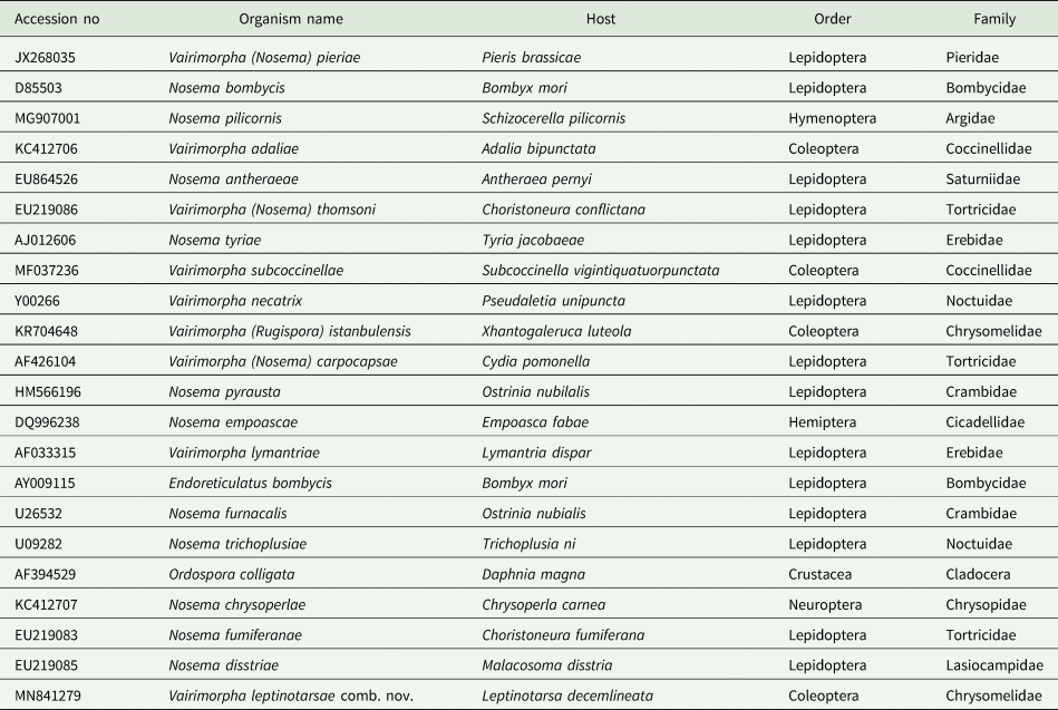
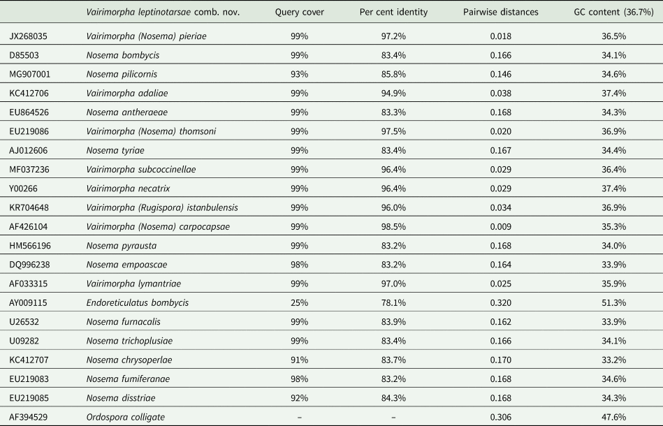
The BioEdit and CLUSTAL_W programs were used for editing and aligning the 16S SSU rRNA gene sequences that were obtained from the NCBI GenBank database according to BLAST search and the literature (Table 1) (Bekircan et al., Reference Bekircan, Bülbül, Güler and Becnel2017b). The GC contents of the sequences were determined with the Fast PCR program. Regions containing gaps and ambiguous sites were removed. In the phylogenetical analysis, the new combinations were used as re-assigned in 2020 by Tokarev et al. Phylogenetic analyses were conducted using the PAUP* program (version 4.0a164 for 32-bit Microsoft Windows). The phylogenetic trees were constructed using the maximum-parsimony (MP) and neighbour-joining (NJ) algorithms and the robustness of the trees was tested using 1000 bootstrap replicates. Bootstrap values were shown at each node.
The list of small subunit (SSU) ribosomal RNA sequences used for phylogenetic analysis

Results
Light microscopy
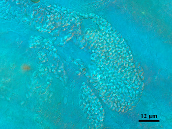
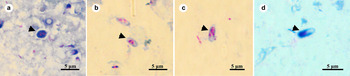
During the observations, 28 beetles were determined to be infected (infection rate 18.66%). An unstained wet smear of midgut tissues from infected beetles showed numerous microsporidian spores and aggregation was also seen often in haemolymph (Fig. 1). Fresh spores, oval in shape, measured 4.36 ± 0.43 μ m (n = 250) in length and 2.49 ± 0.24 μ m (n = 250) in width. Life cycle stages of the current microsporidium were observed in the Giemsa stained smears (Fig. 2). Binucleate ovoid meronts were commonly observed during the observations and measured as 2.70 ± 0.30 μ m × 5.03 ± 0.62 μ m (n = 50) (Fig. 2a). The oval binucleate sporonts were measuring 3.79 ± 0.15 μ m in length and 2.26 ± 0.30 μ m in width (n = 20) (Fig. 2b). The elongated sporoblasts were produced via binary fission (disporous) of sporonts and measured as 2.89 ± 0.15 μ m × 6.09 ± 0.25 μ m (n = 10) (Fig. 2c). Oval mature spores were measured after Giemsa staining [3.56 ± 0.44 μ m × 2.20 ± 0.24 μ m (n = 100)] (Fig. 2d).
Light micrograph of Vairimorpha leptinotarsae comb. nov. in L. decemlineata haemolymph which is heavily infected with binucleate mature spores.

Giemsa stained life cycle stages of the Vairimorpha leptinotarsae comb. nov. (a) Binucleate ovoid meront (midgut); (b) binucleate oval sporont (midgut); (c) elongated sporoblast (midgut); (d) mature spore (haemolymph).

Electron microscopy (TEM)
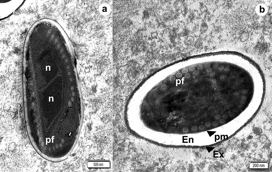
TEM revealed oval mature spore ultrastructure of the current microsporidium within the host gut cell cytoplasm (Fig. 3). The mature spores contained two nuclei in diplokaryotic arrangement (Fig. 3a). The polar filament was comprised of four concentric rings of varying electron density, which was isofilar with 14–15 coils. Coils were 142.85 ± 11.57 nm (n = 30) in diameter. The thick spore wall was trilaminar and measured 242.22 ± 17.48 nm (n = 50) and consisted of a plasma membrane, electron-lucent endospore (140–210 nm) and an electron-dense uniform exospore (51–83 nm) (Fig. 3b).
Transmission electron micrographs of binucleate mature spores. (a) Longitudinal section of mature spores with isofilar polar filaments; (b) cross-section of mature spores. Note the trilaminar structure of the spore wall. En, endospore; Ex, exospore; n, nucleus; pf, polar filament; pm, plasma membrane.

Molecular studies
In this study, phylogeny of the current microsporidium that is isolated from L. decemlineata was based upon partial sequence of the 16S SSU rRNA gene. A partial sequence of the current microsporidium was deposited in GenBank with MN841279 accession code (amplicon length; 1179 bp) and the GC content was 36.7%. The per cent identities of the N. leptinotarsae and other species used in the phylogenetic analysis ranged between 78.1 and 98.5%. Pairwise phylogenetic distances between the N. leptinotarsae and other species ranged from 0.009 to 0.320. Identities between the current microsporidium and the type species of the genera, Vairimorpha necatrix (Pilley, Reference Pilley1976) and Nosema bombycis (Nägeli, Reference Nägeli1857), were 99.4 and 83.4%, respectively (Table 2).
Comparison of 16S SSU rRNA gene sequences: query cover, per cent identity, pairwise distance and GC% content

‘–’ No significant similarity found.
Discussion
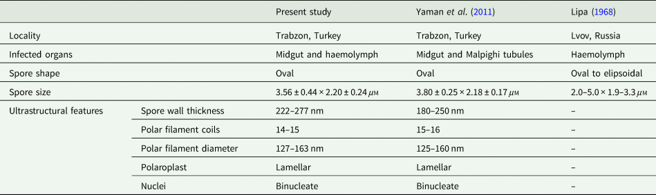
Here the microsporidiosis determined from the Colorado potato beetle (L. decemlineata) was similar to Lipa (Reference Lipa1968) and Yaman et al. (Reference Yaman, Özcan, Radek, Linde and Lipa2011). The microsporidian isolates detected in these two studies are similar to those described as N. leptinotarsae by Lipa. The isolate described here is very similar and almost identical to the previous isolate from Turkey (Yaman et al., Reference Yaman, Özcan, Radek, Linde and Lipa2011) based on light and electron microscopy (Table 3). However, it was not possible to determine the genetic similarity of the three isolates since the molecular characterization of the N. leptinotarsae isolates detected in the first two studies was not performed. Although the detected microsporidiosis from the L. decemlineata was described as a N. leptinotarsae infection previously, the phylogenetical analysis of the present study revealed that it was not a true Nosema species. The isolate from this study was genetically different from all Nosema species in the database and there was 17% difference in the 16S SSU rRNA partial gene sequence between the isolate from this study and that of N. bombycis, the type species of Nosema (Table 2).
Characteristics of the three Nosema leptinotarsae isolates described from Leptinotarsa decemlineata (Coleoptera: Chrysomelidae)

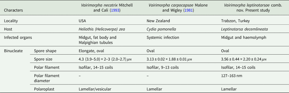
Comparative characteristics of Vairimorpha necatrix, Vairimorpha carpocapsae and Vairimorpha leptinotarsae comb. nov.

Species identification using molecular data in microsporidian taxonomy has been reached to a different dimension. With the identification or re-identification of microsporidian species with molecular data, especially with the SSU rRNA sequences, from the insect species, the genus Nosema and Vairimorpha are currently under extensive revision (Baker et al., Reference Baker, Vossbrinck, Maddox and Undeen1994; Huang et al., Reference Huang, Tsai, Lo, Soichi and Wang2004; Tokarev et al., Reference Tokarev, Huang, Solter, Malysh, Becnel and Vossbrinck2020). In the past, microsporidial taxonomy was constructed based on light and electron microscopic observations and measurements (Kudo, Reference Kudo1924; Weiser, Reference Weiser1977; Canning and Hazard, Reference Canning and Hazard1982; Issi, Reference Issi1986; Larsson, Reference Larsson1986; Sprague et al., Reference Sprague, Becnel and Hazard1992; Voronin, Reference Voronin2001). In the last 25 years, molecular phylogeny has gained importance and the sequencing of the 16S SSU rRNA is now a standard for microsporidial species descriptions. There has been a dilemma from the start been, the classical taxonomy and the new approaches in determining the phylogenetical position of microsporidia (Baker et al., Reference Baker, Vossbrinck, Didier, Maddox and Shadduck1995, Reference Baker, Vossbrinck, Becnel and Maddox1997). In 2020, Tokarev et al. took a serious step to overcome this problem. They revised the Nosematidae family, Nosema, and Vairimorpha genera using molecular data. This revision eliminates the taxonomically unacceptable formation of polyphyletic taxa created by the Nosema and Vairimorpha clades. The present study determined the phylogenetic position of N. leptinotarsae using the approaches of Tokarev et al. (Reference Tokarev, Huang, Solter, Malysh, Becnel and Vossbrinck2020).
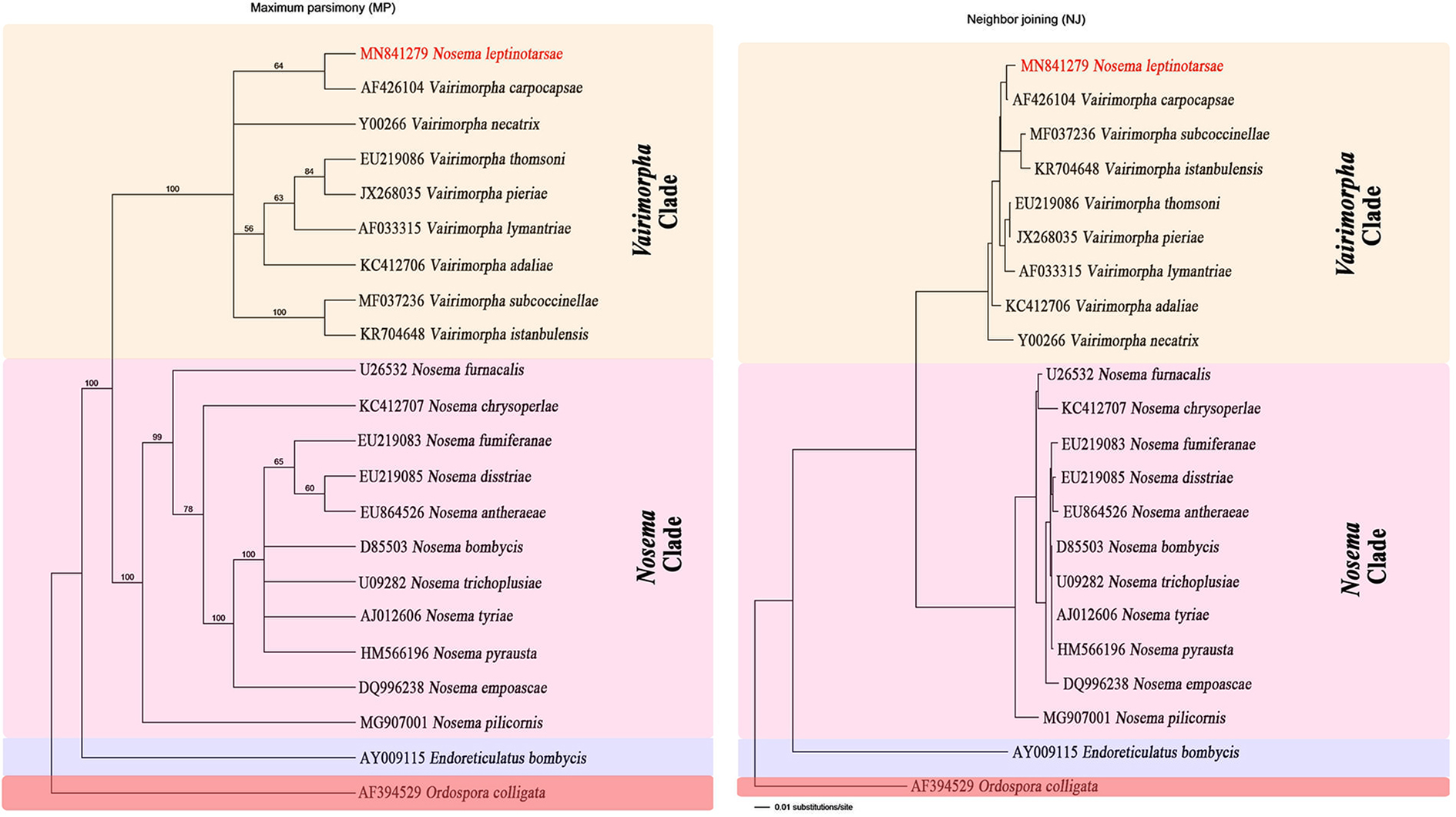
The phylogenetical trees, constructed according to MP and NJ in this study, presented three distinct clades: Vairimorpha, Nosema and Endoreticulatus (Fig. 4). Based on the analyses of the 16S SSU rRNA, the microsporidium (N. leptinotarsae) presented in this study is a member of the genus Vairimorpha and grouping with V. necatrix, the type species for the genus and branches with Vairimorpha carpocapsae (AF426104), a pathogen described from Cydia pomonella (Lepidoptera; Tortricidae) in both MP and NJ trees. Prior to molecular analysis, the genus Vairimorpha was characterized by polymorphic sporulation, meiospores (octospores), and the presence of sporophorous vesicles. None of these key diagnostic characters were observed in this study or in the former studies carried out by Lipa and Yaman et al. This inconsistency could be caused by different factors such as weather conditions and host species. For example, the original description of V. necatrix (type species of Vairimorpha genus) by Kramer (Reference Kramer1965) demonstrated that octospores were produced at 20°C but not at 25°C.
Phylogenetical relationships of the Nosema leptinotarsae with other microsporidian sequences based on the 16S SSU rRNA gene. Ordospora colligata (Microsporidia: Ordosporidae) was used as an outgroup in the analysis. Phylogenetic trees based on maximum-parsimony (MP) and neighbour-joining (NJ) algorithms showed identical topologies for the Nosema and Vairimorpha genera.

The microsporidium described here differs from V. necatrix and V. carpocapsae in regard to several taxonomic features. Phenotypic characteristics used to define microsporidian species included the spore shape and dimensions (Pilley, Reference Pilley1976; Mitchell and Cali, Reference Mitchell and Cali1993; Becnel et al., Reference Becnel, Jeyaprakash, Hoy and Shapiro2002; Bekircan et al., Reference Bekircan, Bülbül, Güler and Becnel2017b). The spores of the microsporidium presented in here are (3.56 ± 0.44 × 2.20 ± 0.24 μ m) smaller than mature binucleate spores of V. necatrix [4.3 (3.9–5.0) × 2–3 (2.0–2.7) μ m] but similar in size to those of V. carpocapsae dimensions (2.4–3.9 × 1.3–3.1 μ m) (Malone and Wigley, Reference Malone and Wigley1981), its closest relative. Additionally, ultrastructural characteristics are very important taxonomic characters for discriminating the microsporidian species (Canning and Vávra, Reference Canning, Vávra, Lee, Leedale and Bradbury2000; Becnel et al., Reference Becnel, Jeyaprakash, Hoy and Shapiro2002; Ovcharenko et al., Reference Ovcharenko, Swiatek, Ironside and Skalski2013). The polar filament number and structure are widely used and an important feature for compared the microsporidian species (Larsson and Voronin, Reference Larsson and Voronin2000; Sokolova et al., Reference Sokolova, Senderskiy and Tokarev2016). The binucleate mature spores of the current microsporidium have an isofilar polar filament with 14–15 coils. While that number is 9–13 coils in V. carpocapsae, it varies from strain to strain in V. necatrix, ranging from 10–15 to 13–17 coils (Table 4) (Maddox and Sprenkel, Reference Maddox and Sprenkel1978; Luo et al., Reference Luo, Liu, Pan, Li, Li, Dang, Liu and Zhou2014).
Molecular characterization of the current microsporidium demonstrated a genetic difference from V. necatrix and V. carpocapsae with 2.9 and 0.93% values, respectively. All structural and phylogenetic analyses support the idea that the present microsporidium is the same as the one described by Lipa as N. leptinotarsae and unique species among the Vairimorpha complex. In conclusion, on the basis of the developmental, structural and molecular evidence, this study suggests a new combination V. leptinotarsae comb. nov. for N. leptinotarsae Lipa (Reference Lipa1968).
Taxonomic description
Vairimorpha leptinotarsae comb. nov.
Phylum Microsporidia (Balbiani, Reference Balbiani1882), Nosematidae (Labbe, Reference Labbe, Schulze and Butschli1899), Vairimorpha (Pilley, Reference Pilley1976).
Host: Leptinotarsa decemlineata Say, 1824 (Coleoptera: Chrysomelidae).
Site of infection: The infection can be seen in the midgut and haemolymph of the host.
Interface: Life cycle stages in direct contact with host cell cytoplasm.
Spores: Fixed binucleate mature spores are oval shaped, measured 3.56 ± 0.44 μ m (2.61–3.56) × 2.20 ± 0.24 μ m (1.59–2.74; n = 100). Fresh spores measured 4.36 ± 0.43 μ m (3.41–5.58; n = 250) in length and 2.49 ± 0.24 μ m (1.89–3.04; n = 250) in width. Spores contained an isofilar polar filament with 14–15 coils and coils were 142.85 ± 11.57 nm (127.02–163.02; n = 30) in diameter. The spore wall has the typical trilaminar structure and is relatively thick (242.22 ± 17.48 nm).
Locality: Specimens described here were collected from Kaşüstü (Trabzon), Turkey.
Deposition of specimens: The samples for light and electron microscopy are preserved in the personal collection of Çağrı BEKİRCAN with the Catalog No. ÇVL-01. Vairimorpha leptinotarsae 16S SSU rRNA gene sequences from samples were deposited to GenBank with MN841279 accession code.
Acknowledgements
The TEM analysis was carried out in Eskişehir Osmangazi University (ESOGU) Central Research Laboratory, Research and Application Centre (ARUM). The author is grateful to the research team; Ph.D. Hilal Yıldırım and Ph.D. Onur TOSUN for reading the manuscript and for their valuable contributions.
Financial support
This research received no specific grant from any funding agency, commercial or not-for-profit sectors.
Conflict of interest
The authors declare that there are no conflicts of interest.
Ethical standards
Not applicable.