Introduction
The role of stereotactic ablative body radiotherapy (SABR) in early stage lung cancer management has been established as the gold standard treatment where the patient is medically inoperableReference Vansteenkiste, De Ruysscher and Eberhardt 1 – Reference Baumann, Nyman and Hoyer 3 with local control rates of up to 92% being reported.Reference Baumann, Nyman and Hoyer 3 In addition SABR has benefit in the management of lung metastases with a local control rate of up to 80% at 1 year being reportedReference Wulf, Haedinger, Oppitz, Thiele, Mueller and Flentje 4 with minimal toxicity.
Motion management of lung tumours is essential in delivering SABR and one method of motion management is deep inspiration breath hold (DIBH). DIBH delivered SABR has been shown to be dosimetrically desirable, over an internal target volume (ITV) technique where tumour motion overall respiratory phases is accounted for.Reference Kontrisova, Stock, Dieckmann, Bogner, Pötter and Georg 5 It was reported that lung organ at risk (OAR) dose (all dose volume constraints), were reduced by 20% using a DIBH technique compared with a free breathing technique. Furthermore, with the addition of DIBH a reduction in planning target volume (PTV) margins was possible, which lead to a 40% reduction in dose to OARsReference Kontrisova, Stock, Dieckmann, Bogner, Pötter and Georg 5 compared with a free breathing technique.
However, within DIBH inter-fraction variability of tumour position, relative to bony anatomy, occurs and this variation is observed on the daily-acquired cone beam computed tomography scans (CBCTs).Reference Zhang, Yu and Stevens 6 , Reference Kimura, Murakami and Kenjo 7 The magnitude of this positional variation has been reported by a number of studies and has been quantified as 3·3 mm,Reference Zhang, Yu and Stevens 6 , Reference Kimura, Murakami and Kenjo 7 5·4±2·5 mmReference Peng, Vedam and Chang 8 or even 6·7 mmReference Berson, Emery and Rodriguez 9 with various methods being used to achieve a breath hold scan.
In order to account for this inter-fraction gross target volume (GTV) displacement, image guided soft tissue tumour matches (away from the bony alignment) are necessary to ensure full dose coverage of the lesion.
Within this institution DIBH SABR is facilitated by the Varian Real-Time Position Management (RPM) system with audio-visual coaching. This has been shown to be a reliable and reproducible method of motion management.Reference Peng, Sadagopan and Vedam 10 , Reference Glide-Hurst, Gopan and Hugo 11
Patients are immobilised supine, arms up in a BodyFIX® system (Elekta, Stockholm Sweden), with a full length evacuated cushion for the patient to lie in and immobilisation is further assisted by a coversheet suctioned around the patients contour.
The RPM system utilises an external gating block, which is placed at a stable point on the patient’s chest (inside the suctioned coversheet) and this is used to quantify the BH amplitude. This process is aided by providing the patients with video goggles showing live video feedback of the BH allowing the patient to monitor their own BH during treatment. This is reproduced for each treatment fraction and a CBCT is acquired to verify both bony and soft tissue anatomy.
SABR patients attend for a breathing assessment appointment, before CT simulation, to ensure they can consistently achieve a reproducible BH as visualised on the RPM system before acquiring a planning CT scan. This appointment consists of a number of steps. The immobilisation device is constructed and evaluated for both stability and comfort. The patient is assessed for any medical requirements before simulation, such as analgesia. The patient is then given an opportunity to practice DIBH as well as coached breathing techniques. They then return for CT simulation 48 hours later, having had experience of the process and practiced the technique.
This study aimed to quantify our institutions inter-fraction GTV displacement based on our process and evaluate if this displacement relates to minor changes in BH or the external gating block. A secondary aim was to evaluate the dosimetric impact of this tumour motion on the surrounding OARs.
Materials and methods
This study was approved by our institutional research committee. Data collection was in accordance with local protocol.
Data collection and evaluation methods
Lung tumour patients treated with DIBH SABR in our institution were retrospectively reviewed. A convenience sampling strategy was utilised with the following criteria:
Included were SABR lung patients treated with a BH technique from 1 January 2012 to 30 April 2013 and tumour location in the lower lobe. Excluded were SABR patients treated with a free breathing or coached breathing technique, breath hold delivered SABR to abdominal lesions, lesions in the upper or middle lobes of the lung and patients not treated as per standard departmental protocol. Within this institution, DIBH is indicated over alternative motion management techniques if the tumour motion is >5 mm.
This yielded a study population of 22 patients, 16 males and six females. Their ages ranged from 50 to 85 years old with an average age of 71·59 years. Of these patients 11 had primary lung tumours and 11 had metastases from a variety of primaries including melanoma, pancreatic cancer, caecal cancer, colon cancer, colorectal cancer and rectal cancer.
Patients were treated with between 3 and 10 fractions to a total dose ranging from 48 to 60 Gy with the most common fractionation being 60 Gy in 5 fractions.
12 patients had right lower lobe tumours and 10 had left lower lobe tumours.
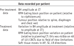
A total of 121 fractions were delivered and a range of data (Table 1) was recorded from each patient who met the inclusion criteria.
Data recorded per patient

Abbreviations: BH, breath hold; CT, computed tomography; RPM, Real-Time Position Management; CBCTs, cone beam computed tomography scans; AP, anterior-posterior; SI, superior-inferior; LR, left-right.
A single investigator carried out all measurements. The intra-observer variability was quantified, where appropriate and was noted to be 0·2 cm when measuring the daily BH amplitude.
Inter-fraction variation in tumour position
At each fraction a CBCT was acquired by a full rotation of the gantry in a 90 second acquisition time. The patient achieved this using three to four breath holds depending on individual ability. The acquired CBCT dataset was first matched to the vertebral column and then to the soft tissue tumour GTV in the planning scan to quantify the inter-fraction GTV displacement. Soft tissue moves in anterior-posterior (AP), superior-inferior (SI) and left-right (LR) planes and were recorded and applied to correct for this displacement.
Absolute values were generated and an overall magnitude of tumour displacement relative to bony anatomy was generated using the Euclidean distance formula for each fraction delivered.
A Pearson product-moment correlation coefficient (r) was generated to investigate the relationship between the various factors and inter-fraction tumour displacement. The coefficient of determination was also generated.
OARs evaluation
The relevant OARs were deemed to be lung, spinal cord, heart, oesophagus and chest wall in accordance with local protocol.
All soft tissue moves recorded at treatment were retrospectively applied to the original planning scans in EclipseTM Treatment Planning System, by moving the isocentre to the on-treatment position for each fraction delivered. The dose distribution was recalculated for each fraction, using the same monitor units. For each patient a cumulative plan was generated by the summation of all the delivered fractions. Cumulative dose volume histograms (DVHs) were generated and then compared with the original approved DVH.
The GTV was not moved in tandem with the soft tissue moves. The significance of not moving the GTV was evaluated on Patient 1. As well as the standard replan, a second replan was carried out where the GTV contour was moved in conjunction with the treatment beams. The new GTV was assigned a Hounsfield unit (HU) value in line with the original lesion as measured on the planning scan. The original lesion, if outside the new GTV, was assigned a HU value in keeping with surrounding lung tissue. This was replicated for each fraction where soft tissue moves were applied. The new plans were recalculated using the original monitor units and a composite plan generated. The relevant OARs were compared and only the ipsilateral lung maximum dose changed by 1·4 Gy. All other OAR dose volume constraints were unchanged. It was therefore deemed acceptable not to move the GTV in tandem with the beams.
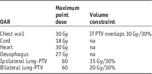
The mean, maximum and minimum dose to each OAR was reviewed as well as the bilateral lung volume receiving 20 Gy (V20), the ipsilateral lung volume receiving 15 Gy (V15) and the chest wall volume receiving 30 Gy (V30). In addition, specific dose constraints used in the researchers institution were reviewed (Table 2).
Organ at risk (OAR) dose constraints

Abbreviation: PTV, planning target volume.
The significance of the observed changes were evaluated using a two-tailed, Type 1 t-test to generate a p value, with a value of <0·05 considered statistically significant.
Results
Inter-fraction GTV displacement
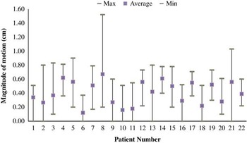
The overall magnitude of tumour displacement relative to the bony anatomy ranged from 0 to 1·52 cm, with a mean overall magnitude of displacement of 0·41 cm with a standard deviation of ±0·28 cm. See Figure 1 for summary of displacement per patient. The largest displacement was observed in the SI plane with an average displacement of 0·31±0·26 cm compared with 0·16±0·18 cm in the AP plane and 0·07±0·12 cm in the LR direction.
Summary of inter-fraction GTV displacement per patient.

The BH amplitude recorded at CT ranged from 1·2 to 2·8 cm with mean amplitude of 1·7 cm within the sample population.
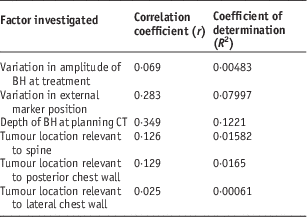
The variation of BH amplitude at treatment measured on the RPM system retrospectively was found to range from 0 to 0·4 cm per fraction with a mean variation of 0·13±0·1 cm. The magnitude of tumour displacement was found not to correlate significantly with the variation in BH amplitude giving a correlation coefficient (r) of 0·069.
Variation in the daily placement of the external gating block was observed on a number of the CBCTs and this ranged from 0 to 3·8 cm, mean discrepancy of 1·02±0·85 cm., no correlation was observed between this and GTV displacement. The influence of the block position on skin was also considered by evaluating if there was any correlation between tumour variability within BH and the distance the marker is placed from the xiphisternum. No significant relationship was observed. No correlation was observed between tumour location and inter-fraction displacement.
See Table 3 for correlation coefficient values for each factor investigated.
Correlation coefficient values

Abbreviations: BH, breath hold; CT, computed tomography.
OARs evaluation
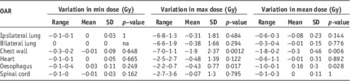
Review of the cumulative plans’ DVHs revealed a range of OAR dose variation from −7·0 to +3·6 Gy. Evaluating the maximum point dose, the minimum point dose and the mean dose to each OAR revealed that only the chest wall and the oesophagus maximum and mean doses were significantly affected (Table 4).
Organ at risk (OAR) evaluation

The remainder of changes observed were not statistically significant. A number of patients having lesions treated in both lungs did not have the ipsilateral lung minus PTV contoured and one patient did not have oesophagus contoured. Where ipsilateral lung minus PTV was not contoured the patients had bilateral lung minus PTV volumes to allow for a more complete view of the lung dose. In the case where the oesophagus was not contoured the GTV was located distal to the oesophagus and it was not deemed an OAR.
In addition the bilateral lung minus PTV V20 varied on average by −0·04±0·30% (p=0·545). The ipsilateral lung minus PTV V15 varied by −0·29±0·75% (p=0·122) and the chest wall V30 had a mean variation of −0·67±0·89% (p=0·002).
Discussion
BH reproducibility and inter-fraction motion
Patient ability to reproduce a stable BH amplitude on treatment was excellent. This data shows a mean variation of 0·13±0·1 cm measured over 121 CBCTs and 22 patients.
This compliance shows that a stable breath hold is reproducible once coaching is utilised and the patient has sufficient time to adapt to the technique. In our experience the initial breathing assessment visit aids this process for the patients.
It should be noted that a single BH amplitude was selected to represent the amplitude for the entire CBCT, which is generally attained over three to four breaths. However, the data showed that compliance to BH amplitude was excellent with a mean variation of 0·13 cm recorded which reassures that a single BH is an adequate representation of amplitude over the scan acquisition.
Following on from this we also found that the range of inter-fraction displacement observed at this institution (0·41±0·28 cm) using a DIBH technique was in keeping with the published literature across a range of breath hold techniques.Reference Peng, Vedam and Chang 8 , Reference Berson, Emery and Rodriguez 9
The data has shown that the motion of lower lobe lung tumours can be comfortably controlled within 5 mm. This facilitates margin reduction and significant lung sparing compared with an ITV technique, the dosimetric benefits to the lung have been well established.Reference Kontrisova, Stock, Dieckmann, Bogner, Pötter and Georg 5 , Reference Rosenzweig, Hanley and Mah 12
Patients 8 and 21 were noted to have a larger range of motion despite no larger variation in the measured breath hold amplitude. Patient 8 required large moves in all directions for one fraction (0·9 cm AP, 1·0 cm SI and 0·7 cm LR), which accounted for the overall larger range of motion. Discounting this single fraction this patient’s maximum magnitude of motion was 0·71 cm, which was similar to the remainder of the data. Patient 21 also had a slightly larger range of motion of 1·03 cm but again this was accounted for in a single fraction where a 0·9 cm SI shift was required.
Correlation with factors investigated
It must also be acknowledged, however, that the data also established that a level of inter-fraction displacement does occur within BH despite the reproducibility of the BH achieved.
This study has shown that the factors investigated show no statistically significant correlation with inter-fraction displacement within DIBH. These factors included minor amplitude changes during daily treatment, positioning of the gating block relative to xiphisternum, minor discrepancies in the gating block positioning at daily treatment and tumour location within the lung. The mean variation in the daily positioning of the gating block was found to be 1·02 cm relative to the planning CT. Clinically, this block is placed at a reference point on skin and a patient’s the external contour may vary from day to day, as is the case with all skin marks. As such the variation observed here might well be a combination of block placement discrepancy and skin position discrepancy.
A recent study by Renming et al.,Reference Renming, Wang and Zhou 13 which was published since this research was carried out, also evaluated the inter-fraction variation produced with lung DIBH. They found a significant correlation between vertebral bone moves required and the inter-fractional soft tissue reproducibility, which was not examined in this research. This study had a large sample size but utilised an active breathing control system and a single breath hold CBCT so results may not be comparable.
This lack of correlation observed could be explained by the fact that in the main there was little variation observed in these factors and as such they had only a modest influence on the inter-factional soft tissue moves required.
Some trends in the data were perceived but they did not reach a level of statistical significance. As expected there was a weak negative correlation between the motion observed and the distance from the diaphragm indicating less motion in tumours located more superiorly in the lower lobes.
This research has shown that there is no change in the stability or reproducibility of BH achieved in all areas of the lower lobe. This corresponds well with the data published by RenmingReference Renming, Wang and Zhou 13 who also reported no significant correlation between relative tumour position within the lung and inter-fractional soft tissue displacement.
OARs evaluation
Overall the application of soft tissue moves had little impact on the surrounding OAR doses.
Two OARs were affected significantly by applying soft tissue moves, with only one statistically significant increase in OAR dose observed which was not clinically significant (oesophagus mean dose increasing by 0·16 Gy). The remainder of dose increases did not correlate significantly with soft tissue moves being applied. This verified that within the sample of 121 CBCTs reviewed, applying soft tissue moves (within the range observed of 0·41±0·28 cm) was safe and did not adversely affect OAR dose. However the limitation of assessing OAR dose on the initial planning scan is acknowledged as OARs may displace in tandem with the tumour displacement on a daily CBCT.
In the case of four patients the spinal cord maximum point dose was increased to above the prescribed tolerance of 18 Gy. In Patients 6 and 14 this was a marginal increase from 18·4 to 18·5 Gy and 18·1 to 19·3 Gy, respectively. Both tumours were located close to the spinal cord and the physician approved cord maximum point dose was also slightly above the tolerance level as these were point doses to a minimal volume. Patient 4 showed a 2·1 Gy increase from 16·7 to 18·8 Gy increasing the cord dose above the approved tolerance. Analysis of the OAR dose changes was limited to evaluation of the maximum, minimum and mean doses and three planning constraints. Review of dose points alone can be misleading without entire review of the DVH curve. ICRU 83Reference Grégoire and Mackie 14 recommends reporting of D2% as a more representative maximum dose.
Conversely to these results it has been reportedReference van Rooijen, van Wieringen, Stippel, Crezee, Koning and Bel 15 that OARs can be clinically affected with statistically significant variations of up to +10% being seen in OAR max points. However this study matched the soft tissue tumour without a threshold and the magnitude of variation from bone is not reported so it is difficult to assess if these results are comparable.
This study has highlighted areas for further research. As no variable investigated has been found to correlate with the soft tissue displacement observed, an area for further research could be to evaluate the role of the vacuum coversheet in the patient set up as this was not considered in this study, as well as patient related factors such as pre-existing lung disease. In addition, the evaluation of the intra-fraction consistency of breath hold and soft tissue positioning would be of interest.
Conclusions
This study has found that producing a reproducible BH is achievable across a range of patients. Our process has been shown to be a reliable and stable technique for inter-fraction motion management in lower lobe tumours.
It has shown that using this robust technique, the magnitude of inter-fraction GTV displacement can easily be controlled in line with a range reported in the literature with a mean variation of 0·41±0·28 cm being measured. As a result of this stringent motion management the application of the remainder of the soft tissue shifts has little effect on OAR dose.
No statistical correlation was found between tumour displacement and the variables investigated. From this data we can conclude that within this patient cohort, this small level of inter-fraction displacement may be somewhat inherent in BH and thus we cannot reduce it by adjusting any of the factors examined in this study. Soft tissue matching based on CBCT image guidance has been shown to be safe and can compensate for this motion ensuring adequate target coverage.
Acknowledgements
None.
Financial support
This research received no specific grant from any funding agency, commercial or not-for-profit sectors.
Conflicts of Interest
None.
Ethical Standards
The authors assert that all procedures contributing to this work comply with the ethical standards of the relevant national guidelines on human experimentation and with the Helsinki Declaration of 1975, as revised in 2008, and has been approved by the institutional committees of the Beacon Hospital, Sandyford, Dublin 18.