Eosinophilic granulomatosis polyangiitis is a rare multisystemic small and medium vessel vasculitis. Classically, it progresses through three phases, prodromal allergic, eosinophilic, and vasculitis phase. Reference Fina, Dubus and Tran1 This disease is well described in adults but there is a paucity of literature in children. Reference Fina, Dubus and Tran1–Reference Gendelman, Zeft and Spalding3 Cardiac involvement may be present in up to 55% of paediatric cases and necessitates early aggressive treatment. Reference Zwerina, Eger, Englbrecht, Manger and Schett4 Cardiac presentation can many times be subclinical and should be evaluated appropriately in all suspected cases. Reference Cereda, Pedrotti, De Capitani, Giannattasio and Roghi5
Case report
A 14-year-old girl presented to the emergency department with recent history of headache, vomiting, visual disturbances, and limb weakness. She also had class IV NYHA dyspnoea. Past history was significant for bronchial asthma and sinusitis. On examination, she was conscious and oriented. There was pallor, petechiae on bilateral thighs, tachycardia, tachypnoea, and hypoxia. Cardiovascular examination revealed elevated jugular venous pulse, cardiomegaly, loud second pulmonary component, and pan-systolic murmur at apex. Bilateral basal crepitations were present. There was hypotonia and decreased power in all limbs.
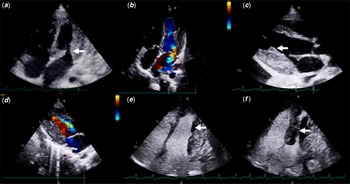
Blood investigations revealed anaemia of chronic disease, leukocytosis with eosinophilia – 16,400/mm3 (normal < 500), and thrombocytosis. IgE levels were high – 2500 international units/ml (normal < 100). Renal, liver function tests, inflammatory markers, and complete urine analysis were normal. Chest radiography showed pulmonary oedema. ECG showed sinus tachycardia with diffuse T wave inversions. Cardiac biomarkers were elevated. Transthoracic echocardiography showed severe mitral regurgitation, posterior mitral leaflet plastered to left ventricle posterior wall, severe tricuspid regurgitation, pulmonary hypertension, and normal biventricular systolic function. There was a mural thrombus in left ventricle which caused systolic obliteration and flow turbulence in left ventricle mid-cavity. Mural thrombus was involving the papillary muscles and posterior mitral leaflet (Fig 1 and Supplementary Videos S1–S4).
Transthoracic echocardiography with accompanying left ventricle (LV) contrast sequence demonstrating endomyocardial fibrosis, LV mural thrombus and severe mitral regurgitation (MR). ( a ) apical four chamber view shows LV hypertrophy primarily involving posterior wall, posterior mitral leaflet plastered to posterior wall (solid white arrow), right atrium, and right ventricle are dilated. ( b ) Corresponding view with colour flow shows severe MR with eccentric posteriorly directed jet. ( c ) Parasternal long-axis view shows mural thrombus attached to LV posterior wall (solid white arrow) and involving the papillary muscles and posterior mitral leaflet. ( d ) Corresponding view with colour flow shows flow turbulence across LV mid-cavity due to systolic obliteration by mural thrombus. ( e ) LV contrast echo shows filling defect corresponding to mural thrombus seen in image ( c ) (solid white arrow). ( f ) LV contrast echo shows systolic obliteration of LV mid-cavity (solid white arrow).

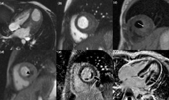
CT chest and pulmonary angiogram showed bilateral ground glass opacities, smooth interlobular septal thickening with no evidence of pulmonary thromboembolism. Cardiac MR images confirmed our echocardiography findings and in addition showed myocardial oedema and diffuse uniform endocardial late gadolinium enhancement (Fig 2). MR brain showed multifocal subacute infarcts. MR angiogram and MR venogram were normal. MR also showed mucosal thickening of maxillary and ethmoid sinuses with left maxillary sinus mucosal polyp. Serial chest radiographs showed migratory pulmonary infiltrates. Thrombophilia work up, antiphospholipid antibodies, and antineutrophil cytoplasmic antibodies were negative. Antinuclear antibodies immunofluorescence was positive. Electromyography and nerve conduction velocity were normal. A diagnosis of eosinophilic granulomatosis polyangiitis was entertained. Petechiae on the lower limbs had disappeared, hence biopsy was deferred.
Cardiac MR images demonstrating features of endomyocardial fibrosis and left ventricular (LV) mural thrombus causing systolic obliteration of LV mid-cavity. ( a ) Cine four chamber and ( b ) short axis views show eccentric wall thickening and LV mid cavity narrowing. ( c ) Short axis pre-contrast T1 black blood image shows isointense LV myocardium and mildly hyperintense thrombus along the endocardium (white arrow). ( d ) Post contrast first pass perfusion shows enhancing myocardium and non-enhancing LV thrombus (black arrowhead). Delayed enhancement short axis ( e ) and four chamber ( f ) views show diffuse uniform endocardial enhancement with non-enhancing layered thrombus in the LV cavity (black solid arrow) involving and encasing the papillary muscles.

The patient was started on pulse dose methylprednisolone, Rituximab, Lisinopril, Metoprolol, diuretics, and Acitrom. She clinically improved. Eosinophils, IgE levels, and cardiac biomarkers normalised. Steroids were tapered and stopped after 3 months.
On follow-up, she was in NYHA class I dyspnoea but continued to have severe mitral regurgitation with pulmonary hypertension. Cardiac MR after 6 months showed resolution of left ventricle thrombus and myocardial oedema but persistence of papillary muscle and posterior mitral leaflet adherence to endocardium. There was also persistent endocardial late gadolinium enhancement for which she was started on mycophenolate mofetil.
She had a relapse with exacerbation of asthma and high eosinophil counts which subsided with a short course of oral steroids. She also developed chronic headache due to cerebral venous thrombosis inspite of therapeutic INR. Acitrom was changed to Dabigatran and her headache improved. She continued to remain in NYHA class I dyspnoea and was on regular follow up; however, there was no improvement in her echocardiogram findings. Two years after diagnosis, she presented with septic shock and died due to multiorgan failure.
Discussion
Eosinophilic granulomatosis polyangiitis is the rarest of vasculitis in children. There are three paediatric series but each have less than 15 cases. Reference Fina, Dubus and Tran1−Reference Gendelman, Zeft and Spalding3 To our knowledge, less than 100 paediatric cases have been reported. Reference Fina, Dubus and Tran1 On review of literature, it is evident that children have more cardiorespiratory and less neurological and renal manifestations than adults. Reference Fina, Dubus and Tran1−Reference Gendelman, Zeft and Spalding3 Cardiac involvement in eosinophilic granulomatosis polyangiitis leads to poor prognosis and mortality. Cardiac manifestations commonly include pericarditis, myocarditis, hypertension, arrhythmias, myocardial infarction, cardiomyopathy, and valvular regurgitation. Reference Fina, Dubus and Tran1
To our knowledge, this combination of myocarditis, cardioembolic stroke due to intracardiac thrombus, and endomyocardial fibrosis has not been previously reported. Reference Fina, Dubus and Tran1−Reference Zwerina, Eger, Englbrecht, Manger and Schett4,Reference Amaddeo, Ventura and Marchetti6−Reference Albahri, Minxová, Lukeš, Mawiri and Štefáčková8 Albahri et al. described a 17-year-old girl with myocardial fibrosis and mitral regurgitation whose cardiac findings recovered on follow-up, unlike our patient where endomyocardial fibrosis and severe mitral regurgitation were persistent until death. Reference Albahri, Minxová, Lukeš, Mawiri and Štefáčková8 Intracardiac thrombus is a rare manifestation in paediatric eosinophilic granulomatosis polyangiitis, to our knowledge, having been reported in only one case by Eleftheriou et al. Reference Eleftheriou, Gale, Pilkington, Fenton, Sebire and Brogan2 Moreover, intracardiac thrombus, causing systolic obliteration of left ventricular mid-cavity, has not been previously reported in children. A similar case has been reported in an adult where there was systolic obliteration of left ventricular cavity with high pressure gradient. This was thought to be a high-risk factor for releasing thrombi into the systemic circulation. Reference Ito, Fujita, Kanzaki, Sohmiya and Hoshiga9 Paediatric eosinophilic granulomatosis polyangiitis presenting with myocarditis is also rare. The prevalence of myocarditis was 22 and 40% according to two adult series. Reference Moradinejad, Rezaei and Ziaee7
We deferred histopathological confirmation because our patient satisfied four out of six criteria of American College of Rheumatology – asthma, eosinophilia >10%, non-fixed pulmonary infiltrates, and paranasal sinus abnormality. Also, biopsy would require invasive pulmonary or cardiac tissue in our patient. Histopathology is generally the gold standard for diagnosis. Histopathological findings include extravascular eosinophils, granulomas, and necrotising vasculitis. Reference Fina, Dubus and Tran1 All these findings are not commonly seen in paediatric EGPA biopsy samples. Eosinophilic infiltrates are commoner when compared to vasculitis and granulomas. This is explained by the theory that children usually have a shorter period between the onset of allergic symptoms and systemic manifestations leading to earlier diagnosis during the eosinophilic phase when compared to adults who are usually diagnosed during the vasculitis phase. This explains the different presentations in children. Reference Fina, Dubus and Tran1
Our patient’s cardiac findings failed to improve inspite of early aggressive treatment. Pathological studies suggest that myocardial fibrosis can develop early and rapidly and lead to poor long-term prognosis in children Reference Guillevin, Cohen, Gayraud, Lhote, Jarrousse and Casassus10 like in our case. Though she had an aggressive form of the disease with extensive cardiac involvement, she was in NYHA class I for almost 2 years after diagnosis. She unexpectedly developed a severe form of sepsis possibly contributed by immunosuppressive medication, a known complication.
Cardiac involvement can often be subclinical. Hence, it is our opinion that early detection by appropriate investigations including echocardiography and cardiac MR is mandatory in all suspect cases. This is paramount in children who often have cardiac involvement, higher chance of relapse, and mortality. If detected early before the myocardial fibrosis stage, aggressive treatment can significantly alter the course of the disease and hopefully lead to a better outcome.
Supplementary material
To view supplementary material for this article, please visit https://doi.org/10.1017/S104795112200155X
Acknowledgements
None.
Financial support
This research received no special grant from any funding agency, commercial, or not-for-profit sectors.
Conflicts of interest
None.
Ethical standards
Institutional ethics committee approved of the manuscript.