Implications
Ochratoxin A (OTA) is one of the most common mycotoxins detected in poultry feedstuff, and one of the major intestinal pathogenic mycotoxins. Curcumin alleviates the negative effects caused by enteropathogenic bacteria and mycotoxins. However, the possible effects of curcumin as well as the mechanism involved in alleviating the enterotoxicity induced by OTA are unknown. The results obtained in this experiment resolved these two questions, thereby helping to understand the molecular mechanism that allows curcumin to alleviate the destruction of the intestinal barrier by mycotoxins, and providing a natural additive to prevent poor performance and reduce the economic losses associated with mycotoxins.
Introduction
Mycotoxins, the secondary metabolites of fungal species, are the most frequently occurring natural food contaminants in human and animal feeds. Ochratoxin A is a well-known and widely spread mycotoxin that is mainly produced by Aspergillus and Penicillum molds and found in cereals, feeds, meat, vegetables, fruits, wine, beer, coffee, etc. (Kőszegi and Poór, Reference Kőszegi and Poór2016). Ochratoxin A inhibits protein synthesis, and promotes membrane peroxidation, and disrupts calcium homeostasis, as well as mitochondrial dysfunctions and DNA adducts (Ringot et al., Reference Ringot, Chango, Schneider and Larondelle2006). The main target organ for OTA toxicity in poultry and other species is identified as the kidney, while its well-known inhibition of protein synthesis and production of reactive oxygen (ROS) and nitrogen species reveal that liver and gastrointestinal tract may be a possible target for OTA as well (Grenier and Applegate, Reference Grenier and Applegate2013). High concentrations of OTA have been found to damage the intestinal mucosa in chickens (Solcan et al., Reference Solcan, Pavel, Floristean, Chiriac, Şlencu and Solcan2015), resulting in a rapid inflammatory effect associated with mucosal necrosis and sloughing. Furthermore, OTA also reduces local immunity, which leads to higher susceptibility to colibacillosis and salmonellosis (Śliżewska and Pitotrowska, Reference Śliżewska and Piotrowska2014). In Caco-2 cells, OTA exhibited an increase in permeability of tight junctions due to altered distribution of specific claudin isoforms, which were integral membrane components of the tight junctions (Maresca et al., Reference Maresca, Mahfound, Pfohl-Leszkowicz and Fantini2001; McLaughlin et al., Reference McLaughlin, Padfield, Burt and O’Neill2004). Also, Ranaldi et al. (Reference Ranaldi, Mancini, Ferruzza, Sambuy and Perozzi2007) found that OTA caused faster and irreversible increase in tight junctions permeability, intracellular delocalization of claudin-4 and extensive apoptosis. These studies indicate that OTA is able to alter the intestinal barrier function.
Curcumin is a polyphenolic compound isolated from the rhizome of Curcuma longa Linn which has a variety of pharmacological activities, including anti-oxidative, anti-inflammatory, antibacterial and antifungal properties (Moghadamtousi et al., Reference Moghadamtousi, Kadir, Hassandarvish, Tajik, Abubakar and Zandi2014). In vivo studies showed that curcumin could increase the expression of various detoxifying enzymes in mouse liver, small intestine and kidney tissues of mice (Shen et al., Reference Shen, Xu, Hu, Jain, Gopalkrishnan, Nair, Huang, Chan and Kong2006). Wang et al. (Reference Wang, Wang, Hao, Ma, Wang, Jiang and Jiang2012) concluded that curcumin could protect human intestinal epithelial cells against H2O2-induced disruption of tight junctions via the heme oxygenase 1 pathway. Supplementation of curcumin to broiler feed increased the villus absorptive area of the small intestine (Rajput et al., Reference Rajput, Muhammad, Yan, Zhong and Wang2013). Muhammad et al. (Reference Muhammad, Sun, Wang, Li, Wang, Cheng, Li, Zhang and Hamid2017) reported that curcumin could prevent the harmful effects of aflatoxin B1 in broilers.
The young animals and ducks or turkeys are usually the most sensitive species with regard to their response to mycotoxin-contaminated feed (Stoev, Reference Stoev2016). Because OTA was shown to affect organization and permeability of tight junctions in the intestine, but the related mechanisms were not clear, it was considered important to investigate whether an adequate intake of curcumin might alleviate the enterotoxicity of OTA. The main objective of this study was to determine the protective effects of added curcumin in an OTA-contaminated diet on performance, antioxidant status, intestinal barrier and mitochondrial function in young ducks.
Material and methods
Preparation of moldy corn
Aspergillus ochraceus AS3.3876 isolate was purchased from Guangdong Institute of Microbiology (Guangzhou, Guangdong, China). The fungus was grown on Czapek Dox Medium (CDM) at 28°C for 7 days, mycelia were then picked with an inoculation loop and inoculated into six Erlenmeyer flasks (1 l), each containing 500 ml CDM liquid medium and incubated at 28°C for 7 days to prepare the conidial suspension. Conidial concentration was 8×10⁵/ml. Ground corn (200 kg) was sterilized in a dryer at 105°C for 48 h, and was uniformly spread on the floor of a sterilization room, then inoculated with the suspension of A. ochraceus conidia and incubated for 7 days at 29°C to 30°C. Twice daily (at 0800 and 2000 h), 3 l of autoclaved CDM medium was added to the inoculated corn meal and thoroughly mixed. All operations were carried out using protective clothing in a sterile environment except for the A. ochraceus. The incubation was terminated on day 7 of incubation, when the contaminated corn was collected and autoclaved at 121°C for 15 min. Ochratoxin A was extracted with a mobile phase of acetonitrile : water : acetic acid (99 : 99 : 2, v/v/v) and quantified by HPLC (Rao et al., Reference Rao, Ramana, Girisham and Reddy2013). The moldy ground corn used here was shown to contain 12.7 mg/kg OTA.
Experimental design, animals, housing and diets
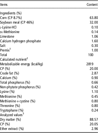
The experimental protocol was approved by the Animal Care and Use Committee of South China Agricultural University. All animal care procedures throughout the study followed Experimental Animal Care and Use Guidelines of China. A total of 540 mixed-sex 1-day-old White Pekin ducklings (from Guiliu Poultry Co., Ltd, Foshan, China) with initial BW of 43.4±0.1 g were randomly assigned to three dietary treatments, using six replicate plastic wire-floor pens (length 300 cm×width 200 cm) each with 30 birds and they were studied for 21 days. The birds were provided with ad libitum access to water and pelleted diets. Daylight was eliminated but 18-h/day lighting was provided from incandescent bulbs. The temperature of the room was maintained at 32°C to 34°C for the first 3 days and then reduced by 2°C to 3°C per week to a final temperature of 26°C. Control birds, not exposed to OTA, were fed a corn–soybean meal basal diet formulated to meet the nutritional requirements for starter ducks (National Research Council, 1994), as shown in Table 1. Birds exposed to OTA were fed the same diet with 16% moldy corn (described above) replacing fresh corn providing a total content of 2 mg of OTA/kg. The third group (CUR+OTA) was fed this diet with 2 mg/kg OTA, further supplemented with 400 mg/kg curcumin. Natural curcumin (>98%, by HPLC analysis) used in the experiment was provided by Nanjing Nutri-herb Biotech Co., Ltd (Nanjing, Jiangsu, China).
Ingredient composition and nutrient content of the basal diet (%, as-fed basis)

1 The premix provided per kilogram diet: vitamin A 10 000 IU, vitamin D3 3000 IU, vitamin E 20 mg, vitamin K3 2.0 mg, vitamin B1 2.0 mg, vitamin B2 8 mg, vitamin B6 4.0 mg, vitamin B12 0.02 mg, choline chloride 500 mg, nicotinic acid 50 mg, d-pantothenic acid 20 mg, folic acid 1 mg, biotin 0.2 mg, Fe 60 mg, Cu 10 mg, Mn 80 mg, Zn 60 mg, I 0.2 mg, Se 0.3 mg, Co 0.25 mg.
2 Values were calculated based on the data provided by Feed Database in China (2004).
3 CP was analyzed by Kjeldahl method; ether extract was analyzed by Soxhlet extraction.
Performance variables
Feed intake was recorded daily on a per replicate basis. Birds were weighed at the start and the end of 3-week test period to determine the average daily gain (ADG). The feed consumed from day 1 to day 21 was measured on a pen basis to calculate average daily feed intake (ADFI) and feed : gain (F/G). Mortality was checked daily and dead birds were weighed in order to adjust F/G.
Tissue sampling and storage
After an overnight (12 h) feed withdrawal at day 21, 12 ducks per treatment (two per replicate) with BW close to the replicate average were selected. Individual blood samples were collected from the wing vein; plasma samples were separated by centrifugation of blood at 1200×g for 10 min at 4°C and stored at −20°C for analysis. These birds were slaughtered quickly after blood collection; the entire liver was then collected and weighed, and the midpoints of jejunum of each bird were harvested and rinsed quickly with cold PBS. Portions were fixed in 10% formalin or 2.5% glutaraldehyde plus 2% paraformaldehyde and mucosa was scraped from the remainder, snap frozen in liquid nitrogen, and stored at −80°C.
Biochemical variables and measurement of jejunal mucosa cytokines
The levels of plasma malondialdehyde (MDA), glutathione peroxidase (GSH-Px), superoxide dismutase (SOD) and total antioxidant capacity (T-AOC) were assayed using a commercial kit (Nanjing Jiancheng Bioengineering Institute, Nanjing, China) according to the manufacturers’ instructions. The activity of diamine oxidase (DAO) in plasma was determined according to the method of previous assay (Li et al., Reference Li, Yu, Hao, Jin and Xu1996).
Jejunal mucosa samples (1 g) were homogenized on ice in 9 ml ice-cold PBS (pH 7.4) using an Ultra-Turrax T8 homogenizer (IKA Labortechnik, Staufen, Germany) and centrifuged at 3000×g for 20 min at 4°C to collect the supernatants. The levels of interleukin-1β (IL-1β), interleukin-8 (IL-8), interleukin-10 (IL-10) and tumor necrosis factor-α (TNF-α) were determined using an ELISA kits (Beijing Fangcheng Biotechnology Co., Ltd, Beijing, China). The mucosal content of MDA, and activities of SOD, and GSH-Px were measured with kits, as described above. All samples were measured in duplicate, at appropriate dilutions, to give activities of the enzymes in the linear range of standard curves constructed with pure enzymes. The protein content of supernatants was determined using the Coomassie Brilliant Blue G250 from Sigma Chemical Co. (St Louis, MO, USA) with bovine serum albumin as the standard.
Histopathological analysis of jejunum
Jejunal segments were carefully oriented with regard to the crypt to villus axis during embedding in paraffin. Sections of 5-μm thickness were dewaxed, rehydrated and stained with hematoxylin–eosin. Tissue sections were scanned with the Pannoramic MIDI (3D Histech, Budapest, Hungary). Thereafter, they were analyzed, quantified and photographed with the virtual microscopy software (Pannoramic Viewer version 1.15.2, 3D Histech) to determine histomorphometric variables: villus height and crypt depth. The morphometric analysis of the sections was performed on 10 randomly selected, well-oriented villi and crypts per duck.
Ultrastructure of tight junctions and mitochondria in jejunal enterocytes using transmission electron microscopy
Jejunal samples were processed for transmission electron microscopy (TEM) following standard procedures. Ultra-thin sections were mounted on copper grids, contrasted with standard uranyl acetate and lead citrate double-staining, and examined with a TEM (Hitachi HT7700, Tokyo, Japan) at 80 kV with an Ultrascan CCD camera. To evaluate changes in the ultrastructure of tight junctions, only sections with longitudinally oriented microvilli were examined in each sample. Ultrastructure morphometric assessments of mitochondria were conducted on every sixth epithelial cell CCD micrographs using NIH ImageJ software. Examinations were performed independently in a blinded manner per duck, using non-overlapping fields.
RNA isolation and real-time PCR
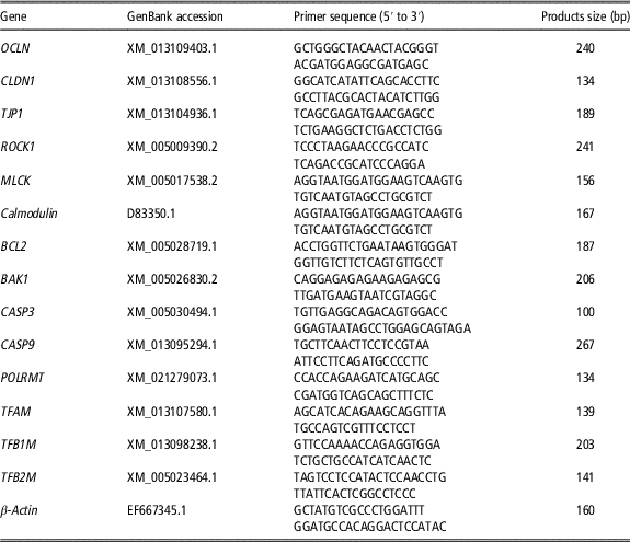
Total RNA was isolated from the frozen jejunal mucosa samples using Trizol reagent (Invitrogen Biotechnology, Carlsbad, CA, USA), and dissolved in RNase-free water. The concentration of extracted RNA was qualified by the NanoDrop ND-1000 spectrophotometer (Thermo Fisher Scientific, Waltham, MA, USA) and the integrity of RNA was verified using denatured RNA electrophoresis. The complementary DNA (cDNA) was synthesized using a PrimeScriptTM RT reagent kit with gDNA Eraser (Takara Biotechnology, Dalian, China), and was stored at −20°C. The cDNA was diluted with nuclease-free water (1 : 9) directly before quantitative real-time PCR analysis. The primers in the study, as shown in Table 2, were designed based on duck gene sequences using Primer Premier 5.0 and prepared from Shanghai Shenggong Biological Company (Shanghai, China).
Primers used for quantitative real-time PCR

OCLN=occludin; CLDN1=claudin1; TJP1=tight junction protein 1; ROCK1=ρ-associated protein kinase 1; MLCK=myosin light-chain kinase; BCL2=B-cell CLL/lymphoma 2; BAK1=BCL2-antagonist/killer 1; CASP3=apoptosis-related cysteine peptidase 3; CASP9=apoptosis-related cysteine peptidase 9; POLRMT=RNA polymerase mitochondrial; TFAM=mitochondrial transcription factor A; TFB1M=mitochondrial transcription factor B1; TFB2M=mitochondrial transcription factor B2.
Quantitative real-time PCR was performed using a iTaqTM Universal SYBER® Green Supermix (Takara Biotechnology, Dalian, China) in CFX96 Real-Time PCR Detection System (Bio-Rad, Hercules, CA, USA). The protocol of PCR was as follows: denaturation at 95°C for 3 min followed by 39 cycles at 95°C for 15 s, 30 s at the annealing temperature (TA) and 72°C for 30 s. Specificity of PCR products was evaluated by the analysis of melting curve. All analyses were performed in triplicate, each in a volume of 20 μl, and the average values were obtained. The
 $${\rm 2}^{{{\minus}\Delta \Delta C_{t} }} $$
method was used to calculate the relative mRNA expression of intestinal genes. The expression of β-actin was selected as an internal control to normalize the expression of targeted genes.
Western blotting analysis
Western blotting for detection of occludin (OCLN), tight junction protein 1 (TJP1), ρ-associated protein kinase 1 (ROCK1) and myosin light-chain kinase (MLCK) proteins was performed followed previously described methods with minor modifications (Bradford, Reference Bradford1976). In brief, 1 g of Jejunal mucosa tissue was sampled from each duck (one duck per replicate, six ducks per treatment), 0.5 g of tissue was homogenized on ice in lysis buffer (3 ml/g) with a protease inhibitor enzyme for 5 min. The homogenized mixture was centrifuged at 12 000×g for 20 min at 4°C, and 20 μl of supernatant was boiled for 5 min before being loaded into the gels. The protein concentrations were determined using the BCA protein assay kit (ThermoFisher Scientific, Rockford, IL, USA) according to the manufacturer’s instructions. Protein samples (50 μg) were subjected to 12% SDS-PAGE (25 min at 60 V and after that 60 min at 120 V). Then, the separated proteins were transferred on a polyvinylidene fluoride (PVDF) membrane. PVDF membranes were blocked using tris buffered saline with tween (TBST) containing 5% (w/v) non-fat milk powder for 1 h at room temperature, followed by overnight incubation at 4°C with the following primary antibodies at the indicated dilutions: anti-OCLN (rabbit monoclonal antibody, 1 : 10000 dilution, ab167161, Abcam, MA, USA); anti-TJP1 (rabbit IgG antibody, 1 : 1000 dilution, 13663, CST, MA, USA); anti-ROCK1 (rabbit monoclonal antibody, 1 : 1000 dilution, ab45171, Abcam); anti-MLCK (rabbit monoclonal antibody, 1 : 3000 dilution, ab76092, Abcam); anti-GAPDH (rabbit polyclonal antibody, 1 : 1500 dilution, AB-P-R, Hangzhou Xianzhi, Hangzhou, China). Next, the membranes were washed three times with TBST and incubated with HRP-conjugated secondary antibody (1 : 2500 dilution, Boster, Wuhan, China) for 1 h at room temperature. Analysis of blot images was performed by ImageJ program (National Institutes of Health, MD, USA) to measure the densitometry value. The relative densitometry value, D, was calculated as D=Dg/Da, where Dg is the densitometry value of target protein and Da the densitometry value of GAPDH.
Statistical analysis
Replicate (n=6) was taken as the experimental unit. Except where otherwise noted, two sampled birds per replicate were used. Statistical analysis was performed using the one-way ANOVA procedure of SAS 9.3 (SAS Institute, Cary, NC, USA). Where appropriate, differences among treatments were tested using Duncan’s multiple-range test at a significance level of P<0.05.
Results
Growth performance
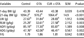
The results for performance of the ducks are summarized in Table 3. Compared with controls, ducks fed OTA diet had significantly lower 21-day BW, ADFI and ADG, and higher absolute and relative liver weight (P<0.05). Addition of curcumin to the OTA diet (CUR+OTA) significantly increased 21-day BW, ADFI and ADG to the values of the controls, while partially ameliorated the increase in liver weight (P<0.05).
Effect of ochratoxin A and curcumin on growth performance of ducks from day 1 to day 21

OTA=Ochratoxin A (2 mg/kg); CUR+OTA=curcumin (400 mg/kg)+ochratoxin A (2 mg/kg); LW=liver weight; RLW=relative liver weight; ADFI=average daily feed intake; ADG=average daily gain; F/G=feed : gain.
a,b,cValues within a row with different superscript letters differ significantly at P<0.05.
Antioxidant parameters and jejunal cytokines
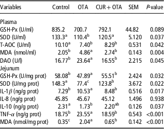
Table 4 shows the effect of the OTA-contaminated diet on antioxidant indices in plasma and jejunal mucosa cytokines in the ducks at 21 days. The plasma activities of SOD and T-AOC in ducks fed the OTA-contaminated diet decreased and DAO increased along with levels of MDA (P<0.05). In jejunal mucosa, contents of inflammatory cytokines like IL-1β and TNF-α increased and IL-10 decreased, compared with the controls (P<0.05); mucosal activities of GSH-Px and SOD decreased whereas the content of MDA was increased to 483%. Additional supplementation with curcumin significantly improved antioxidant status in jejunum in terms of GSH-Px and SOD (P<0.05), decreased jejunal IL-1β and TNF-α levels (P<0.05), and further decreased both plasma and jejunal content of MDA (P<0.01). There were no differences in jejunal levels of IL-8 among dietary treatments (P>0.05).
Effect of ochratoxin A and curcumin on biochemical indices and jejunal immunity of 21-day ducks

OTA=Ochratoxin A (2 mg/kg); CUR+OTA=curcumin (400 mg/kg)+ochratoxin A (2 mg/kg); GSH-Px=glutathione peroxidase; SOD=superoxide dismutase; T-AOC=total antioxidant capacity; MDA=malondialdehyde; DAO=diamine oxidase; IL-1β=interleukin-1β; IL-8=interleukin-8; IL-10=interleukin-10; TNF-α=tumor necrosis factor-α.
a,b,cValues within a row with different superscript letters differ significantly at P<0.05.
Histomorphological changes in jejunum
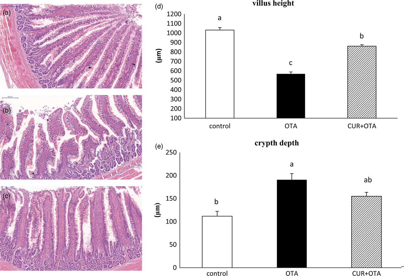
Representative photomicrographs of jejunal villi in are depicted in Figure 1. Compared with the controls, ducks fed the diet containing OTA showed villous blunting and epithelial denudation along with a related decrease in villous length and increased the depth of crypts. Supplementation of the OTA diet with curcumin partly restored villus length in jejunum.
Representative photomicrographs of hematoxylin–eosin stained of villi in jejunum (magnification, 200×; scale bars, 200 μm). (a) The jejunal mucosa of the controls showed normal villous structure. (b) Ducks fed the diet with 2 mg/kg ochratoxin A (OTA) showed villous blunting and epithelial denudation in the jejunum, compared with the controls. (c) Supplementation of the 2 mg/kg OTA diet with 400 mg/kg curcumin (CUR) improved villous height and width in jejunum. (d) The villous height was significantly decreased in the jejunum of ducks exposed to 2 mg/kg OTA compared with the controls while supplementation of the OTA diet with 400 mg/kg CUR increased villus height. (e) Crypt elongation was significantly increased in the jejunum of ducks fed the diet with 2 mg/kg OTA compared with the controls and supplementation of the OTA diet with 400 mg/kg CUR decreased crypt elongation. Results are expressed as means±SEM; n=6 animal/group. a,b,cMeans with different letters were significantly different (P<0.05).

Transcript abundance of tight junction-related proteins in jejunal mucosa
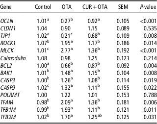
Jejunal mucosa expression of genes related to tight junctions were quantified at day 21. As shown in Table 5, transcripts of OCLN and TJP1 in ducks fed the diet with OTA were significantly decreased than those in controls (P<0.01); and those of ROCK1 and MLCK were increased (P<0.05). The expression of OCLN and TJP1 in ducks fed curcumin in addition to OTA were significantly higher than with the toxic diet alone (P<0.01); simultaneously, ROCK1 and MLCK transcripts were reduced (P<0.05). There were no differences in the jejunal relative expression of claudin 1 or Calmodulin among dietary treatments (P>0.05).
Effects of ochratoxin A and curcumin on relative jejunal mucosa expression of genes related to tight junctions and mitochondrial function in day 21 ducks

OTA=Ochratoxin A (2 mg/kg); CUR+OTA=curcumin (400 mg/kg)+ochratoxin A (2 mg/kg); OCLN=occludin; CLDN1=claudin1; TJP1=tight junction protein 1; ROCK1=ρ-associated protein kinase 1; MLCK=myosin light-chain kinase; BCL2=B-cell CLL/lymphoma 2; BAK1=BCL2-antagonist/killer 1; CASP3=apoptosis-related cysteine peptidase 3; CASP9=apoptosis-related cysteine peptidase 9; POLRMT=RNA polymerase mitochondrial; TFAM=mitochondrial transcription factor A; TFB1M=mitochondrial transcription factor B1; TFB2M=mitochondrial transcription factor B2.
a,b,cValues within a row with different superscript letters differ significantly at P<0.05.
Relative expression of genes related to apoptosis and mitochondrial transcription factors in jejunal mucosa
Table 5 also shows the relative abundance of apoptosis and mitochondrial transcription factors transcripts in jejunal mucosa. The transcript abundance of BCL2-antagonist/killer 1 (BAK1) and apoptosis-related cysteine peptidase 3 (CASP3) increased over controls in jejunal mucosa of ducks fed OTA while there were no changes in these transcripts between CUR+OTA ducks and controls (P>0.05). The expression of B-cell CLL/lymphoma 2 (BCL2) mRNA in ducks fed the diet with OTA was significantly lower (P<0.01) than in the controls and was intermediate when curcumin was given, in addition to OTA. Ducks fed the diet containing OTA showed increases (P<0.01) in mitochondrial transcription factor A (TFAM), mitochondrial transcription factor B1 and B2 (TFB1M, TFB2M). In all cases, the addition of curcumin to the OTA diet attenuated the increased expression of these transcription factors.
Protein expression of tight junction-related proteins in jejunal mucosa
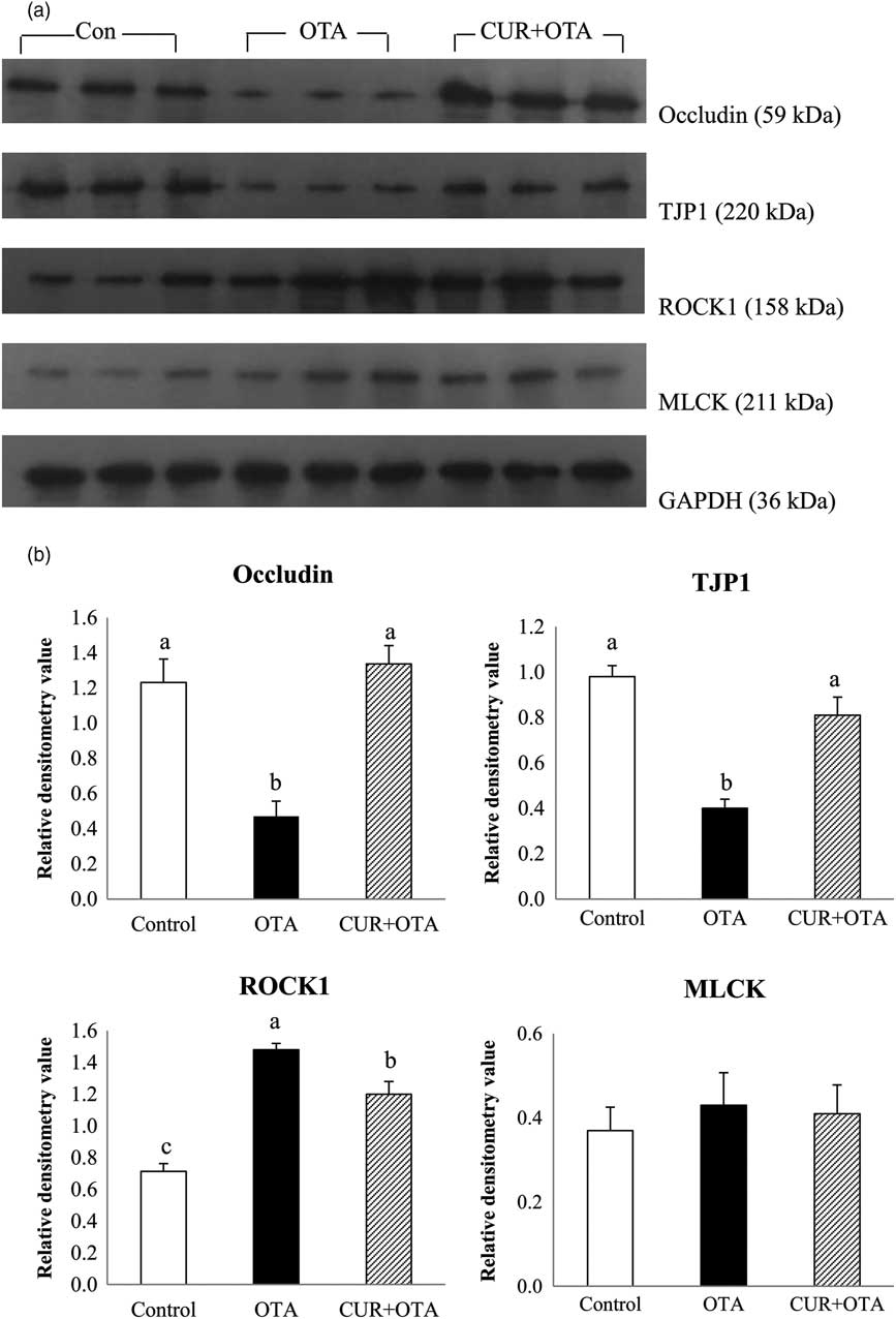
The full gels with molecular weight ladder and the appropriate controls are presented in Supplementary Figure S1. Immunoblotting (Figure 2a) and subsequent densitometry (Figure 2b) demonstrated that OCLN and TJP1 protein expression were significantly decreased in ducks fed the diet with OTA compared with the controls (P<0.05); and those of ROCK1 was increased (P<0.05). Occludin and TJP1 expression were decreased by 1.6-fold and 1.5-fold, respectively, whereas ROCK1 expression was increased by 1.2-fold. The protein expression of OCLN and TJP1 in ducks fed curcumin in addition to OTA were significantly higher than with the toxic diet alone (P<0.01); simultaneously, ROCK1 protein expression was reduced (P<0.05).
Western blotting analysis of jejunal mucosa tight junction and cytoskeleton-related protein expression in the control, 2 mg/kg ochratoxin A (OTA) group and co-administration of 400 mg/kg curcumin (CUR) group of ducks after 21 days of feeding. (a) A representative immunoblot of three replicates is shown. (b) Densitometry value of protein expression levels is shown. Data are expressed as means±SEM of all determinations. TJP1=tight junction protein 1; ROCK1=ρ-associated protein kinase 1; MLCK=myosin light-chain kinase protein. a,b,cColumns with different letters are considered significant (P<0.05).

Ultrastructure of tight junctions and mitochondria in jejunal mucosa
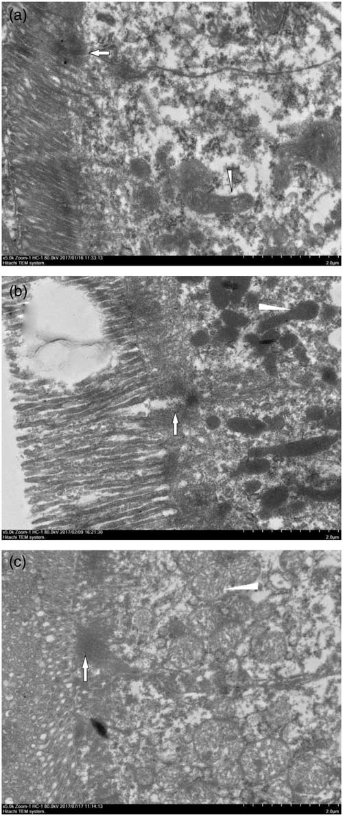
The ultrastructure of tight junctions and mitochondria in jejunal mucosa, visualized by TEM (magnification 5000×), is shown in Figure 3. Enterocytes of control ducks demonstrated unaffected tight junctions and normal mitochondria with clear cristae. In contrast, enterocytes from ducks exposed to OTA had obviously damaged microvilli, widened intercellular spaces, condensed cytoskeleton and mitochondria appeared to be swollen and suffering from the disappearance of the inner and outer membrane and there were no visible cristae. These diverse morphological alterations of tight junctions and mitochondria suggest intestinal barrier and mitochondrial dysfunctions in the enterocytes. These adverse effects were alleviated in ducks fed curcumin along with the OTA-contaminated corn.
Ultrastructure of tight junctional complex and mitochondria in jejunal mucosa. These panels show representative micrographs of the jejunal epithelium (original magnification 5000×; scale bars, 2.0 μm). (a) Enterocyte of the control exposed to vehicle with unaffected tight junction region (arrow) and normal mitochondria (arrowhead). (b) Widening of the intercellular space (arrow), absence of mitochondria membrane with fuzziness and derangement of the cristae (arrowhead) after being fed the diet with 2 mg/kg ochratoxin A (OTA). (c) Normal intercellular space (arrow), visible mitochondria membrane and regular cristae (arrowhead) after feeding with the diet containing 2 mg/kg OTA plus 400 mg/kg curcumin (CUR).

Discussion
Ochratoxin A, as a secondary metabolite of Penicillium and Aspergillus microfungi, is a well-known and widely spread mycotoxin all over the world, which exerts hazardous effects on human and animal health (Kőszegi and Poór, Reference Kőszegi and Poór2016). This study used an in vivo exposure model to study the harmful effects of OTA in White Pekin ducklings. Feed containing 2 mg/kg OTA significantly decreased ADFI, ADG, and increased F/G between day 1 and day 21, all of which agrees with earlier reports of performance-depressing effects of OTA (Prior et al., Reference Prior, Oneil and Sisodia1980; Hoehler et al., Reference Hoehler and Marquardt1996; Zhong, Reference Zhong2008). The additional supplementation with 400 mg/kg curcumin improved the performance, or offset the toxicity of OTA, of the ducks studied here. No information appears to be available in the literature regarding the effect of curcumin on ameliorating the decrease in BW of ducks fed OTA-contaminated feed. There have been such effects in broilers fed aflatoxin B1-contaminated rations, where the co-administration of curcumin, in a dose-dependent manner partially ameliorated the decrease in BW (Gowda et al., Reference Gowda, Ledoux, Rottinghaus, Bermudez and Chen2009; Muhammad et al., Reference Muhammad, Sun, Wang, Li, Wang, Cheng, Li, Zhang and Hamid2017). Additional evidence was obtained here for the improved performance of ducks fed the diet containing 400 mg/kg curcumin with the OTA being attributable to the high antioxidant properties claimed of curcumin.
Numerous mycotoxins are able to alter intestinal barrier function and the intestinal mucosa represents the first site of interaction between noxious agents of dietary origin and the organism. In the present experiment, the enterotoxicity of OTA was observed in terms of deleterious modifications in villus architecture, grossly evident in the shortened villi and elongated crypts. These changes were largely reversed by co-exposure to curcumin (Figure 1). Mucosal architecture injury is initiated by increasing number of intraepithelial lymphocytes. Crypt elongation is a compensatory feature occurring in response to villous shortening, which may be caused by releasing various growth factors and cytokines from inflammatory cells due to the stromal–epithelial interactions. Then villous atrophy associated with crypt hyperplasia occurred (Solcan et al., Reference Solcan, Pavel, Floristean, Chiriac, Şlencu and Solcan2015). Therefore, it can be speculated that curcumin might control the number of intraepithelial lymphocytes, and inhibit secretion of inflammatory cytokines to protect intestinal integrity. Contact between intestinal epithelial cells includes three ultrastructural components: desmosomes, adherens junctions and tight junctions. The latter is the most important structures in intestinal barrier function, and play a decisive role in intestinal mucosa permeability. Proteins of tight junctions include transmembrane proteins (OCLN, CLDN, tricellulin, etc.) and intracellular proteins (zonula occludens-1, -2, -3, cingulin, etc.). These tight junction proteins interact with F-actin and MLC and anchor them to the cytoskeleton structure. The kinase ROCK plays an important role in maintaining the structure of tight junctions by regulating the phosphorylation status of downstream MLC to influence overall cytoskeletal structure (Du et al., Reference Du, Kim, Shen and Dai2016). In the present study, ducks fed the OTA-contaminated diet had significantly reduced OCLN and TJP1 transcripts in the jejunal mucosa, but increased expression of ROCK1 and MLCK mRNA. Western Blot analyses also showed a significant decrease in OCLN and TJP1 protein mass, and increased protein expression of ROCK1 (Figure 2) without any changes in MLCK in OTA diet, the addition of curcumin to the OTA diet attenuated the modifications of those proteins. Furthermore, histomorphological and ultrastructural examination of the jejunum showed villous blunting and epithelial denudation along with damaged microvilli and widened intercellular spaces in ducks fed the diet containing OTA. These findings indicate that OTA may have affected the phosphorylation of MLC through the ROCK pathway, thereby destroying the cytoskeletal structure, and leading to delocalization of tight junction proteins. Other studies have reported that human enterocytes exposed to OTA reduced the barrier function, by removal of specifical claudin isoforms and affecting of lipid rafts in biological membranes (McLaughlin et al., Reference McLaughlin, Padfield, Burt and O’Neill2004). Mainly involved in protein synthesis inhibition and oxidative stress-related cytotoxicity could also participate in the effect of OTA on intestinal permeability (Ranaldi et al., Reference Ranaldi, Mancini, Ferruzza, Sambuy and Perozzi2007; Maresca et al., Reference Maresca, Mahfound, Pfohl-Leszkowicz and Fantini2001; Grenier and Applegate, Reference Grenier and Applegate2013). In addition, the intestinal barrier dysfunction is associated with intestinal inflammation, which is based on the destruction of tight junctions (Turner, Reference Turner2009). Tumor necrosis factor-α which is secreted by activated T cells increases intestinal permeability, and may directly or indirectly inhibit the expression of tight junction proteins (Chung et al., Reference Chung, Hwang, Park and Kim2002). Al-Anati et al. (Reference Al-Anati, Essid, Stenius, Beuerlein, Schuh and Petzinger2010) reported that only macrophages and macrophage-derived cells respond to OTA and were considered to be sources of the TNF-α, released by exposure to OTA. In the present study, the contents of IL-1β, and TNF-α in jejunal mucosa were increased in ducks given OTA-contaminated feed but were decreased by co-supplementation with curcumin. These results showed that curcumin largely protected T cells from OTA-induced impairment. The protective role of curcumin is mainly due to its antioxidant and anti-inflammatory properties.
The mitochondrion is the predominant site for intracellular production of ROS production and a prime target for oxidative damage, especially of the exposed mitochondrial DNA and membrane unsaturated fatty acids (Venditti et al., Reference Venditti, Stefano and Meo2013). Several studies indicate that both in vivo and in vitro exposure to OTA results in the overproduction of ROS. In the present experiment, OTA-induced lipid peroxidation in jejunum epithelial cells, and was offset by dietary curcumin. As a liposoluble substance, it may use lipophilic integration into the epithelial cell’s membrane and mitochondrial membrane phospholipids, thereby directly neutralizing ROS in its role of an antioxidant, thus significantly alleviating OTA-induced oxidative damage. Previous study pointed out that the natural flavonoids could be specifically enriched in mitochondria, thereby playing a role in protecting the body (Dorta et al., Reference Dorta, Pigoso, Mingatto, Rodrigues, Prado, Helena, Uyemura, Santos and Curti2005). Curcumin, as a plant-derived polyphenolic substance, may achieve its antioxidant activity by similar subcellular localization like natural flavonoids, but this remains to be demonstrated. The mitochondrial genome is transcribed by specialized machinery that includes RNA polymerase mitochondrial (POLRMT), TFAM, TFB1M and TFB2M paralogues (Asin-Cayuela and Gustafsson, Reference Asin-Cayuela and Gustafsson2007). The TFAM, which was cloned as a transcription factor for mitochondrial DNA (mtDNA), may play a crucial role in maintaining mtDNA as a main component of the nucleoid (Kang et al., Reference Kang, Kim and Hamasaki2007). A strong negative effect of OTA on cellular energy (ATP) production (Poór et al., Reference Poór, Veres, Jakus, Antus, Montskó, Zrínyi, Vladimir-Knežević, Petrik and Kőszegi2014) is consistent with OTA penetrating into mitochondria and most probably binding to proteins participated in maintenance of membrane potential and oxidative phosphorylation via interfering with phosphate transport along with suppression of electron transport (Wei et al., Reference Wei, Lu and Wei1985). This may resulted in depletion of mtDNA and mutation-mediated mitochondrial toxicity. Increased expression of TFAM, TFB1M and TFB2M was observed here in ducks given OTA-contaminated feed with no marked difference in POLRMT. We hypothesized that the enhanced expression of genes for these factors may represent a compensatory mechanism in response to oxidative stress-related mitochondrial dysfunctions, including deficient electron transport system, incomplete mitochondrial biogenesis and excessive cell apoptosis. Taha et al. (Reference Taha, Seidman, Mailhot, Boudreau, Gendron, Beaulieu, Menard, Delvin, Amre and Levy2010) reported that iron–ascorbate-mediated lipid peroxidation raised the gene expression and protein mass of TFAM, TFB1M and TFB2M in Caco-2/15 cells. Accordingly, TFAM was found to be upregulated in response to lipopolysaccharide-induced oxidative damage to mitochondria, presumably to enhance mtDNA levels and oxidative phosphorylation activity (Suliman et al., Reference Suliman, Carraway, Welty-Wolf, Whorton and Piantadosi2003). Ikeda et al. (Reference Ikeda, Ide, Fujino, Arai, Saku, Kakino, Tyynismaa, Yamasaki, Yamada, Kang, Suomalainen and Sunagawa2015) found that overexpression of TFAM increased mtDNA copy number, suppressed mitochondrial ROS, and facilitated cardioprotection associated with limited mitochondrial oxidative stress. It was demonstrated that a low level of curcumin was able to elicit mitochondrial biogenesis in cells and tissues through induction of peroxisome proliferator-activated receptor γ co-activator 1-α, modulating mitochondrial transcription factors (Rainey et al., Reference Rainey, Moustapha, Parker and Petit2017). This suggests the existence of a retrograde signaling pathway from mitochondria to the nucleus, to precisely regulate the related factors expression. The mechanism involved needs further investigation. The ultrastructure of jejunum epithelial cells of ducks oxidative stressed by the OTA-contaminated feed showed that inner and outer mitochondrial lipid bilayer and cristae were obscure whereas nuclei did not seem to be affected (Figure 3); this is again consistent with mtDNA being more vulnerable than nuclear DNA to oxidative damage (Kanki et al., Reference Kanki, Nakayama, Sasaki, Takio, Alam, Hamasaki and Kang2004).
In conclusion, ducklings fed OTA had depressed performance, exhibited intestinal lipid peroxidation, disrupted tight junctions, cell apoptosis and caused mitochondrial dysfunction, likely related to oxidative stress. Addition of dietary curcumin effectively prevented OTA-induced toxicity: caused less lipid peroxidation, improved intestinal barrier and mitochondrial function, mainly by affecting tight junctions, cytoskeleton proteins, apoptotic proteins and mitochondrial transcription factors. These results may be helpful in revealing the molecular mechanism of curcumin in alleviating the destruction of the intestinal barrier caused by mycotoxins.
Acknowledgments
The authors sincerely thank W. Bruce Currie (Emeritus Professor, Cornell University, Ithaca, NY) for his excellent technical assistance in the presentation of the manuscript. This work was financially supported by the Modern China Agricultural Research System Program (CARS-42-15 and CARS-42-13), President Foundation of Guangdong Academy of Agricultural Sciences (201619), National Natural Science Foundation of China (31500045), the National Key Research Program (2016YFD0500509-07), Science and Technology Program of Guangdong Province (2013A061401020), Operating Funds for Guangdong Provincial Key Laboratory of Animal Breeding and Nutrition (2014B030301054), Supporting Program for the Research of State Key Laboratory of Livestock and Poultry Breeding, and Supporting Program for Guangdong Agricultural Research & Development Center of Livestock and Poultry Healthy Breeding.
Declaration of interest
The authors declare that there are no conflicts of interest.
Ethics statement
The experimental animal protocol for this study was approved by the Animal Care and Use Committee of South China Agricultural University. All animal care procedures throughout the study followed Experimental Animal Care and Use Guidelines of China.
Software and data repository resources
The authors declare that the data of this research are not deposited in any official repository.
Supplementary Material
To view supplementary material for this article, please visit https://doi.org/10.1017/S1751731118000678