Atrial septal defect and ventricular septal defect are the two most common forms of congenital heart disease that occur as an isolated anomaly with a prevalence of 2.6 and 1.6 per 1000 live births, respectively.Reference van der Linde, Konings and Slager 1 These septal defects vary in size, ranging from small defects without haemodynamic significance to large shunts. Pathophysiologically, the intracardiac shunt will impose a haemodynamic burden on the heart, which can lead to atrial and ventricular dilatation and increased pulmonary vascular resistance. Surgical or percutaneous closure of the defect is used as standard treatment when patients with atrial septal defect and ventricular septal defect develop conditions such as intractable chronic cardiac failure, failure to thrive, pulmonary hypertension, or significant left-to-right shunt.
Brain natriuretic peptide and its inactive precursor N-terminal pro-brain natriuretic peptide are cardiac markers that are released into the circulation after pressure overload, volume expansion, and increased myocardial wall stress.Reference Levin, Gardner and Samson 2 The active fragment brain natriuretic peptide has natriuretic, vasodilatory, and diuretic effects. Both markers have proven their ability to detect cardiac impairment in cardiac failure in the general population.Reference Jessup, Abraham and Casey 3
Although both brain natriuretic peptide and N-terminal pro-brain natriuretic peptide have proven to be useful for assessing cardiac function in acquired heart disease, their role in the diagnostic approach and clinical decision making in patients with simple congenital heart diseases such as atrial septal defect and ventricular septal defect is not well defined. The aim of this systematic review was to evaluate the recent literature on B-type natriuretic peptide activation in patients with atrial or ventricular septal defects and clarify the relationship of these markers with cardiac function and haemodynamic changes after defect closure.
Methods
Search strategy, selection criteria, and data extraction
On January 20th, a systematic literature search using MEDLINE and EMBASE, with focus on atrial septal defect and ventricular septal defect, was executed. The following Medical Subject Headings and text keywords were used: “natriuretic peptide, brain” or “pro-brain natriuretic peptide” and “heart septal defects” or “ventricular septal defect”, or “atrial septal defect”.
All article titles and abstracts were screened to identify relevant studies. Articles had to be written in English and involved human subjects. Brain natriuretic peptide levels and patients’ age at the time of brain natriuretic peptide measurement had to be reported clearly per cardiac diagnosis. Therefore, articles describing brain natriuretic peptide levels for a group of patients with diagnoses of congenital heart disease were excluded. We focused on isolated atrial and ventricular septal defect as these diagnoses are two of the most common congenital heart defects, and especially ventricular septal defect is often associated with cardiac failure at young age. Patients with septal defects as part of a more complex congenital heart disease were excluded because natriuretic peptide levels could be influenced by other components of the complicated anatomy. Articles on Eisenmenger syndrome due to isolated ventricular septal defect or atrial septal defect were included to provide a complete overview, including this rare but severe complication. In all selected articles, references were cross-checked using the same inclusion and exclusion criteria to identify articles missed by the initial search strategy.
We extracted data on the type of congenital heart disease, age, plasma brain natriuretic peptide levels, and brain natriuretic peptide immunoassay method. When reported in the article, plasma brain natriuretic peptide levels of healthy controls, moment of brain natriuretic peptide measurement – when sequential measurement was reported – and correlations between plasma brain natriuretic peptide and cardiac function parameters measured with echocardiography, cardiac magnetic resonance imaging, cardiac catheterisation, exercise test, or New York Heart Association classification were collected. For all potentially relevant articles, eligibility was assessed by two authors. Discrepancies were resolved by discussion. Both brain natriuretic peptide and N-terminal pro-brain natriuretic peptide will further be referred to as brain natriuretic peptide in this article, unless a separate use is needed for clarification.
Results
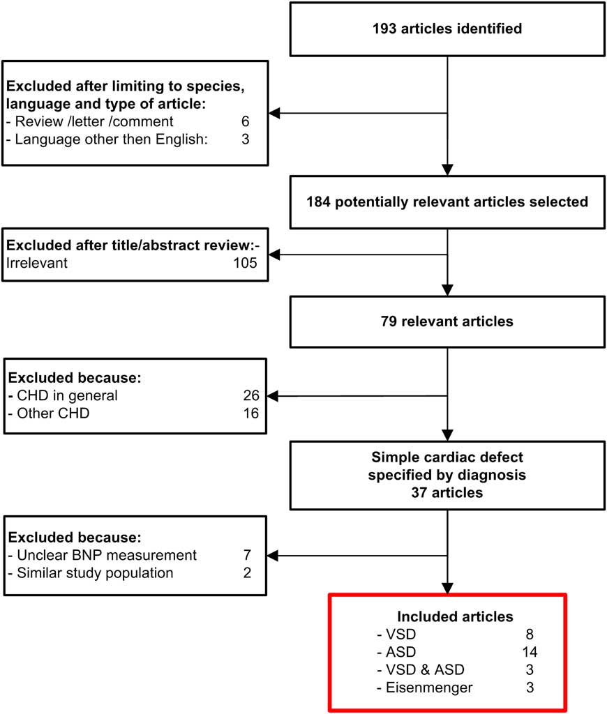
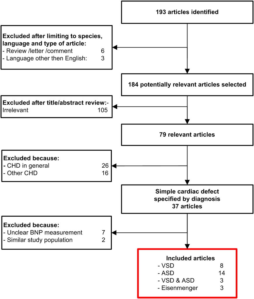
The literature search yielded 193 potential eligible studies (Fig 1). First, 114 articles were excluded as they described topics irrelevant for this systematic review. We excluded 41 articles because of their focus on other congenital heart diseases or congenital heart disease in general. In all, 38 articles met our inclusion criteria. After critical review of the complete article, nine more articles were excluded. In seven studies, brain natriuretic peptide values were not clearly reported. Furthermore, two articles were excluded as patient populations were described for the second time in a subgroup analyses. Eventually, 28 articles were included in this systematic literature review, describing atrial septal defect (n = 14), ventricular septal defect (n = 8), both atrial septal defect and ventricular septal defect (n = 3), and Eisenmenger syndrome (n = 3). In 11 articles, longitudinal data were available, describing sequential brain natriuretic peptide measurements before and after septal defect closure.
Literature search and selection. Numbers of articles for each step of the process are indicated. CHD = congenital heart disease; BNP = brain natriuretic peptide; VSD = ventricular septal defect; ASD = atrial septal defect.
Atrial septal defect
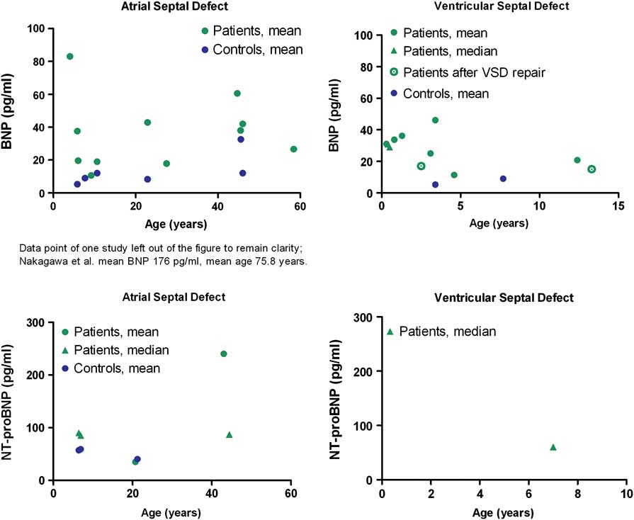
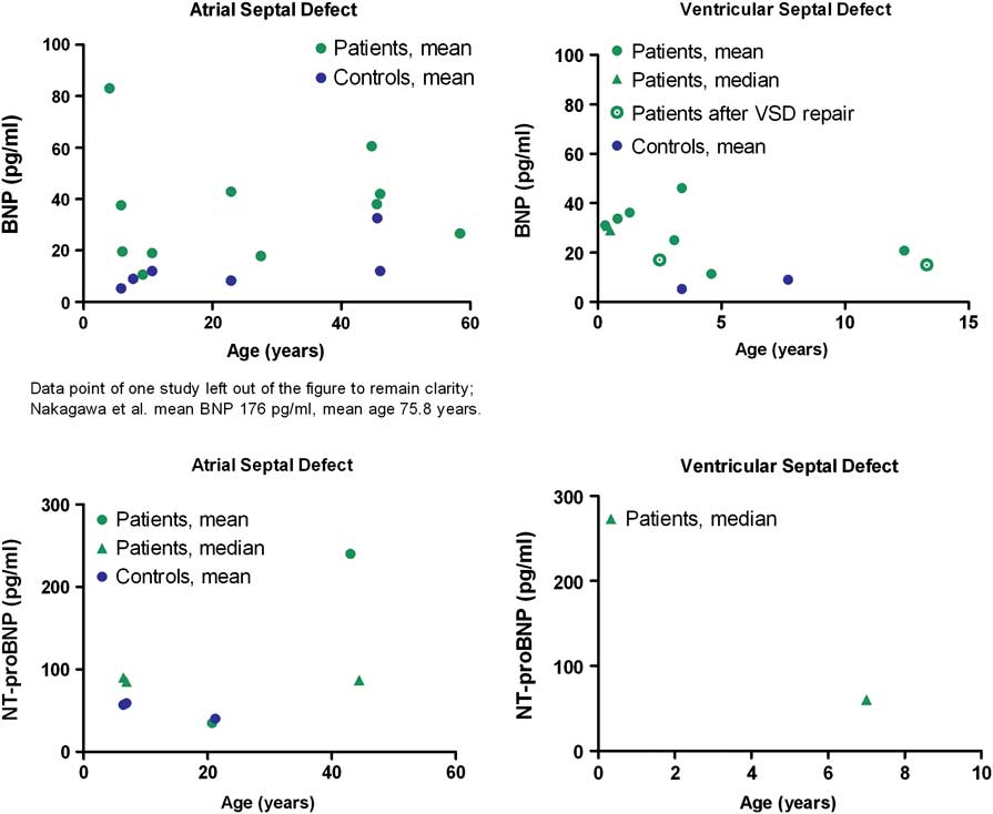
We included 17 articles reporting data on brain natriuretic peptide values in patients with unrepaired atrial septal defect, describing a total of 429 patients with a mean/median age ranging from 4 to 75.8 years.Reference Uz, Aparci and Acar 4 – Reference Wu, Akagi and Taniguchi 20 No studies describing brain natriuretic peptide in repaired atrial septal defect patients were found. A significant difference was observed for all ages in brain natriuretic peptide levels between patients with atrial septal defect (mean/median values of brain natriuretic peptide and N-terminal pro-brain natriuretic peptide ranging from 10.6 to 175.9 picograms per millilitre and 35.2 to 240 picograms per millilitre, respectively) and healthy age-matched controls (mean/median values of brain natriuretic peptide and N-terminal pro-brain natriuretic peptide ranging from 5.3 to 32.6 picograms per millilitre and 4.04 to 59 picograms per millilitre, respectively; Fig 2). Symptomatic patients in New York Heart Association class III revealed significantly higher N-terminal pro-brain natriuretic peptide values when compared with asymptomatic patients in New York Heart Association functional class I (142 plus or minus 72 picograms per millilitre versus 285 plus or minus 101 picograms per millilitre, p-value less than 0.05).Reference Schoen, Zimmermann and Kittner 16
Brain natriuretic peptide and N-terminal pro-brain natriuretic peptide measurements per cardiac diagnosis. Mean/median values of brain natriuretic peptide, N-terminal pro-brain natriuretic peptide and age for patients and controls per cardiac diagnosis. Each symbol reflects one study patient population or control population.
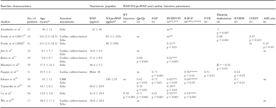
A total of 11 articles presented correlations between plasma brain natriuretic peptide and cardiac function parameters measured by echocardiography, cardiac magnetic resonance imaging, and/or cardiac catheterisation (Table 1). In five out of seven studies, a positive correlation was observed between brain natriuretic peptide levels and right ventricular end-diastolic dimensions and/or volume.Reference Uz, Aparci and Acar 4 , Reference Attenhofer Jost, Oechslin and Seifert 7 , Reference Eerola, Jokinen and Pihkala 9 – Reference Kunii, Kamada and Ohtsuki 11 , Reference Schoen, Zimmermann and Kittner 16 , Reference Trojnarska, Szyszka and Gwizdala 18 In contrast, left ventricular end-diastolic dimensions did not relate to brain natriuretic peptide and N-terminal pro-brain natriuretic peptide values.Reference Eerola, Jokinen and Pihkala 9 , Reference Eerola, Pihkala, Boldt, Mattila, Poutanen and Jokinen 10 In three studies, measures of diastolic dysfunction correlated with brain natriuretic peptide values.Reference Masutani, Taketazu and Mihara 6 , Reference Attenhofer Jost, Oechslin and Seifert 7 , Reference Eerola, Pihkala, Boldt, Mattila, Poutanen and Jokinen 10 Right ventricular end-diastolic pressure measured during cardiac catheterisation also related to brain natriuretic peptide values.Reference Nagaya, Nishikimi and Uematsu 14 , Reference Schoen, Zimmermann and Kittner 16 When focusing on left ventricular end-diastolic pressure this relation was not observed.Reference Nagaya, Nishikimi and Uematsu 14 In three out of four studies, pulmonary artery pressure correlated strongly with natriuretic peptide values.Reference Uz, Aparci and Acar 4 , Reference Nagaya, Nishikimi and Uematsu 14 , Reference Schoen, Zimmermann and Kittner 16 In contrast, in one study the presence of pulmonary hypertension was not related to brain natriuretic peptide.Reference Jan, Fu, Hwang and Lin 5
Atrial septal defect.

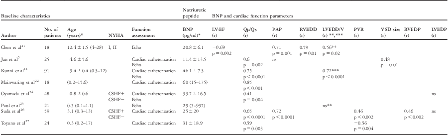
BNP = brain natriuretic peptide; CMR = cardiac magnetic resonance imaging; LVESV = left ventricular end-systolic volume; NS = not significant; NT-proBNP = N-terminal proBNP; PAP = pulmonary artery pressure; PVR = pulmonary vascular resistance; RV-function = right ventricular function; RVEDP = right ventricular end-diastolic pressure
β: beta coefficient; (r): correlation coefficient; +: positive correlation, not further specified; Qp/Qs: pulmonary-to-systemic flow ratio
BNP levels were determined by Triage BNP immunoassay (Biosite Diagnostics, La Jolla, California, United States of America), IRMA (Shionoria, Shionogi, Japan). NT-proBNP was evaluated with ECLIA (Elecsys, Roche Diagnostics, Basel, Switzerland)
*Values are presented as mean ± SD or median (range)
**RVEDD: right ventricular end-diastolic diameter
***RVEDV: right ventricular end-diastolic volume
****RAV: right atrial volume
*****RAP: right atrial pressure
Correlations between brain natriuretic peptide and the ratio of pulmonary to systemic blood flow (Qp/Qs) were assessed in seven studies and ranged from non-significant up to highly significant (r equals 0.71, p-value less than 0.001).Reference Uz, Aparci and Acar 4 However, atrial septal defect size was not or only weakly related to brain natriuretic peptide measures in four studies.Reference Jan, Fu, Hwang and Lin 5 , Reference Eerola, Jokinen and Pihkala 9 , Reference Schoen, Zimmermann and Kittner 16 , Reference Wu, Akagi and Taniguchi 20 In one study, an exercise test was performed, which revealed a negative correlation between maximum oxygen uptake and brain natriuretic peptide values.Reference Trojnarska, Szyszka and Gwizdala 18
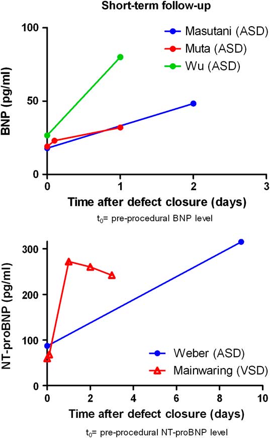
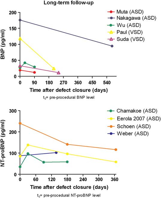
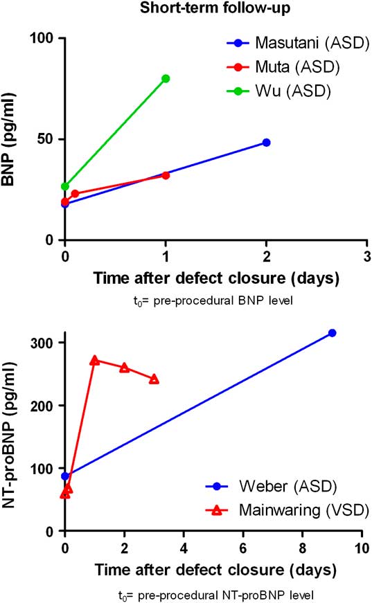
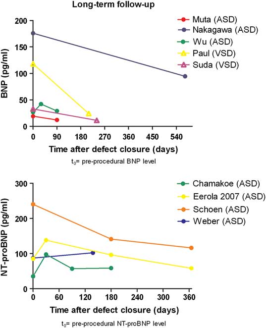
In eight studies, longitudinal data were available, describing sequential brain natriuretic peptide measurement before and after percutaneous or surgical atrial septal defect closure.Reference Masutani, Taketazu and Mihara 6 , Reference Chamakou, Dede and Moutafi 8 , Reference Eerola, Pihkala, Boldt, Mattila, Poutanen and Jokinen 10 , Reference Muta, Ishii, Maeno, Akagi and Kato 13 , Reference Nakagawa, Akagi and Taniguchi 15 , Reference Schoen, Zimmermann and Kittner 16 , Reference Weber, Dill and Deetjen 19 , Reference Wu, Akagi and Taniguchi 20 Initially, brain natriuretic peptide levels increased notably within days after atrial septal defect closure (Fig 3). A few months post procedure, a decrease in brain natriuretic peptide levels was observed and they became comparable to pre-procedural levels or even lower (Fig 4).Reference Chamakou, Dede and Moutafi 8 , Reference Eerola, Pihkala, Boldt, Mattila, Poutanen and Jokinen 10 , Reference Muta, Ishii, Maeno, Akagi and Kato 13 , Reference Nakagawa, Akagi and Taniguchi 15 , Reference Schoen, Zimmermann and Kittner 16 , Reference Weber, Dill and Deetjen 19 , Reference Wu, Akagi and Taniguchi 20 When brain natriuretic peptide values were measured 1 year after closure, levels in percutaneously treated patients were comparable to those of control patients. However, surgically treated patients still revealed elevated brain natriuretic peptide levels 1 year after the procedure.Reference Eerola, Pihkala, Boldt, Mattila, Poutanen and Jokinen 10 In one study, the decrease in right ventricular systolic pressure and right ventricular end-diastolic volume 12 months after atrial septal defect closure correlated with the changes in plasma brain natriuretic peptide (r equals 0.6, p-value less than 0.01, r equals 0.63, p-value less than 0.01, respectively).Reference Schoen, Zimmermann and Kittner 16
Short-term follow-up of brain natriuretic peptide/N-terminal pro-brain natriuretic peptide after defect closure. Data points within the same study are connected to maintain clarity. However, no linear interpolation should be assumed. ASD = atrial septal defect; VSD = ventricular septal defect.
Long-term follow-up of brain natriuretic peptide/N-terminal pro-brain natriuretic peptide after defect closure. Data points within the same study are connected to maintain clarity. However, no linear interpolation should be assumed. ASD = atrial septal defect; VSD = ventricular septal defect.
Ventricular septal defect
A total of 11 articles concerning patients with ventricular septal defect were included, describing a total of 328 patients with unrepaired ventricular septal defect and 43 with repaired ventricular septal defect.Reference Jan, Fu, Hwang and Lin 5 , Reference Kunii, Kamada and Ohtsuki 11 , Reference Takaya, Ikemoto, Teraguchi, Nogi and Kobayashi 17 , Reference Chen, Wei and Wang 21 – Reference Westerlind, Wahlander, Lindstedt, Lundberg and Holmgren 28 All studies comprised children with mean/median age ranging from 4 months to 13.3 years, whereas no studies in adult patients were found. None of the studies reported separate results for muscular or (peri)membranous ventricular septal defect. Compared with age-matched controls, brain natriuretic peptide values were higher in both unrepaired and repaired ventricular septal defect patients (mean/median plasma brain natriuretic peptide and N-terminal pro-brain natriuretic peptide values ranging from 11.4 to 46.1 picograms per millilitre and 60 picograms per millilitre, respectively; Fig 2). Correlations between age and brain natriuretic peptide levels differed widely from non-significant to highly significant negative correlations.Reference Chen, Wei and Wang 21 , Reference Oyamada, Toyono and Shimada 24 , Reference Toyono, Harada and Tamura 27 Patients with clinical signs of cardiac failure revealed higher brain natriuretic peptide values than those without clinical signs.Reference Suda, Matsumura and Matsumoto 26 , Reference Westerlind, Wahlander, Lindstedt, Lundberg and Holmgren 28
In all, eight articles described correlations between brain natriuretic peptide and cardiac function parameters (Table 2). Echocardiographic measured left ventricular end-diastolic dimensions correlated significantly with brain natriuretic peptide levels in two out of three studies.Reference Kunii, Kamada and Ohtsuki 11 , Reference Chen, Wei and Wang 21 , Reference Paul, Backer and Binns 25 The same observation was made for right ventricular end-diastolic dimensions.Reference Chen, Wei and Wang 21 In contrast, left ventricular end-diastolic pressure measured with cardiac catheterisation was not related to brain natriuretic peptide.Reference Oyamada, Toyono and Shimada 24 , Reference Suda, Matsumura and Matsumoto 26 In six studies, the amount of left-to-right shunt (Qp/Qs) was measured by echocardiography or cardiac catheterisation. In all studies, a significant positive correlation between brain natriuretic peptide levels and Qp/Qs was observed.Reference Jan, Fu, Hwang and Lin 5 , Reference Kunii, Kamada and Ohtsuki 11 , Reference Mainwaring, Parise, Wright, Juris, Achtel and Fallah 22 , Reference Oyamada, Toyono and Shimada 24 , Reference Suda, Matsumura and Matsumoto 26 , Reference Toyono, Harada and Tamura 27
Ventricular septal defect.

BNP = brain natriuretic peptide; CSHF = clinical signs of heart failure; LVEDP = left ventricular end-diastolic pressure; LV-EF = left ventricular ejection fraction; ns = not significant; NYHA = New York Heart Association; PAP = pulmonary artery pressure; PVR = pulmonary vascular resistance; RVEDD = right ventricular end-diastolic diameter; RVEDP = right ventricular end-diastolic pressure; RVEDV = right ventricular end-diastolic volume
(r): correlation coefficient; Qp/Qs = pulmonary-to-systemic flow ratio
BNP levels were determined by Triage BNP immunoassay (Biosite Diagnostics, La Jolla, California, United States of America), IRMA (Shionoria, Shionogi, Japan). NT-proBNP was evaluated with ECLIA (Elecsys, Roche Diagnostics, Basel, Switzerland)
*Values are presented as mean ± SD or median (range)
**LVEDD: left ventricular end-diastolic diameter
***LVEDV: left ventricular end-diastolic volume
A significant correlation between brain natriuretic peptide and pulmonary artery pressure measured by echocardiography or cardiac catheterisation was found in two out of three studies.Reference Jan, Fu, Hwang and Lin 5 , Reference Chen, Wei and Wang 21 , Reference Suda, Matsumura and Matsumoto 26 In patients with ventricular septal defect and severe pulmonary hypertension, a significant negative correlation between brain natriuretic peptide and pulmonary vascular resistance was observed.Reference Toyono, Harada and Tamura 27 In contrast, another study that included patients with both normal and elevated pulmonary pressures found a positive correlation between pulmonary vascular resistance and brain natriuretic peptide levels.Reference Suda, Matsumura and Matsumoto 26
Longitudinal data were provided in three studies, describing a total of 55 patients.Reference Mainwaring, Parise, Wright, Juris, Achtel and Fallah 22 , Reference Paul, Backer and Binns 25 , Reference Suda, Matsumura and Matsumoto 26 Brain natriuretic peptide values were measured before and after surgical closure of the defect. With sequential measurement, pre- and post-operatively, a fivefold increase in brain natriuretic peptide levels was observed 1 day after the procedure (Fig 3).Reference Mainwaring, Parise, Wright, Juris, Achtel and Fallah 22 Brain natriuretic peptide values measured 8 months after ventricular septal defect repair were significantly lower than pre-procedural levels (Fig 4).Reference Paul, Backer and Binns 25 , Reference Suda, Matsumura and Matsumoto 26 Brain natriuretic peptide was not related with blood pressureReference Chen, Wei and Wang 21 or right atrial pressure.Reference Suda, Matsumura and Matsumoto 26
Eisenmenger syndrome
Results on patients with ventricular septal defect or, less frequently, atrial septal defect leading to Eisenmenger syndrome were reported in three articles, including a total of 56 patients with a mean/median age ranging from 40.8 to 45.3 years.Reference Toyono, Harada and Tamura 27 , Reference Trojnarska, Gwizdala and Katarzynski 29 – Reference Williams, Houser, Miner and Aboulhosn 31 Mean brain natriuretic peptide ranged from 110.1 to 115.7 picograms per millilitre.Reference Trojnarska, Gwizdala and Katarzynski 29 , Reference Williams, Houser, Miner and Aboulhosn 31 Median N-terminal pro-brain natriuretic peptide was 709 picograms per millilitre.Reference Iversen, Jensen, Jensen, Vejlstrup and Sondergaard 30 Correlations between brain natriuretic peptide and exercise capacity were evaluated by treadmill test or 6-minute walk distance. When the treadmill test was used, a negative correlation between brain natriuretic peptide and maximum oxygen uptake (r equals 0.39, p equals 0.006) was observed.Reference Trojnarska, Gwizdala and Katarzynski 29 Brain natriuretic peptide levels were negatively correlated with 6-minute walk distance (r equals minus 0.50, p-value less than 0.01).Reference Iversen, Jensen, Jensen, Vejlstrup and Sondergaard 30 Furthermore, a strong significant relationship between brain natriuretic peptide and oxygen saturation was seen (r equals minus 0.45, p-value less than 0.0001).Reference Trojnarska, Gwizdala and Katarzynski 29
Discussion
This systematic review demonstrates that both brain natriuretic peptide and N-terminal pro-brain natriuretic peptide are potential markers for functional status, cardiac function, and haemodynamic status in patients with atrial or ventricular septal defect. Although patient numbers were small and most studies were originally not designed to asses the relationship between brain natriuretic peptide levels and cardiac function, some interesting results were gathered. Brain natriuretic peptide values in both patients with unrepaired atrial or ventricular septal defect were slightly increased when compared with age-matched controls. All studies that included both symptomatic and asymptomatic patients observed higher brain natriuretic peptide levels in patients with clinical symptoms than in asymptomatic patients.
In both congenital heart defects, a strong correlation between brain natriuretic peptide levels and the severity of left-to-right shunt and pulmonary artery pressure was found. These findings may have important clinical implications. They indicate that brain natriuretic peptide assessment may serve as an additional tool next to cardiac imaging to evaluate shunt severity and may help identify those patients in need of early intervention. However, these correlations were only investigated cross-sectionally, and therefore no firm conclusions on the prognostic value of brain natriuretic peptide and its contribution to timing of intervention can be drawn.
In six atrial septal defect studies, only children were included. Some of these patients were already in need of an intervention,Reference Jan, Fu, Hwang and Lin 5 which indicates that these patients possibly had a large, haemodynamically significant shunt with symptoms at earlier age. Nevertheless, no significant differences were observed between the mean/median brain natriuretic peptide levels of these studies describing children and those focusing on adults.
Several studies in this review concluded that no age-related differences were observed in the brain natriuretic peptide values,Reference Masutani, Taketazu and Mihara 6 , Reference Oyamada, Toyono and Shimada 24 but patient numbers could have been too small to reveal a true relation, as we know from larger studies focusing on other patient groups that brain natriuretic peptide levels are age dependent.Reference Redfield, Rodeheffer, Jacobsen, Mahoney, Bailey and Burnett 32 This could explain the interestingly high mean brain natriuretic peptide values reported by Nakagawa et al,Reference Nakagawa, Akagi and Taniguchi 15 as their study population comprised geriatric patients.
Brain natriuretic peptide is a well-established biomarker, which has proven its usefulness in acquired heart disease several years ago, and therefore it is surprising to see that not one study has reported on brain natriuretic peptide in adult patients with (corrected) ventricular septal defect. The only study with a longer follow-up is reported by Man et al, describing brain natriuretic peptide values for children with repaired ventricular septal defect 9.2 years after closure. They found brain natriuretic peptide values to be higher than those of healthy controls used in other studies. The fact that brain natriuretic peptide values do not normalise completely might be because of the residual scarring after surgery or the presence of the ventricular patch, which could influence ventricular function.
The findings on correlations between brain natriuretic peptide and pulmonary vascular resistance differed strongly between studies. This could be explained by the difference in study population as Suda et alReference Suda, Matsumura and Matsumoto 26 included mainly patients with normal pulmonary artery pressures and none of their patients revealed pulmonary vascular obstructive disease, whereas Toyono et alReference Toyono, Harada and Tamura 27 only included patients with severe pulmonary hypertension.
Of the 24 patients that Toyono et al included in their study, four showed apparent Eisenmenger physiology. They found much lower brain natriuretic peptide levels (7.1 plus or minus 1.1 picograms per millilitre) in the Eisenmenger patients than brain natriuretic peptide levels reported for the three Eisenmenger syndrome studies.Reference Toyono, Harada and Tamura 27 Possibly, the difference could be explained by the fact that Toyono et al included only children, whereas the Eisenmenger syndrome studies included adult patients of similar age. All adult patients with Eisenmenger syndrome had higher brain natriuretic peptide levels than atrial and ventricular septal defect patients of similar age without Eisenmenger physiology.
Pre- and post-procedural brain natriuretic peptide measurement
Initially, in the first few hours and days after defect closure, a rise in brain natriuretic peptide is noticed followed by a significant decrease a few months later, resulting in natriuretic peptide levels almost comparable to those of healthy age-matched controls. This remarkable fluctuation in brain natriuretic peptide levels possibly mirrors the acute intracardiac haemodynamic changes for which the heart needs time to compensate or could be seen as a result of the direct influence of the surgical or percutaneous closure of the defect on the myocardium.
Furthermore, Eerola et alReference Eerola, Pihkala, Boldt, Mattila, Poutanen and Jokinen 10 reported significantly higher brain natriuretic peptide levels in patients with atrial septal defect 1 year after surgical closure compared with percutaneous closure. This could be explained either by faster haemodynamic improvement after percutaneous treatment or by differences in septal defect size, as larger defects will more frequently be closed with surgery.
Limitations
Most studies that reported data on the relationship between brain natriuretic peptide and cardiac function parameters were cross-sectional and used multiple measures for cardiac function. Several studies reported follow-up data of brain natriuretic peptide measurements without focusing on relationships with cardiac function parameters. Although atrial and ventricular septal defect are two of the most common types of congenital heart disease, the investigated patient numbers were very small. Furthermore, a great variety in brain natriuretic peptide levels was observed in all studies, which indicates that conclusions for individual patients should be drawn with caution.
Conclusion
Brain natriuretic peptide levels in asymptomatic patients with unrepaired atrial septal defect or ventricular septal defect are mildly increased compared with controls. After defect closure, brain natriuretic peptide levels initially rise significantly in the first few hours and days after the intervention, whereas after longer follow-up brain natriuretic peptide levels become comparable to pre-procedural values. Brain natriuretic peptide measurement might be a useful additional tool in the diagnostic work-up of patients with atrial or ventricular septal defect as brain natriuretic peptide levels are related to shunt severity and pulmonary artery pressure. Nevertheless, larger, prospective studies are clearly warranted to elucidate the true prognostic value of brain natriuretic peptide in patients with simple congenital heart disease.