Introduction
Various helminth parasites of dogs that have been reported to develop or live in humans include: Toxocara spp., Ancylostoma caninum, Dipylidium caninum and Echinococcus spp. According to Mignet et al. (Reference Mignet, Bresson-Hadni, Vuitton, McIntyve, Benhamon, Bircher, Rizetto and Rhodes1991), Ferraris et al. (Reference Ferraris, Reverso, Parraricini and Ponzone1993), Kayes (Reference Kayes1997) and Wolfe & Wright (Reference Wolfe and Wright2003), some of these helminth parasites are capable of causing very serious pathological conditions in humans. Of the human infections acquired through close association with dogs in the USA, England and other parts of the world, perhaps next to Echinococcus, the most significant, widespread and damaging is visceral larva migrans caused by Toxocara canis (Stehr-Green & Schantz, Reference Stehr-Green and Schantz1987). Visceral larva migrans is primarily a disease of young children who ingest soil polluted with the eggs of Toxocara. Since Toxocara does not mature in humans (a paratenic host), no eggs are produced for easy diagnosis. The most effective laboratory diagnostic approach for confirming Toxocara as the aetiological agent of the disease in clinically suspected cases is through serology (Smith, Reference Smith, Lewis and Maizels1993; Magnaval et al., Reference Magnaval, Glickman, Dorchies and Morassin2001). In humans, migrating larvae can invade visceral tissues, causing eosinophilia and non-pathogenic malaise, as well as the eye, causing ocular larva migrans (Schantz, Reference Schantz1989; Good et al., Reference Good, Holland, Taylor, Larragy, Moriarty and O'Regan2004). There is also evidence of central nervous system invasion (Magnaval et al., Reference Magnaval, Galindo, Glickman and Clanet1997) which might extend to the brain (Holland & Hamilton, Reference Holland, Hamilton, Holland and Smith2006).
Various surveys conducted in Nigeria and other parts of the world have shown that T. canis is widely distributed in dogs, particularly in puppies (Ugochukwu & Ejimadu, Reference Ugochukwu and Ejimadu1985; O'Lorcain, Reference O'Lorcain1994; Traub et al., Reference Traub, Robertson, Irwin, Mencke and Thompson2002; Habluetzel et al., Reference Habluetzel, Traldi, Ruggieri, Attili, Scuppa, Marchetti, Menghini and Esposito2003; Onyenwe & Ikpegbu, Reference Onyenwe and Ikpegbu2004). Comparison of data with those obtained from other similar coprological studies of canine populations indicates that the prevalence of T. canis infections in stray dogs in temperate countries were comparable to those obtained in dog populations in tropical countries. In a study conducted in Poland, Luty (Reference Luty2001) reported a prevalence of 32% for T. canis infections among 445 dogs examined. In another study in Germany, Barutzki & Schaper (Reference Barutzki and Schaper2003) reported a prevalence of 22.4% for T. canis infections among 8438 dogs examined. The 39% prevalence of T. canis infections reported by Dada & Belino (Reference Dada and Belino1979) in 166 stray dogs examined in Zaria, Nigeria was similar to the prevalence of 33.6% reported in 295 dogs examined in the Marche region of Italy (Habluetzel et al., Reference Habluetzel, Traldi, Ruggieri, Attili, Scuppa, Marchetti, Menghini and Esposito2003). In another study in USA, a prevalence of 14.54% for T. canis infections among 6458 dogs examined was reported by Blagburn (Reference Blagburn2001). This is similar to the findings of Ugochukwu & Ejimadu (Reference Ugochukwu and Ejimadu1985) who reported a prevalence of 14.96% for T. canis among 254 dogs examined in Calabar, south-eastern Nigeria. Unfortunately, previous studies have excluded intensity from their investigations and, in addition, no work had been carried out on the prevalence and intensity of T. canis in Ile-Ife, which is an important university town in south-western Nigeria. The present study attempts to determine the prevalence and intensity of T. canis in dogs in Ile-Ife in a bid to consider the potential public health significance of the findings.
Materials and methods
Area of study
Ile-Ife is located within latitudes of 07°26′N–07°33′N and longitudes 004°30′E– 004°35′E. The town is about 200 km north-east of Lagos, about 120 km north of the Atlantic coast, and about 600 km south-west of Abuja, (the Federal Capital of Nigeria) (Olabanji & Adeniyi, Reference Olabanji and Adeniyi2005). With a population of about 403,000 (Adamu, Reference Adamu2000), Ile-Ife is the only town in Osun State that belonged to the group of 18 towns in Nigeria with a human population of more than 400,000 people.
The climate of the area is typically tropical, with a characteristic dry season of about 6 months (October–March) and a wet season of about 6 months (April–September) (Akinbuwa & Adeniyi, Reference Akinbuwa and Adeniyi1996). The mean annual rainfall ranges between 1000 and 1250 mm (Oguntoyinbo, Reference Oguntoyinbo, Barbour, Oguntoyinbo, Onyemelukwe and Nwafor1982), the mean annual relative humidity from 75 to 100% (Ayoade, Reference Ayoade, Barbour, Oguntoyinbo, Onyemelukwe and Nwafor1982) and the mean annual temperature is about 30 °C (Ndifon & Ukoli, Reference Ndifon and Ukoli1989). The vegetation of the area is tropical rainforest, characterized by large and tall trees. The inhabitants of the community are a mixture of people from different ethnic groups in Nigeria, although the majority are the Yoruba-speaking people of the south-west. The people of Ile-Ife are mainly peasant farmers growing cocoa, vegetables, maize and cassava. Traders, civil servants (especially teachers), hunters, artisan workers (e.g. mechanics) and transport workers are also found in smaller numbers. These people share a close relationship with semi-domesticated dogs, often allowing them into their houses. Most dogs were observed to roam freely within and outside the premises of their owners and some were seen on the public walkways and gardens of neighbouring dwellings.
Collection of faecal samples and laboratory procedure
Reconnaissance visits to identify 400 dog-owning households in Ile-Ife were carried out between November and December 2003 for exploratory discussion on the purpose of this study. Based on proximity and geographic location, 269 dogs were selected for the study. Between January and December 2004, faecal samples were collected from the selected dogs into clean 30 ml universal bottles.
About 10 g of faecal samples collected from each dog was mixed thoroughly with 10% aqueous formaldehyde for preservation. Information was obtained for each dog on the approximate age, sex, breed and mode of life, as well as the occupation of the dog's owner. Samples were examined for T. canis eggs in the laboratory by means of the modified Kato–Katz procedure (Forrester & Scott, Reference Forrester and Scott1990) and, in addition to qualitative diagnosis, an indirect measure of helminth intensity was obtained by counting eggs expressed as eggs/g of faeces (epg).
Statistical analysis
χ2 tests were used to study the relationship between parasite prevalence and host age and sex. Differences in egg output were determined using Mann–Whitney U tests for dichotomous variables and Kruskal–Wallis tests for explanatory variables with more than two levels. Non-parametric tests were used in the analysis of egg counts because eggs in the analysed faecal samples were not normally distributed.
Results
Prevalence of T. canis infection relative to host age and gender
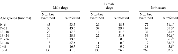
An overall prevalence of 33.8% was recorded for T. canis infection in the dogs examined (table 1). The result showed that young dogs (6 months old) were more frequently infected than older ones, with the highest prevalence of T. canis infection observed in the 0–6 months age group, which is significantly higher (P < 0.05) than in dogs of age group 25–36 months and in those older than 48 months. Analysis also showed that there was no statistically significant difference (P>0.05) observed in the prevalence of T. canis infection in both sexes of dog belonging to the older age groups (7–12, 13–18, 19–24 and 37–48 months). The results also showed that there was a significant difference in the overall prevalence of T. canis infection between male (41.0%) and female dogs (26.2%) (χ2 = 6.621, df = 1, P < 0.05).
Prevalence of Toxocara canis by age and sex in dogs examined between January and December, 2004.
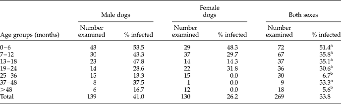
Values followed by different lower-case letters in each category are significantly different.
For both sexes χ2 = 26.554, df = 6, P < 0.05.
Intensity of T. canis infection relative to host age and gender
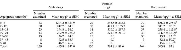
The mean intensities of T. canis infection of dog, determined by eggs/g (epg) of faeces, are shown in table 2. The overall mean intensity recorded for T. canis infection of the dogs was 393.8 ± 83.4. The results of intensity of infection of dogs with T. canis were similar to those of prevalence, with intensity of infection being highest in the dogs aged 0–6 months which subsequently declined gradually with the age of the dogs. The overall intensity was significantly higher among males than female dogs (U = 7582.0, df = 1, P < 0.05).
Intensity of Toxocara canis by age and sex in dogs examined between January and December, 2004.
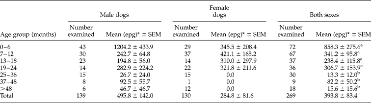
* (epg), eggs/g of faeces.
Values followed by different lower-case letters in each category are significantly different.
Seasonal variations in the prevalence and intensity of T. canis in the host
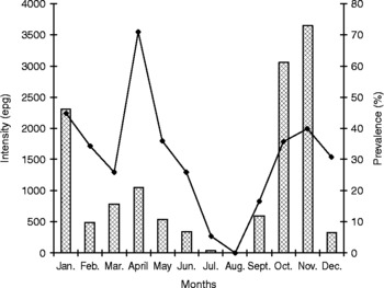
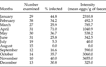
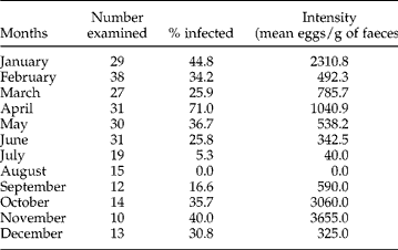
The seasonal prevalence and intensity of T. canis in the dogs examined during the period of study are shown in fig. 1. It was observed that the highest prevalence of T. canis occurred in April, followed by a gradual decline until it reached zero level in August. The prevalence then increased between September and November, while it decreased between January and March during the period of study. The differences in prevalence of T. canis in dogs between the dry months (35.1%) and wet months (32.6%) were found not to be statistically significant (P>0.05). The intensity of T. canis infection was observed to be highest in November, followed by October and January, and lowest between July and August during the period of study. The pattern of intensity also showed that between February and June the intensity of T. canis in dogs had a very small amplitude of variation (between 342.5 and 1040.9 epg). The seasonal differences in intensity between the dry (N = 131) and wet (N = 138) months were also found not to be significantly different (P>0.05) (table 3).
Seasonal variation in the prevalence of Toxocara canis in dogs examined during the period of study. Bars, intensity (eggs/g faeces); solid line, prevalence (%).

Seasonal variation in the prevalence and intensity of T. canis in dogs examined between January and December, 2004.
Discussion
The prevalence of T. canis observed in this study (33.8%) was similar to the reported prevalence (39%) from 166 stray dogs examined from Zaria, Kaduna State, Nigeria (Dada & Belino, Reference Dada and Belino1979). Toxocara canis, which is more prevalent in younger puppies, as observed in this study, is of utmost public health importance because of its zoonotic implication in the pathogenesis of visceral larva migrans (VLM) in humans, especially among children who are traditional playmates with puppies (Sprent, Reference Sprent1958; Ezeokoli, Reference Ezeokoli1984; Omudu et al., Reference Omudu, Amuta, Unoqur and Okoye2003).
The prevalence of T. canis in dogs aged 0–6 months (65.8%) as reported by Jalaya (Reference Jalaya1969) was comparable with the prevalence recorded (51.4%) for dogs of the same age group in this study. Oldham (Reference Oldham1965) reported a higher prevalence with T. canis in male dogs of age group 0–6 months compared to females. A similar trend was observed in this study, where the prevalences for male and female dogs of age 0–6 months were 53.5% and 48.3% respectively. Jacobs et al. (Reference Jacobs, Woodruff, Shah and Prole1977) observed that male dogs in general had higher infection rates than females, which is in agreement with the present study where the overall prevalence of T. canis in male dogs was higher than that of female dogs. For older male and female dogs, Jalaya (Reference Jalaya1969) reported prevalences of 26.2% and 19.9%, and for the present study they were 35.4% and 19.8%, respectively. In both Jalaya's and the present study, the prevalences were higher in older male dogs than female dogs. A suggested explanation for this phenomenon is that infective eggs ingested by adult female dogs end up mainly as resting larval stages in their somatic tissues and that these larvae infect their offspring in future. On the other hand, Toxocara infection in male dogs develops to adult worms in their intestinal tract, thereby leading to higher prevalence in male dogs (Overgaauw, Reference Overgaauw1997).
The intensity of T. canis in the present study revealed that dogs of age group 0–6 months have higher intensities compared to dogs of older age groups. The implication of this result is that dogs less than 6 months old contribute more to the potential dissemination of T. canis ova into the environment than older dogs. Furthermore, the egg output of the older dogs indicates that they are not always free of Toxocara worms and should not be overlooked with regard to routine anthelminthic treatment. Similar observations were reported by Jacobs et al. (Reference Jacobs, Woodruff, Shah and Prole1977) and O'Lorcain (Reference O'Lorcain1994). Studies have shown, however, that analysis of egg counts will underestimate the numbers of dogs with adult T. canis infection by 25–35%, due to the presence of male worms in some dogs (Jalaya, Reference Jalaya1969; Jacobs & Prole, Reference Jacobs and Prole1976).
The overall prevalence of 33.8% for T. canis infection recorded in this study, presents a potentially serious condition that might increase the possibility of many children in the study area harbouring T. canis. Studies on the prevalence of Toxocara and other intestinal parasites of dogs in various areas of the USA and other parts of the world have shown that a prevalence of 7% for T. canis infection among a population of dogs should be considered hazardous to children. This is because of the daily shedding of many thousands of Toxocara eggs into the environment, leading to contamination and thereby exposing children to accidental ingestion of the eggs as a result of their play habits (Hindman & Baker, Reference Hindman and Baker1936; Ehrenford, Reference Ehrenford1957; Vaughn & Jordan, Reference Vaughn and Jordan1960; Williams & Menning, Reference Williams and Menning1961).
In a study conducted on the epidemiology of Ascaris lumbricoides at Ile-Ife, (Holland et al., Reference Holland, Asaolu, Crompton, Stoddart, Macdonald and Torimiro1989), it was reported that 88.5% of the schoolchildren were infected with the roundworm. Dent (Reference Dent1960) reported that the hazard of concomitantly acquired infections with Ascaris ova on the hypersensitive or immune status of an individual suffering from visceral larva migrans is not known, but that young children sensitized by even minimal numbers of Toxocara or Ascaris suddenly develop severe and occasionally fatal bronchial asthma. It should be emphasized, that the occurrence of Ascaris ova in the stools suggests that the individual had ingested ‘dirt’ and perhaps with it Toxocara ova. Thus, the potential hazard to small children playing in areas where dogs are permitted to roam freely, as in Ile-Ife, are very great.
Seasonal variations in the prevalence of T. canis infections observed in this study were not well pronounced, although the highest prevalence of T. canis infections was recorded during the wet months, there was no significant difference in the prevalence between the wet and dry months during the period of study. This finding was similar to the observation of Ehrenford (Reference Ehrenford1957), who recorded the highest prevalence of T. canis infection in male and female dogs during the winter months and the lowest during the summer months.
Acknowledgements
The author would like to thank dog owners from Ile-Ife for their help and cooperation during faecal collections from the dogs. The author would also like to thank Dr A.I. Akinpelu for helpful comments. This research was partly funded by the Obafemi Awolowo University Postgraduate Research Grant.