Introduction
Chronic rhinosinusitis is a common inflammatory condition of the nose and paranasal sinuses, characterised by persistent symptoms for more than 12 weeks, together with objective evidence on nasal endoscopy and/or computed tomography.Reference Fokkens, Lund, Hopkins, Hellings, Kern and Reitsma1 Chronic rhinosinusitis is clinically heterogeneous; it is commonly described as chronic rhinosinusitis with or without nasal polyps, as these groups often differ in inflammatory pattern and clinical behaviour.Reference Fokkens, Lund, Hopkins, Hellings, Kern and Reitsma1 Nasal polyps typically represent oedematous mucosa with inflammatory cell infiltration, and eosinophil-predominant inflammation is frequently observed, particularly in chronic rhinosinusitis with nasal polyps.Reference Enache, Ioni, Mitroi, Anghelina, Mogoant and Ciolofan2
Eosinophils have been widely studied as a marker of disease severity and recurrence risk in chronic rhinosinusitis. In several cohorts, higher tissue eosinophil density has been associated with poorer disease control and higher rates of post-operative recurrence, supporting the concept of ‘eosinophilic chronic rhinosinusitis’ as a clinically relevant endotype.Reference Toro, Antonio, Alves Dos Reis, de Assumpcao and Sakano3–Reference Lou, Meng, Piao, Wang, Zhang and Bachert5 However, tissue sampling is invasive and is not always available for ongoing monitoring. This has led to interest in systemic markers that may reflect underlying inflammation and help guide prognostication. Peripheral blood eosinophilia has been explored as one such marker, but correlations between peripheral eosinophils and local tissue inflammation are variable and may differ by population and co-morbid inflammatory disease.Reference Drake, Rafaels and Kim6
Immunoglobulin E (IgE) is another immune marker implicated in chronic rhinosinusitis, particularly in patients with allergic sensitisation and type 2 inflammation. Local IgE production in sinonasal tissue has been described, and IgE-related pathways may contribute to inflammatory amplification in chronic rhinosinusitis.7 Even so, serum IgE is influenced by multiple factors and may not consistently mirror tissue inflammation or post-operative mucosal behaviour in individual patients.Reference Toro, Antonio, Alves Dos Reis, de Assumpcao and Sakano3, Reference Baba, Kondo, Toma-Hirano, Kanaya, Suzukawa and Ushio7 Taken together, these uncertainties mean that early post-operative endoscopic findings still require careful clinical interpretation rather than reliance on biomarkers alone.
Functional endoscopic sinus surgery (FESS) remains an established option for chronic rhinosinusitis that is refractory to appropriate medical therapy, improving sinus ventilation and access for topical treatment.Reference Fokkens, Lund, Hopkins, Hellings, Kern and Reitsma1 Nonetheless, polyp recurrence after FESS is well recognised, particularly in patients with eosinophilic inflammation.Reference DeConde, Mace, Levy, Rudmik, Alt and Smith4,,Reference Lou, Meng, Piao, Wang, Zhang and Bachert5 The early post-operative period is also a time of active mucosal healing, during which oedema and polypoid changes can appear and may be difficult to distinguish from true early recurrent disease.Reference Xu, Jiang, Li, Shi and Chen8 In routine practice, these early polypoid lesions are sometimes described as ‘reactionary’ polyps, but their histopathological profile and relationship with systemic inflammatory markers have not been well characterised.
In this study, we investigated reactionary nasal polyps developing during early post-operative follow up after FESS. Patients with chronic rhinosinusitis undergoing FESS at Universiti Malaya Medical Centre were followed endoscopically at defined intervals (including at one week, three weeks and three months). Early post-operative reactionary nasal polyps were defined as polypoid lesions arising between the 2nd and 12th post-operative week, with tissue sampling performed once only when detected. Histopathological assessment focused on tissue eosinophil density, quantified as the mean eosinophil count across three high-power fields selected from the area of densest cellular infiltration, with assessment performed by a pathologist blinded to clinical data. Pre-operative peripheral eosinophil count and serum IgE were recorded (with ‘high’ thresholds defined as more than 250 cells/mm3 and more than 150 IU/ml, respectively), and correlations between intra-operative polyp histology, early post-operative reactionary polyp histology and systemic markers were explored. This approach aimed to clarify whether these early post-operative lesions resemble recurrent inflammatory polyps or represent a distinct, reactive healing-related phenotype.
Materials and methods
Study design and setting
A prospective observational study was conducted in the Rhinology Clinic, Universiti Malaya Medical Centre, Kuala Lumpur, Malaysia, between November 2023 and March 2025. Adult patients (aged 18 years or over) with a diagnosis of chronic rhinosinusitis who were scheduled to undergo FESS were recruited consecutively.
Eligibility criteria
Patients were excluded if they had isolated sinonasal disease not consistent with chronic rhinosinusitis, autoimmune disease, acute or chronic infectious diseases other than sinusitis, sinonasal malignancy, haematological disorders, or mucocele. A clinical history suggestive of asthma and intolerance to anti-inflammatory drugs was also used as an exclusion criterion.
Pre-operative assessment and laboratory measures
All participants underwent pre-operative blood testing during admission, including a full blood count (for peripheral blood eosinophil count) and measurement of total serum IgE. All samples were processed in the same Universiti Malaya Medical Centre laboratory. Patients received intranasal mometasone furoate 400 μg/day in divided doses with alkaline nasal douching. Systemic corticosteroids were not administered within two weeks prior to surgery. For categorical analyses, eosinophilia was defined as a peripheral blood eosinophil count of more than 250 cells/mm3, and raised total IgE as greater than 150 IU/ml.
Surgical procedure and post-operative care
Functional endoscopic sinus surgery was performed under general anaesthesia. The extent of surgery was individualised and could include uncinectomy, middle meatal antrostomy, ethmoidectomy, sphenoidotomy and/or frontal sinusotomy, depending on disease distribution. Post-operatively, patients continued alkaline nasal irrigation twice daily and intranasal corticosteroid spray as per the pre-operative regimen.
Endoscopic follow up
Rigid nasal endoscopy (0-degree, 2.7 mm or 4 mm endoscope) was performed in clinic at one week, three weeks and three months after FESS. An early post-operative reactionary nasal polyp was defined as a polypoid lesion arising between post-operative week 2 and week 12 following FESS. Where such a lesion was identified, a biopsy sample was obtained once only (at a single time) from one nasal cavity side.
Histopathological assessment and eosinophil quantification
Biopsied tissue from early post-operative reactionary polyps and the corresponding intra-operative polyp tissue were processed for routine histopathology. Eosinophil quantification was performed using light microscopy at ×400 magnification. For each specimen, three high-power fields with the densest inflammatory cell infiltrate (‘hot spots’) were selected, and eosinophils were counted. Eosinophil density was recorded as the mean absolute eosinophil count across the three high-power fields. Counts were restricted to the submucosa; intraepithelial and intravascular eosinophils were not included. All slides were reviewed by an expert pathologist who was blinded to clinical data.
Outcomes
The objectives were to examine associations between (1) eosinophil density in early post-operative reactionary polyps and intra-operative polyp tissue, and (2) peripheral blood eosinophil count and total serum IgE.
Statistical analysis
All analyses were performed using SPSS Statistics (version 29; IBM, Armonk, New York, USA). Descriptive statistics were reported as mean (standard deviation) or median (interquartile range) as appropriate; categorical variables were summarised as frequency (percentage). Normality was assessed using the Shapiro–Wilk test. As data were not normally distributed, non-parametric analyses were used. Spearman’s rank correlation was applied to evaluate relationships between histopathological eosinophil counts (intra-operative and early post-operative reactionary polyp tissue) and peripheral eosinophil count and total serum IgE. Statistical significance was set at p < 0.05.
Ethical approval
Ethical approval was obtained from the Medical Research Ethics Committee, Universiti Malaya Medical Centre (Medical Research Ethics Committee identification number: 2023322-12276). Written informed consent was obtained from all participants.
Results and analysis
Patient characteristics
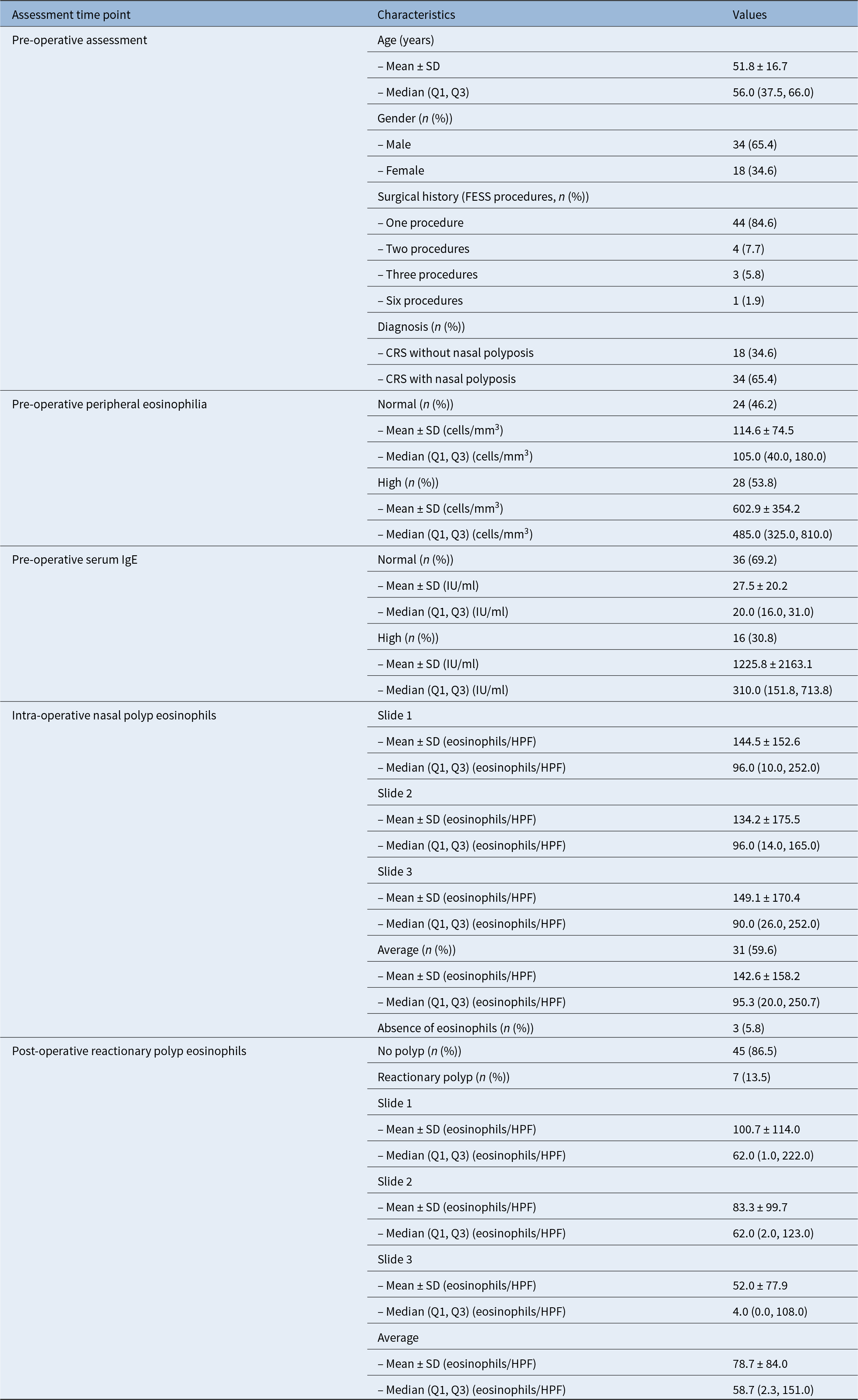
A total of 52 patients (34 male and 18 female) with chronic rhinosinusitis underwent FESS (Table 1). Thirty-four patients (65.4 per cent) had chronic rhinosinusitis with nasal polyposis and 18 (34.6 per cent) had chronic rhinosinusitis without nasal polyposis. Mean age was 51.8 ± 16.7 years. Eight patients underwent revision FESS during the study period.
Participant demographics

Table 1 Long description
Pre-operative demographics and lab measures are summarized for 52 participants, along with intra-operative and post-operative eosinophil findings. Age averaged 51.8 years, with a median of 56.0 years. Gender distribution was 34 male (65.4 percent) and 18 female (34.6 percent). Most had one prior functional endoscopic sinus surgery (44, 84.6 percent), with smaller numbers having two (7.7 percent), three (5.8 percent), or six (1.9 percent) procedures. Diagnosis favored chronic rhinosinusitis with nasal polyps (34, 65.4 percent) over without polyps (18, 34.6 percent). Pre-operative peripheral eosinophilia was classified as high in 28 (53.8 percent) and normal in 24 (46.2 percent), with much higher eosinophil counts in the high group. Pre-operative serum IgE was normal in 36 (69.2 percent) and high in 16 (30.8 percent), and the high group showed very large variability. Intra-operative nasal polyp eosinophil counts were high on average across three slides, and eosinophils were absent in 3 participants (5.8 percent). Post-operatively, 41 (78.8 percent) had no polyp and 11 (21.2 percent) had a reactionary polyp; among those with reactionary polyps, eosinophils were present in 7 (13.5 percent of all participants) and absent in 4 (7.7 percent of all participants), with average eosinophil counts lower than intra-operative values.
SD = standard deviation; Q1 = first quartile; Q3 = third quartile; FESS = functional endoscopic sinus surgery; CRS = chronic rhinosinusitis; IgE = immunoglobulin E; HPF = high-power field
Pre-operative systemic inflammatory markers
Pre-operatively, 28 out of 52 patients (53.8 per cent) had elevated peripheral eosinophil counts (median, 485.0 cells/mm3; interquartile range = 325.0–810.0), while 24 of 52 (46.2 per cent) had normal eosinophil levels (median, 105.0 cells/mm3). Serum IgE was elevated in 16 of 52 patients (30.8 per cent) (median, 310.0 IU/ml; interquartile range = 151.8–713.8) and was normal in 36 of 52 (69.2 per cent) (median, 20.0 IU/ml).
Intra-operative histopathology
Intra-operative histological assessment demonstrated eosinophilic infiltration in 31 of 52 specimens (59.6 per cent), with a median polyp eosinophil density of 95.3 eosinophils per high-power field (interquartile range = 20.0–250.7). Eosinophils were absent in 3 of 52 specimens (5.8 per cent).
Post-operative early reactionary nasal polyps
Early post-operative reactionary nasal polyps were detected in 11 patients during endoscopic follow up; of these, 9 had a pre-operative diagnosis of chronic rhinosinusitis with nasal polyps and 2 had chronic rhinosinusitis without nasal polyposis. Biopsy was performed in all 11 patients; histopathological assessment revealed eosinophilic infiltration in 7 of the 11 specimens (63.6 per cent), with a median eosinophil density of 58.7 eosinophils per high-power field (interquartile range = 2.3–151.0). The remaining four specimens (36.4 per cent) showed no eosinophilic infiltration on histopathological assessment.
Intra-operative and reactionary polyp eosinophil density association
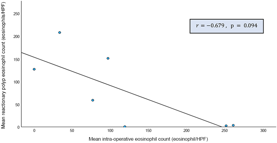
Objective one examined whether eosinophil density in intra-operative polyp tissue was associated with eosinophil density in early post-operative reactionary polyp tissue. Spearman’s rank correlation demonstrated a moderate inverse correlation between intra-operative and reactionary polyp eosinophil counts (ρ = −0.679, p = 0.094) (Table 2), and this relationship is illustrated in Figure 1.
Spearman’s correlation between average eosinophil counts in intra-operative versus reactionary nasal polyp slides

Table 1 Long description
Spearman rank correlation assessing how average eosinophil counts in intra-operative nasal polyp slides relate to counts in reactionary nasal polyp slides is reported. The correlation indicates a moderately strong negative relationship, meaning higher averages in one slide type tend to align with lower averages in the other. The reported correlation value is minus 0.679. The p-value is 0.094, which does not meet a common threshold for statistical significance. Results should be interpreted cautiously because the analysis uses a rank-based method suited to small samples and data that may not follow a normal distribution.
Note: Spearman’s rank correlation was used due to small sample size and possible non-normal distribution; p < 0.05 is considered statistically significant.
Scatter plot showing the correlation between eosinophil counts in intra-operative and early post-operative reactionary nasal polyp tissue following functional endoscopic sinus surgery. HPF = high-power field.

Tissue eosinophil density and systemic markers association
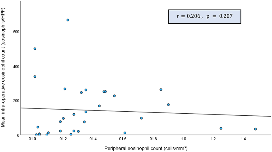
Objective two evaluated associations between tissue eosinophil density and systemic inflammatory markers (peripheral eosinophil count and serum IgE), assessed separately for intra-operative polyp tissue and post-operative reactionary polyp tissue. Intra-operative polyp eosinophil counts demonstrated weak positive correlations with peripheral eosinophil level (ρ = 0.206, p = 0.207; Figure 2) and serum IgE level (ρ = 0.213, p = 0.259).
Scatter plot showing the correlation between intra-operative eosinophil count and peripheral eosinophil level. HPF = high-power field.

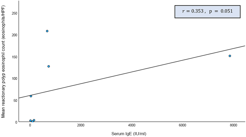
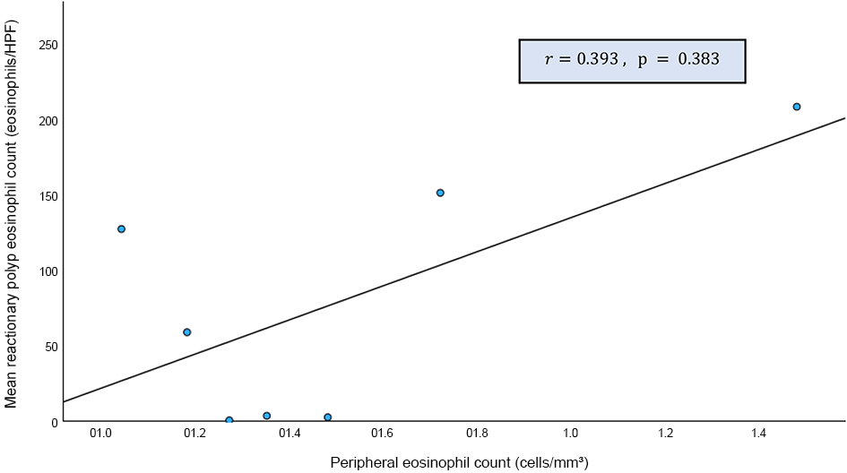
For post-operative reactionary polyps, eosinophil counts demonstrated a weak positive correlation with peripheral eosinophil level (ρ = 0.393, p = 0.383) and a weak positive correlation with serum IgE level (ρ = 0.353, p = 0.051) (Table 3). These relationships are illustrated in Figures 3 and 4.
Spearman’s correlation between reactionary polyp eosinophil count and inflammatory markers

Table 3 Long description
Spearman rank correlations are reported between reactionary polyp eosinophil count averaged per high-power field and two inflammatory markers. For peripheral blood eosinophil level in cells per cubic millimeter, the correlation is weakly positive at about 0.39 with a p-value of 0.383. For serum immunoglobulin E in international units per milliliter, the correlation is also weakly positive at about 0.35 with a p-value of 0.051. The association with immunoglobulin E is closer to statistical significance than the association with peripheral eosinophils, but it still does not meet the stated significance threshold. Overall, higher tissue eosinophil counts tend to align with higher blood eosinophils and higher immunoglobulin E, but the evidence is not strong based on these p-values.
Note: p < 0.05 is considered statistically significant. HPF = high-power field; IgE = immunoglobulin E
Scatter plot showing the correlation between reactionary nasal polyp eosinophil count and peripheral eosinophil level. HPF = high-power field.

Scatter plot showing the correlation between reactionary nasal polyp eosinophil count and serum immunoglobulin E level. HPF = high-power field; IgE = immunoglobulin E.

Discussion
Overview of key findings
This prospective observational study describes a distinct early post-operative endoscopic phenomenon – early post-operative reactionary nasal polyps – occurring between post-operative weeks 2 and 12 after FESS, with tissue sampled once at detection. In the present cohort, reactionary nasal polyps were seen endoscopically in 11 patients, while histopathological eosinophil quantification was available in 7 cases. The reactionary nasal polyp specimens demonstrated eosinophilic infiltration but with lower eosinophil density than the corresponding intra-operative polyp tissue. Together, these findings support the concept that early post-operative ‘polypoid’ lesions may represent a reactive healing-related phenotype rather than established recurrent chronic rhinosinusitis with nasal polyps, and that tissue eosinophilia within an early post-operative lesion should be interpreted in the context of timing and clinical course rather than assumed to indicate irreversible recurrence.
Challenge of interpreting early post-operative endoscopy
The early post-operative period following FESS is characterised by dynamic mucosal change, including oedema, crusting, granulation and remodelling, and endoscopic appearances may fluctuate substantially during this phase.8 In routine practice, early polypoid mucosal protrusions are not uncommon and may be labelled as ‘reactionary’ polyps; however, this entity is poorly characterised in terms of histopathology and relationship to systemic biomarkers. The present study provides initial tissue-based evidence that reactionary nasal polyps can show eosinophilic inflammation, yet differ quantitatively from intra-operative polyps, supporting the idea that they may reflect post-surgical mucosal reactivity rather than mature recurrent polyposis.
This distinction is clinically relevant. If early polypoid changes are misclassified as recurrence, patients may undergo premature escalation (including repeated systemic corticosteroids, earlier consideration of revision surgery, or unnecessary referral for biologics). Conversely, under-recognition of early inflammatory signals in high-risk patients could delay optimisation of post-operative care. Evidence-based post-operative strategies following FESS emphasise consistent saline irrigation, topical corticosteroids and appropriate debridement to aid healing and improve delivery of topical therapy.Reference Rudmik, Soler, Orlandi, Stewart, Bhattacharyya and Kennedy9, Reference Orlandi, Kingdom, Smith, Bleier, DeConde and Luong10 In this context, the present data support a pragmatic approach: early post-operative polypoid lesions should be interpreted using serial endoscopy and trajectory, rather than a single early time point, while ensuring that standard post-operative anti-inflammatory therapy is optimised.
Interpretation of objective one
Objective one examined whether eosinophil density in the intra-operative nasal polyp specimen was associated with eosinophil density in post-operative reactionary nasal polyp tissue. In the analysable reactionary nasal polyp subset (n = 7), a moderate inverse correlation was observed between intra-operative and reactionary nasal polyp eosinophil counts (ρ = −0.679), although this did not reach statistical significance (p = 0.094) (Table 2 and Figure 1).
Several factors may explain this pattern. Firstly, the small number of reactionary nasal polyp specimens with eosinophil quantification limits the stability of correlation estimates; in small samples, correlation co-efficients are particularly sensitive to outliers and may not reflect a reproducible biological relationship. Secondly, intra-operative polyps represent established chronic type-2-skewed inflammation, in which tissue eosinophilia has been associated with refractory disease and post-operative recurrence risk in multiple cohorts and systematic reviews.Reference Toro, Antonio, Alves Dos Reis, de Assumpcao and Sakano3–Reference Lou, Meng, Piao, Wang, Zhang and Bachert5, Reference McHugh, Snidvongs, Xie, Banglawala and Sommer11 By contrast, reactionary nasal polyps in this study were defined within the early healing window (weeks 2–12), a period characterised by dynamic mucosal repair and evolving inflammatory infiltrates after FESS.8 The inflammatory milieu during healing (epithelial injury or repair, local oedema, granulation and remodelling) may therefore generate early reactive polypoid lesions whose cellular profile does not necessarily mirror the pre-operative polyp endotype.
Thirdly, methodological factors may contribute to apparent discordance between paired tissues. Eosinophil quantification was based on a ‘hot spot’ approach (mean of three densest high-power fields), which is commonly used but can accentuate spatial heterogeneity – particularly in smaller post-operative biopsy samples – thereby increasing variability when comparing intra-operative polyp specimens with early post-operative lesions. These considerations support a cautious interpretation: the observed inverse correlation should be treated as hypothesis-generating and reinforces the broader concept that early post-operative polypoid change is not synonymous with established recurrence, even when eosinophils are present.Reference Xu, Jiang, Li, Shi and Chen8, Reference McHugh, Snidvongs, Xie, Banglawala and Sommer11
Tissue eosinophilia as a marker of recurrence
Tissue eosinophilia has repeatedly been associated with disease severity and recurrence risk in chronic rhinosinusitis with nasal polyps, and several cohorts support eosinophilic chronic rhinosinusitis as a clinically relevant endotype.Reference Toro, Antonio, Alves Dos Reis, de Assumpcao and Sakano3–Reference Lou, Meng, Piao, Wang, Zhang and Bachert5, Reference Stevens, Schleimer and Kern12 However, the key message from this study is that eosinophils can be present in early post-operative reactionary nasal polyps, yet the lesions may still resolve with time. This is consistent with the broader literature that eosinophils are an important marker of type 2 inflammation but are not, by themselves, a definitive surrogate for clinical ‘recurrence’ at an early time point.
Meta-analytic work suggests that tissue eosinophil cut-offs (commonly reported per high-power field) can help identify eosinophilic chronic rhinosinusitis and may predict recurrence; for example, one systematic review and meta-analysis proposed a threshold of more than 55 eosinophils per high-power field to predict the likelihood of recurrence in eosinophilic chronic rhinosinusitis.Reference McHugh, Snidvongs, Xie, Banglawala and Sommer11 However, such thresholds were developed largely in contexts of established disease and longer-term outcomes, rather than the immediate healing window. Applying recurrence thresholds to a lesion arising within weeks of FESS risks conflating reactive post-operative inflammation with true polyp regrowth, particularly when the clinical definition of recurrence typically implies persistence or progression beyond expected healing and ongoing disease activity.
Therefore, the present findings support a time-sensitive interpretation: early post-operative polypoid change with eosinophils should prompt close follow up and optimisation of topical therapy, rather than immediate classification as recurrence, unless accompanied by persistent symptoms, worsening endoscopic appearance over time or objective evidence of progressive disease.
Interpretation of objective two
Objective two examined relationships between tissue eosinophil density and systemic inflammatory markers (peripheral eosinophils and serum IgE), assessed for both intra-operative polyps and post-operative reactionary nasal polyps. In the present study, correlations between tissue eosinophils and systemic markers were weak, with a trend towards a stronger association between reactionary nasal polyp eosinophils and serum IgE (ρ = 0.353, p = 0.051).
These results align with the wider concept that systemic biomarkers may be imperfect proxies for local sinonasal eosinophilia. Peripheral blood eosinophilia has been evaluated as a clinically accessible marker, but correlations with tissue eosinophilia vary across populations and may be influenced by co-morbid airway disease and atopy.6 Likewise, serum IgE is shaped by multiple factors (atopy, parasitic exposure, environmental sensitisation) and may not consistently mirror local IgE-driven mucosal inflammation.Reference Toro, Antonio, Alves Dos Reis, de Assumpcao and Sakano3, Reference Baba, Kondo, Toma-Hirano, Kanaya, Suzukawa and Ushio7 The present dataset suggests that even when eosinophils are demonstrable within tissue, systemic measures may not reliably predict the degree of local eosinophilic infiltration in early post-operative lesions.
A possible explanation for the observed trend with IgE is the growing recognition of local IgE production and IgE-related inflammatory pathways within sinonasal tissue, including evidence supporting local class-switch recombination.7 In such a framework, serum IgE may correlate weakly with local processes overall, but could appear more relevant in subsets where IgE-linked pathways are particularly active during mucosal repair or early post-operative inflammatory amplification. This hypothesis remains speculative in the present study, and would require validation with larger cohorts and more granular phenotyping (e.g. atopic status, specific IgE, asthma status, non-steroidal anti-inflammatory drug (NSAID)-exacerbated respiratory disease assessment and local tissue cytokine profiling).
Implications for post-operative management and escalation
Contemporary guidance emphasises long-term medical therapy and endoscopic follow up after FESS, particularly for chronic rhinosinusitis with nasal polyps and type 2 inflammatory disease.Reference Fokkens, Lund, Hopkins, Hellings, Kern and Reitsma1 In the early post-operative period, evidence-based post-operative care – saline irrigation, topical corticosteroids and debridement when appropriate – aims to optimise healing and reduce inflammatory burden.9 The present findings support a clinical strategy in which early reactionary nasal polyps are managed initially by ensuring adherence and optimisation of topical therapy, combined with scheduled follow-up endoscopy to document resolution versus persistence.
For patients who remain uncontrolled despite appropriate surgery and maximal medical therapy, escalation pathways now include biologic therapies, guided by disease severity and type 2 inflammatory features. The 2023 update from the European Position Paper on Rhinosinusitis and Nasal Polyps (‘EPOS’) and the European Forum for Research and Education in Allergy and Airway Diseases (‘EUFOREA’) provides practical criteria for the indication and evaluation of biologics in chronic rhinosinusitis with nasal polyps.Reference Fokkens, Viskens, Backer, Conti, De Corso and Gevaert13 Randomised trials have demonstrated efficacy for biologics such as dupilumab, omalizumab and mepolizumab in severe chronic rhinosinusitis with nasal polyps.Reference Bachert, Han, Desrosiers, Hellings, Amin and Lee14–Reference Han, Bachert, Fokkens, Desrosiers, Wagenmann and Lee16 However, the present study highlights an important guardrail: a polypoid lesion within weeks of FESS may be reactionary and self-limiting, and escalation decisions should be based on sustained disease activity rather than early reactive appearances alone.
Limitations
Several limitations should be acknowledged. Firstly, while reactionary nasal polyps were detected endoscopically in 11 patients, eosinophil quantification was available in only 7, limiting the statistical power and stability of correlation estimates. Secondly, reactionary nasal polyp biopsies were performed at a single time point, which prevents assessment of how tissue inflammation evolves as lesions resolve. Thirdly, systemic markers were measured pre-operatively; post-operative systemic inflammatory status may differ, and contemporaneous post-operative blood sampling could provide additional information. Key phenotype variables that can influence biomarker relationships – such as objective atopy assessment, asthma status by formal testing and NSAID-exacerbated respiratory disease – were not analysed as modifiers, which may partly explain variability in correlations.
Future work should validate these findings in larger, ideally multicentre, prospective cohorts with standardised endoscopic scoring and longer follow up to distinguish early reactive post-operative change from true recurrent chronic rhinosinusitis with nasal polyps, while clarifying predictors of reactionary nasal polyp development and persistence (e.g. chronic rhinosinusitis with nasal polyps endotype, atopy, asthma or NSAID-exacerbated respiratory disease, revision status, and peri-operative steroid exposure). In addition, repeated sampling or longitudinal biomarker assessment could better define the temporal evolution of inflammation from the intra-operative state through the early healing window, and expanded immunophenotyping (including local IgE measurement, type 2 cytokine signatures such as interleukin (IL)-4, IL-5 and IL-13 pathways, epithelial barrier markers, and microbiome profiling) may help determine whether reactionary nasal polyps represent a benign healing variant, an inflammatory flare, or an early marker of subsequent recurrence and need for escalation, including biologic therapy.
Fourthly, formal endoscopic confirmation of resolution was not obtained in this cohort, and the self-limiting nature of these lesions was not directly documented. Future studies should incorporate serial endoscopic assessment beyond week 12 to establish whether reactionary nasal polyps resolve or progress to established recurrent polyposis. Fifthly, the threshold used to define elevated total serum IgE (greater than 150 IU/ml) was a study-specific convention based on the local laboratory reference range, and differs from the threshold of 100 IU/ml or more referenced in some European Position Paper on Rhinosinusitis and Nasal Polyps 2020 type 2 biomarker criteria; categorical analyses involving IgE elevation should therefore be interpreted in this study-specific context.
• Chronic rhinosinusitis with nasal polyps is associated with type 2 inflammation, and tissue eosinophilia is linked to disease severity and recurrence risk
• Early endoscopy after functional endoscopic sinus surgery (FESS) can show polypoid mucosal changes difficult to distinguish from true early recurrence
• Reactionary nasal polyps can occur during early healing (weeks 2–12) after FESS and may be self-limiting
• Intra-operative polyps showed higher eosinophil density than reactionary polyps; early post-operative ‘polyp-like’ lesions may represent reactive healing rather than established recurrence
• Correlations between tissue eosinophilia and peripheral eosinophils and serum immunoglobulin E were weak; thus, systemic biomarkers may not reliably reflect local inflammation in these early post-operative lesions
Conclusion
Early post-operative reactionary nasal polyps were observed in 11 patients during the early post-operative period after FESS, with histological eosinophil quantification available in 7 of these 11 cases. Intra-operative polyps demonstrated higher eosinophil density than reactionary polyps, and the relationship between intra-operative and reactionary polyp eosinophil counts showed a moderate inverse correlation that did not reach statistical significance. Associations between tissue eosinophilia and systemic markers (peripheral eosinophils and serum IgE) were weak, with a trend towards correlation between reactionary polyp eosinophils and serum IgE.
Overall, these findings suggest that early post-operative polypoid lesions may represent a reactive healing-related phenotype rather than definite early recurrence, and should be interpreted based on the post-operative time course and serial endoscopic trajectory. Larger studies with longer follow up and broader inflammatory profiling are needed to confirm the natural history of these lesions and their relationship to true long-term recurrence.
Data availability statement
The datasets generated and/or analysed during the current study are available from the corresponding author on reasonable request.
Funding statement
This study was supported by Bantuan Khas Penyelidikan Universiti Malaya – Early Career Researcher funding (grant number: BKP 043-2023-ECR).
Competing interests
The authors declare they have no conflicts of interest.
Ethical standards
Ethical approval was obtained from the Medical Research Ethics Committee, Universiti Malaya Medical Centre (Medical Research Ethics Committee identification number: 2023322-12276). Written informed consent was obtained from all participants.