I. INTRODUCTION
Garnet structure oxides are compounds that can be incorporated into the crystal lattice of almost all group IV cations. Nevertheless, among these are quite rare compounds containing cations of titanium, zirconium, and tin. Hisanori and Tetsuya (Reference Hisanori and Tetsuya2011) described the crystal structure of Ca3Sn2,2Ti0,8Al2O12 [a = 12.5309(3) Å, space group Ia
 $\bar 3$
d]. The structural formula is presented as Ca3[Sn0.96Ti0.04]octa[Al0.67Ti0.24Sn0.09]tetraO12. Polycrystalline solid solutions of Ca3Sn3−x
Ti
x
Al2O12 were also prepared by solid-state reaction in air at 1370 °C with nominal titanium contents from x = 0.6 to 1.4. Ferdov et al. (Reference Ferdov, Rute, Lin and Wu
2012) synthesized zirconium germanate with garnet-type structure Na3Zr1.8Ge3O10(OH)2.2 [a = 12.704(7) Å, space group Ia
 $\bar 3$
d]. Blasse and de Blank (Reference Blasse and de Blank1995) described the photoluminescence of Ca4ZrGe3O12, which was prepared from CaCO3, ZrO2, and GeO2 by heating at 1000 and 1100 °C for 1 day with repeated grindings and at 1400 °C for 1 week. They suggested that in garnet-type Ca3
M
2+
M
4+Ge3O12 it is probable that all tetravalent ions other than Ge4+ will prefer the octahedral sites because of their relative size. The effects of Zr-substitution on the dielectric and magnetic properties of yttrium garnet–ferrites were investigated in (Huang et al., Reference Huang, Yang, Qui, Wang and Jin2012). The Y2.6−x
Ca0.4+x
Zr
x
V0.2Fe4.8−x
O12, x = 0–0.5, was prepared by solid-phase synthesis by annealing it for 2 h at 1150 °C. Ceramics for measuring dielectric loss were pressed and annealed at 1320–1420 °C for 4 h. With the increase of Zr4+ content, the dielectric loss (tangents δ
E) decreased.
The garnet Ca4ZrGe3O12 represents a potential solid-state laser. The ability of iso- and heterovalent substitutions in its crystal lattice allows not only to expand the number of optically active material, but also to create new dielectrics. The refinement of the crystal structure and the simplification of the synthesis method contribute to the solution of these problems.
II. EXPERIMENTAL
The starting materials were powders of CaCO3 (99.9%), GeO2 (96.95%), C6H8O7·H2O (99.5%), and solutions of ZrO(NO3)2 (76.4 g dm−3 Zr) and HNO3 (65%). The reaction mixture was calculated to obtain 3 g of the compound. Calculated amounts of calcium carbonate were dissolved in nitric acid. Germanium dioxide was dissolved separately under heating in citric acid at a ratio of GeO2: C6H8O7·H2O = 1:3. Citric acid was also added to the solution of ZrO(NO3)2 at a ratio of ZrO(NO3)2: C6H8O7·H2O = 1:2, and then the three solutions were mixed together. The reaction solution was heated to a viscous mass at a temperature of 80–90 °C. Thereafter, the temperature was raised to 200–250 °C, which resulted in complete dehydration and decomposition of the nitrate groups and reagents of the reaction mass. The precursor was a porous brown mass because of the carbon particles formed as a result of redox processes. The precursor was mixed in an agate mortar and heated in air using an electric furnace to remove carbon particles and obtain a garnet crystal structure. The annealing temperatures were from 850 to 950 °C and the annealing process lasted for ~10 h. Monophasic polycrystalline samples were obtained after annealing at 1100 °C for 5 h.
X-ray powder diffraction (XRD) patterns were collected at room temperature on a STADI P (STOE) diffractometer in transmission geometry with a linear mini-PSD detector using CuKa 1 radiation in the 2θ range from 5° to 120° with a step of 0.02°. Polycrystalline silicon [a = 5.43075(5) Å] was used as an external standard. Possible impurity phases were checked by comparing their XRD patterns with those in the PDF2 (Powder Diffraction File) date bases (2009). International center for Diffraction Date, USA. The crystal structure refinement was carried out with the GSAS program suite using XRD data (Larson and Von Dreele, Reference Larson and Von Dreele2004; Toby, Reference Toby2001). The peak profiles were fitted with a pseudo-Voigt function, I(2θ) = x·L(2θ) + (1 − x)·G(2θ) (where L and G are the Lorentzian and Gaussian parts, respectively). The angular dependence of the peak width was described by the relation (FWHM)2 = Utg2θ + Vtgθ + W, where FWHM is the full line-width at half-maximum. The background level was described by a combination of 36-order Chebyshev polynomials. The absorption correction function for a flat plate sample in transmission geometry was applied.
Density measurements were carried out on the AccuPyc II 1340 Gas Displacement Pycnometry System (Micromeritics, USA) with a sample chamber of 1 cm3. About 50–60% of the chamber volume was filled by the powder sample, and 15 measurements were done for the sample.
III. DISCUSSION
The XRD pattern of Ca4ZrGe3O12 was indexed in cubic symmetry with unit-cell parameter a = 12.71299(3) Å [space group Ia
 $\bar 3$
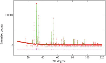
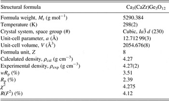
d (230), Z = 8]. The crystal structure of Ca3Sn2SiGa2O12 (Rulmont et al., Reference Rulmont, Tarte, Cartié and Choisnet1993) was used as starting model. Weak reflections of CaZrGeO5 (Malcherek and Elleman-Olesen, Reference Malcherek and Elleman-Olesen2005) were fitted as a second phase during the full-profile fitting; the fraction was calculated to be 1.7 % mass. Figure 1 shows experimental, theoretical, and difference XRD patterns for a Ca4ZrGe3O12 sample. The results of crystal structure refinement and selected interatomic distances for Ca4ZrGe3O12 are listed in Tables I–III, respectively.
Observed (crosses), calculated (solid line), and difference (bottom line) XRD patterns of Ca4ZrGe3O12 with CaZrGeO5 as the impurity phase. Red and black lines show the positions of the peaks of Ca4ZrGe3O12 and CaZrGeO5, respectively.

Crystal data and structure refinement for Ca4ZrGe3O12.

Atomic coordinates and isotropic thermal parameters [U iso × 100, (Å2)] for Ca4ZrGe3O12.

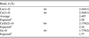
Shortest interatomic distances (d) in Ca4ZrGe3O12.

a The sum of the crystal radii according to Shannon (Reference Shannon1976): Ca+2(VIII) – 1.26 Å, Ca+2(VI) – 1.14 Å, Zr+4(VI) – 0.86 Å, Ge+4(IV) – 0.530 Å, and O−2(IV) – 1.24 Å.
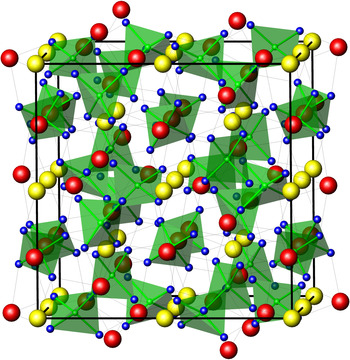
The crystal structure of Ca4ZrGe3O12 shown in Figure 2 is a three-dimensional framework of corner-sharing tetrahedra and octahedra in the interstices of which Ca(1) atoms are placed on the 24c site coordinated by eight oxygen atoms (96h site). Average Ca(1)–O distance, 2.497 Å, is in perfect agreement with the sum of the crystal radii of Ca+2 (VIII) and O−2 (IV), 2.50 Å. Germanium atoms were found to occupy the 16a site in tetrahedral coordination with oxygen atoms with a bond length of 1.779(2) Å; this fits well with the expected 1.77 Å. Zirconium and calcium atoms in a ratio of 0.5/0.5 are located on the 16a site coordinated by six oxygen atoms at a distance of 2.179(2) Å, which is somewhat less than the expected 2.24 Å. The interatomic distances are consistent with those reported for Na3Zr1.8Ge3O10(OH)2,2 germanate with a garnet-type structure (Mateika et al., Reference Mateika, Völkel and Haisma1990) where Zr–O and Ge–O distances were found to be 2.083(5) and 1.754(8) Å, respectively. One should note that Ca4ZrGe3O12 belongs to the garnets with large unit-cell constant a = 12.71299(3) Å. Only lattice parameters of silver–lead garnets Ag2Pb2 M 2 IV3O12, M = Mg, Zn, Cd, Co, Mn3+, and M IPb2 M IIV3O12 (Vorobiev, Reference Vorobiev2006) exceed this value.
The crystal structure of Ca4ZrGe3O12 showing GeO4 tetrahedra (green), Ca(1) (red), and Ca/Zr(2) (yellow).

SUPPLEMENTARY MATERIAL
The supplementary material for this article can be found at http://dx.doi.org/10.1017/S0885715616000506.
ACKNOWLEDGEMENT
The authors acknowledge the access to the user facility for X-ray structure analysis at the Institute of Solid State Chemistry, UB RAS (Ekaterinburg, Russia).