Introduction
Nematode parasitism of the central nervous system is rare in bats. However, there are reports of the infection by Angiostrongylus cantonensis and Molossinema wimsatti (Farina et al., Reference Farina, Lankton, Terio, McAloose and St. Leger2018).
Angiostrongylus cantonensis resides in the pulmonary arteries of rats (definitive host), hence the sobriquet ‘rat lungworm’ ascribed to the parasite (Farina et al., Reference Farina, Lankton, Terio, McAloose and St. Leger2018). The complex life cycle of this nematode includes intermediate, paratenic and accidental hosts (Spratt, Reference Spratt2015). The infection has been reported in a variety of domestic animals, mammalian and avian wildlife, and humans (Spratt, Reference Spratt2015). Neurological disease has been reported in black and grey-headed flying foxes (Reddacliff et al., Reference Reddacliff, Bellamy and Hartley1999; Barrett et al., Reference Barrett, Carlisle and Prociv2002).
The filarioid nematode M. wimsatti was reported in the central nervous system of insectivorous bats. These nematodes reside particularly in the ventricular system of the brain (Nguyen & Myers, Reference Nguyen and Myers1987). Clinical signs are minimal and rarely reported (Lichtenfels et al., Reference Lichtenfels, Bhatnagar, Whittaker and Frahm1981; Nguyen & Myers, Reference Nguyen and Myers1987). The life cycle of this parasite remains unknown (Nguyen & Myers, Reference Nguyen and Myers1987).
The present report describes two cases of M. wimsatti infection in Pallas's mastiff bats (Molossus molossus), which, to the best of our knowledge, is the first report of the condition in Brazil.
Results
Case 1
An adult female Pallas's mastiff bat (M. molossus) was captured and killed by a domestic cat in a suburban area of the municipality of Patos, Paraiba, north-eastern Brazil (7.0192°S, 37.2751°W).
Case 2
The second case was in an adult male Pallas's mastiff bat (M. molossus) that was found at dusk crawling on the ground in the same area. The bat was placed in a metal cage for observation and died a day later.
Pathological findings
At post-mortem examination, both bats were in good body condition and had no gross lesions other than small bite pounds on the wings (case 1). No search for microfilaria in the peripheral blood was performed.
Samples from the skin, central nervous system and organs from the thoracic and abdominal cavities were fixed in 10% neutral buffered formalin, processed routinely for histopathology, embedded in paraffin, cut into 3 μm sections and stained with haematoxylin and eosin.
One hemisphere of each brain was frozen as a laboratory standard protocol. Upon the histological finding of nematodes in the central nervous system, the brains’ frozen halves were reassessed. Nematodes were recovered using stereomicroscopy, fixed in 10% neutral buffered formalin and maintained in alcohol–formalin–acetic acid. They were then cleared with lactophenol and morphologically analysed by light microscopy.
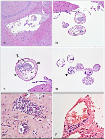
Histopathological examination of the central nervous system revealed filarioid nematodes in the lateral (case 1), third and fourth ventricles of the brain, and cerebellar neuroparenchyma (case 2). There were adults, subadults and eggs, sometimes containing microfilariae (fig. 1a, b).
Molossinema wimsatti in the brain of insectivorous bats. (A) Brain, right lateral ventricle. Filarioid nematodes: adult, subadult and eggs. Haematoxylin and eosin (HE). Objective 5×. (B) Brain, left lateral ventricle. Cross-section of two adults and three subadults (arrow). Filarioid nematodes. HE. Obj. 20×. (C) Cross-section of an adult female. Filarioid nematode showing coelomyarian–polymyarian musculature (M), lateral chords (LC), internal cuticular ridge (CR), pseudocoelom (PC), two uteri (U) and intestine (i). HE. Obj. 40×. (D) Cross-section of subadult forms (arrow) and eggs, one of which containing microfilariae (arrowhead). HE. Obj. 40×. (E) Cerebellar neuroparenchyma. Cyst-like structures (presumably eggs) containing numerous microfilariae. HE. Obj. 40×. (F) Cerebellum, leptomeninges. Perivascular cuffing composed of lymphocytes, plasma cells and rare macrophages. HE. Obj. 40×.

The adult forms, seen in transverse sections, were round, had thin coelomyarian–polymyarian musculature, and were divided into dorsal and ventral fields by lateral cords with internal cuticular ridges. In the pseudocoelom there was a slender intestine and, if female specimens, two uteri (fig. 1c). The diameter of the adult nematodes ranged from 130 to 150 μm. These morphological characteristics are consistent with that of M. wimsatti.
The subadult forms had similar shapes to that of adults but were smaller (up to 70 μm in diameter) and lacked well-defined internal structures. Also, round thin-shelled eggs, measuring ± 50 μm in diameter, and sometimes containing microfilariae, appeared in ventricles (fig. 1d). In case 2, multifocally within the white and grey matter of the cerebellum there were numerous microfilariae within cyst-like structures (presumably eggs) (fig. 1e).
There was no evidence of tissue damage or inflammatory response in the nervous system of bat 1, while bat 2 presented a mild non-suppurative meningoencephalitis characterized by perivascular cuffs composed of lymphocytes, plasma cells and rare macrophages (fig. 1f).
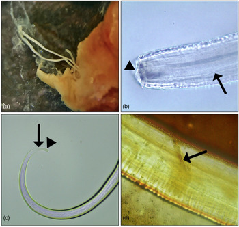
Three filarioid nematodes (two females and a male) were recovered from the brain ventricular system of bat 2 (fig. 2a), and were identified as M. wimsatti according to the morphological features previously described (Georgi et al., Reference Georgi, Georgi, Jiang and Frongillo1987).
Molossinema wimsatti in the brain of insectivorous bats. (A) Adult filarioid nematodes recovered from the brain ventricular system. Stereomicroscopy (SM). Objective 2×. (B) Anterior body portion. Inconspicuous buccal cavity (arrowhead) and a long slender cylindrical oesophagus (arrow). Light microscopy (LM). Obj. 40×. (C) Female, posterior body portion. Small cylindrical tail with phasmids that surmount small hemispherical bases flanking the tip of the tail (arrow). Note the anus almost at the tip of the tail (arrowhead). LM. Obj. 10×. (D) Male, posterior body portion. Arcuate spicule (arrow). Contralateral spicule is not visible due to overlap. LM. Obj. 40×.

Adult nematodes were characterized by an inconspicuous buccal cavity and a long slender cylindrical oesophagus, not divided into muscular and glandular portions (fig. 2b). The females measure approximately 51 mm in total body length and have a small cylindrical tail with phasmids that surmount small hemispherical bases flanking the tail's tip (fig. 2c). The anus is present almost at the tip of the tail (sub-terminal). The male measure approximately 20 mm in total body length. The tail presented two arcuate spicules, unequal in size and shape (fig. 2d). The anus is present at the tip of the tail (terminal).
Discussion
The diagnosis of M. wimsatti infection was based on the histomorphological and parasitological characteristics of the agent and its location in the brain ventricular system of insectivorous bats.
Infections by M. wimsatti have been identified in bats from South America and the Caribbean – in most cases, as an incidental finding. Infection occurred in a supposedly healthy bat from El Colegio, Colombia (Lichtenfels et al., Reference Lichtenfels, Bhatnagar, Whittaker and Frahm1981), and in an outbreak of bat mortality in the island of Trinidad, Republic of Trinidad and Tobago, when more than 60 bats were parasitized; however, the nematodes did not contribute to the death of the bats (Georgi et al., Reference Georgi, Georgi, Jiang and Frongillo1987; Nguyen & Myers, Reference Nguyen and Myers1987).
Pallas’ mastiff bats are insectivores with a diet consisting of mosquitoes and other airborne insects (Esberard & Bergallo, Reference Esberard and Bergallo2010). In the previously reported cases of M. wimsatti infection, the affected bats were also insectivores (Lichtenfels et al., Reference Lichtenfels, Bhatnagar, Whittaker and Frahm1981; Nguyen & Myers, Reference Nguyen and Myers1987). Despite that, the life cycle of this parasite remains unknown but presumably involves an arthropod.
Interestingly, no microfilariae have been found in the blood or tissues of any of the bats previously affected (Nguyen & Myers, Reference Nguyen and Myers1987), making it even more challenging to determine the life cycle of the nematode.
Although clinical signs are rare in bats with brain parasitism by M. wimsatti, bat 2 had neurologic disorders, consistent with ataxia and inability to fly. Similarly, ataxia was present in a previously reported case (Nguyen & Myers, Reference Nguyen and Myers1987). It is fair to assume that in massive infections bats may manifest neurological signs, such as prostration, inability to fly or flying inappropriately low, conditions that may even favour their capture by natural predators.
In these cases, the nematodes were seen mainly in the brain ventricular system. They can be found in the lateral, third and fourth ventricles, and in the subdural space of the cervical and thoracic spinal cord (Nguyen & Myers, Reference Nguyen and Myers1987). In some cases, dilation of the affected ventricle may occur but injury to the periventricular parenchymal tissue was observed only in one case, in which the animal had symptoms (Nguyen & Myers, Reference Nguyen and Myers1987; Farina et al., Reference Farina, Lankton, Terio, McAloose and St. Leger2018). In bat 2, numerous microfilariae were deposited within the cerebellar neuroparenchyma, which may have incited the inflammatory response and the clinical signs.
Georgi et al. (Reference Georgi, Georgi, Jiang and Frongillo1987) provide a detailed description of M. wimsatti morphological features in scanning electron microscopic, histology and art illustrations, and data that provided sufficient features to identify M. wimsatti and differentiate it from other species of bat nematodes by light microscopic examination.
Differential diagnosis should include infections caused by A. cantonensis, Litomosoides and Litosoma spp.
Angiostrongylus cantonensis infections cause eosinophilic and granulomatous meningoencephalitis, usually associated with focal areas of malacia and nematodes in the inflamed meninges and brain parenchyma (larval migration) (Reddacliff et al., Reference Reddacliff, Bellamy and Hartley1999; Barrett et al., Reference Barrett, Carlisle and Prociv2002). In some rare cases, inflammation may be absent (Barrett et al., Reference Barrett, Carlisle and Prociv2002). In these cases, the histomorphological features and location of the agent can assist the identification.
Litomosoides and Litosoma spp. have morphological characteristics that might resemble M. wimsatti but have only been described in the thoracic cavity of bats without affecting the central or peripheral nervous system (Farina et al., Reference Farina, Lankton, Terio, McAloose and St. Leger2018).
In conclusion, infection by M. wimsatti occurs as an incidental finding or as a cause of neurological disorders in Pallas's mastiff bats from north-eastern Brazil. The infection likely occurs in other insectivorous bats from South American and Caribbean countries but maybe overlooked.
Financial support
The fellowship of research productivity granted by the National Council for Scientific and Technological (CNPq) (309460/2017-4). This study was partially funded by the Coordination for the Improvement of Higher Education Personnel (CAPES) (finance code 001).
Conflicts of interest
None.
Ethical standards
We declare that the present paper followed all the ethical procedures present in the International Guiding Principles for Biomedical Research Involving Animals as issued by the Council for the International Organizations of Medical Sciences. In addition, the Laboratory of Animal Pathology of the Federal University of Campina Grande did not carry out animal experiments, it only received tissue samples or performed necropsies.