Atrioventricular septal defect in the setting of a common atrioventricular junction has historical importance for paediatric cardiac surgeons. The first operation performed on cardiopulmonary bypass, carried out by Clarence Dennis, in 1952, at the University of Minnesota, was an attempt to correct this lesion. Early success with surgical repair was subsequently achieved by Walton Lillehei, using cross-circulation.Reference Lillehei1 Surgical results have improved remarkably over recent decades, reflecting increased understanding of the anatomic arrangement and favourable evolution of perioperative management. Despite these advances, the entity remains a challenge from both the anatomic and physiologic points of view, and continues to generate discussion. In this review, we summarise our current thinking with regard to the strategies for surgical repair of atrioventricular septal defect with common atrioventricular valvar orifice, both in isolation and when found in combination with tetralogy of Fallot.
Background
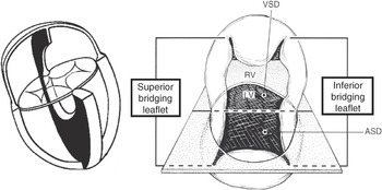
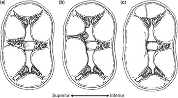
Taking note of extensive anatomical descriptions and classifications, cardiac surgeons rightly regard atrioventricular septal defect as a spectrum of malformations seen in the setting of a common atrioventricular junction. It is the commonality of the atrioventricular junction that is the unifying feature, although this, of necessity, involves absence of the atrioventricular component of the membranous septum, and failure of formation, during development, of the atrioventricular muscular sandwich.Reference Anderson, Wessels and Vettukattil2 The anatomy ranges from the presence of shunting exclusively at the atrial level, with the presence of separate right and left atrioventricular orifices within the common junction, the so-called partial form, to the situation in which both bridging leaflets of the common valve float freely within the septal defect, resulting in unrestricted communication between the atrial and ventricular chambers (Fig 1). Between these extremes are various transitional forms, such as those defined on the basis of a pressure-restrictive interventricular communication, but which retain the phenotypic feature of atrioventricular septal defect with common atrioventricular junction. Another key feature in the understanding of the anatomic arrangement was the Rastelli classification,Reference Rastelli, Kirklin and Titus3 with the underlying arrangement of the leaflets in the spectrum shown in Figure 2.
The cartoon shows the surgically relevant features of atrioventricular septal defect with common atrioventricular valvar orifice. As shown in the left-hand panel, the common atrioventricular junction implies a deficiency in the atrioventricular portions of the normal atrioventricular junctions, with loss of the normal offset of the leaflets of the mitral and tricuspid valves. As shown in the right-hand panel, the anatomy can be conceptualised as the intersection of the plane of the atrioventricular valves with the plane of the septal structures. This visualisation is useful during repair, irrespective of the technique employed (modified from Idriss RF in: Mavroudis C and Backer CL, eds Pediatric Cardiac Surgery. Philadelphia: Mosby, 2003).

Giancarlo Rastelli is probably best remembered for his classification of the atrioventricular valvar arrangement in atrioventricular septal defect, one of the few anatomic schemes that has remained virtually unchanged since it was first proposed. The classification is based on the degree of bridging of the superior leaflet, which remains quite relevant to most cardiac surgeons. From left to right: Types A, B (rarely encountered), and C (always present in cases with features of tetralogy of Fallot). Even in the Type C arrangement, it is possible to recognise a small antero-superior leaflet in the right ventricle. Type C typically carries the greatest potential for shunting at the ventricular level (modified from Idriss RF in: Mavroudis C and Backer CL, eds Pediatric Cardiac Surgery. Philadelphia: Mosby, 2003).

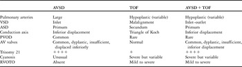
The essence of atrioventricular septal defect is the common atrioventricular junction. It is hardly surprising, therefore, that this lesion can coexist with tetralogy of Fallot, this combination making up perhaps one-tenth of the overall spectrum of atrioventricular septal defect itself. Conversely, those patients with atrioventricular septal defect account for around one-twentieth of the overall group of tetralogy of Fallot. Any variant of tetralogy may be encountered in the setting of an atrioventricular septal defect, including those with pulmonary atresia, double outlet right ventricle, absent pulmonary valve syndrome, and coronary arterial anomalies. The essential phenotypic feature of these patients, over and above the common atrioventricular junction, is the anterior and cephalad displacement of the muscular outlet or infundibular septum. The feature produces obstruction of the subpulmonary outflow tract, with varying degrees of severity. The arrangement of the pulmonary arterial tree is also highly variable. In this setting, the deformity of the left ventricular outflow tract as seen in the isolated lesion, reflecting the unwedged position of the aorta, is exaggerated by the subaortic extension of the interventricular communication and the rightward position of the aorta as seen in tetralogy of Fallot. In Table 1, we compare the features of atrioventricular septal defect and tetralogy of Fallot as seen in isolation, and the findings when the two lesions are combined.
Comparison of features of the isolated tetralogy of Fallot, isolated atrioventricular septal defect, and the two lesions in combination.

ASD=atrial septal defect; AV=atrioventricular; AVSD=atrioventricular septal defect; PVOD=pulmonary vascular obstructive disease; RVOTO=right ventricular outflow tract obstruction; TOF=tetralogy of Fallot; VSD=ventricular septal defect
Trisomy 21 figures importantly in the analysis of patients with atrioventricular septal defect, especially in those with tetralogy of Fallot. Perhaps fortunately, it is difficult to show that trisomy 21 is an independent risk factor for mortality after repair. Up to half of the children born with Down’s syndrome have a cardiac lesion, with atrioventricular septal defect being the most common lesion, followed by atrial and ventricular septal defects, patency of the arterial duct, and tetralogy of Fallot. Of the offspring of mothers with atrioventricular septal defect, one-sixth have congenitally malformed hearts, most frequently atrioventricular septal defect, or tetralogy of Fallot. The patients having tetralogy of Fallot in the setting of trisomy 21 are significantly more likely to have an additional intracardiac lesion, the strongest association being with atrioventricular septal defect. Taken together, more than three-quarters of the patients with both atrioventricular septal defect and tetralogy have trisomy 21, as compared to no more than 0.125% of the general population. The region of chromosome 21 known to be involved in Down’s syndrome contains genes associated with both ventricular septal defect and tetralogy of Fallot. Tetralogy-like lesions and atrioventricular septal defect with common atrioventricular junction are also known to occur together in mice with trisomy 16, although mouse chromosome 16 is syntenic for human chromosomes 21 and 22. Atrioventricular septal defect with common atrioventricular junction has also been observed in a strain of rats known genetically to develop tetralogy of Fallot.
Indications and timing
Pre-operative transthoracic echocardiographic assessment should now suffice to define all relevant structural details, including mechanisms of atrioventricular valve insufficiency. In this respect, nonetheless, intraoperative transoesophageal echocardiography has now become an indispensable adjunct to surgical repair.
Decades of experience and observation of the malignant natural history suggest that infants must undergo surgical correction electively before they are 3 months of age so as to reliably minimise the risk of pulmonary vascular obstructive disease. The second and more immediate consideration is the need for early palliation of failure to thrive related to the high flow of blood to the lungs, especially in syndromic infants. It remains the case that some patients with well-balanced haemodynamics can come to clinical attention later in life, and can still undergo successful surgical repair, even as adults, but they are exceptional. However, repair in the first month of life is usually not required, as most newborns can be managed conservatively with medical therapy for the first 2 or 3 months of life, which according to current opinion may promote a more favourable neurodevelopmental outcome. Post-operative atrioventricular valvar insufficiency, pulmonary insufficiency, and ventricular dysfunction are all more problematic in the neonate than in infants 3 months of age. Delay in repair, nonetheless, is justified in infants with extracardiac problems needing urgent treatment, as well as during infections with respiratory syncytial virus and other agents, as the systemic inflammatory response may be significantly exaggerated in these settings.
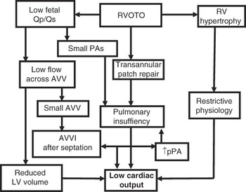
Unlike in those with atrioventricular septal defect in isolation, cyanosis may be the predominant feature in children also having tetralogy of Fallot, the pulmonary arteries in the latter situation being protected by the obstructed right ventricular outflow tract. We summarise the complex physiology of atrioventricular septal defect seen in combination with tetralogy in Figure 3. In those patients with obstruction to the right ventricular outflow tract, construction of a systemic-to-pulmonary arterial shunt may improve saturations of haemoglobin, as well as promoting atrioventricular valvar growth by volume loading, but the physiology created is otherwise suboptimal, especially for those with atrioventricular valvar insufficiency. For this reason, primary repair, whenever possible, is recommended for all those undergoing surgery beyond the neonatal period.
Schematic of the complex physiologic features of tetralogy of Fallot with atrioventricular septal defect. Effective palliation can be difficult to achieve by construction of a systemic-to-pulmonary arterial shunt in patients with atrioventricular valve insufficiency. On balance, therefore, primary repair is usually a better option when possible.

The problems of ventricular imbalance, the additional difficulties imposed by the finding of isomeric atrial appendages, and the potential unsuitability for septation are beyond the scope of our current discussion, but have been considered in detail elsewhere.Reference Kuribayashi4
Surgical technique
The general principles of surgical repair are now widely accepted. We aim not to leave patients with significant residual shunting, especially any combination of interventricular communication and left atrioventricular valvar insufficiency, which predictably leads to post-operative cardiopulmonary dysfunction. Repair can be achieved using a number of established techniques, often determined by the number of patches employed for septation. We favour direct closure of ventricular communication, combined with septation of the atrium using a pericardial patch, the so-called modified one patch, Nunn, or Australian technique. It was Nunn et al, who, while working in Sydney, reported the first large series of unselected cases undergoing operative repair in this manner,Reference Cohen and Spray5 and hence the description as the Australian repair.Reference Nicholson, Nunn and Sholler6 When using this technique, there is no need to divide the atrioventricular valvar leaflets, while entrapment of bridging cords is avoided, since there is no patch to be sutured to the ventricular septum.Reference Nicholson, Nunn and Sholler6 The technique remains suitable for most, if not all, of those patients with balanced ventricles, and as we will discuss, for many patients also having tetralogy of Fallot.
Repair is performed on cardiopulmonary bypass at 32°C, with direct cannulation of the aorta and both caval veins, cold sanguineous cardioplegic arrest, and modified ultrafiltration. Through a right atriotomy, we determine the relation of the bridging leaflets of the common atrioventricular valve relative to the plane of the ventricular septum (Fig 1). Coaptation is assessed by injecting saline into the ventricles, taking care to avoid excessive pressure, with the aortic root vented to prevent entry of air into the coronary arteries.
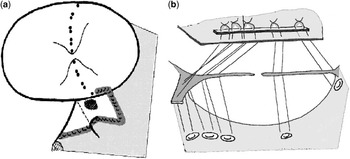
Having determined the appropriate geometry, interrupted pledgeted sutures are placed several millimetres away from the crest on the right ventricular aspect of the muscular ventricular septum (Fig 4). In case of extreme bridging of the superior leaflet, as in the Rastelli C variant, the sutures are placed through the ventricular aspect of the leaflet and brought out through the atrial aspect. In the presence of minimal bridging, as in the Rastelli A variant, sutures are taken through both the superior bridging leaflet and the anterosuperior leaflet of the right ventricle, thus bringing together the zone of apposition between these leaflets above the septal crest. These sutures are then placed through the base of an oval patch of autologous pericardium, reinforced with a strip of polytetrafluorethylene. The sutures are tied, apposing the leaflets to the crest of the ventricular septum, and eliminating the interventricular communication.
The cartoons show the details of the Nunn technique for repair of atrioventricular septal defect. Panel (a) shows the line of attachment of the atrioventricular leaflets to the crest of the ventricular septum, which will close the ventricular communication and partition the common valve into left and right components. Note the site of the atrioventricular node, tendon of Todaro, and coronary sinus. Panel (b) shows placement of sutures on the right side of the septum, through the valvar tissue, and through a pericardial patch reinforced with a strip of polytetrafluoroethylene. The pericardial patch is used to septate the atrium following repair of the atrioventricular valves (modified from NunnReference Nunn7).

Before completion of atrial septation, atrioventricular valvar competence is assessed by a repeated injection of saline. Gaps in the skirt of leaflet tissue guarding the newly created right and left valvar orifices are sutured closed even if the valves themselves are deemed competent in the native state, as long as the valvar orifices are of adequate caliber. There is probably no downside to this practice, and possibly some benefit in preventing post-operative valvar insufficiency. Moreover, use of this approach avoids later uncertainty regarding the relationship of valvar problems to failure to close the zone of apposition between the left ventricular components of the bridging leaflets, the so-called cleft.
In case atrioventricular valvar insufficiency is not eliminated with simple closure of the zone of apposition between the left ventricular components of the bridging leaflets, the question must be posed as to whether septation has allowed appropriate partitioning of the common atrioventricular valve. In case of serious doubt, it may be advisable to re-create the septal suture line, as it is difficult to compensate with any type of leaflet or annular adjustment. Other techniques include plication at the peripheral commissures and circular or semi-circular annuloplasties. These procedures are generally more appropriate at late re-operation for atrioventricular valvar insufficiency, which should be rare using the technique described above for septation.Reference Nicholson, Nunn and Sholler6
The atrial chambers are partitioned using the patch that secures the leaflets of the common atrioventricular valve to the crest of the muscular ventricular septum, leaving the orifice of the coronary sinus draining to the right atrium. There may be some disadvantage to placing the orifice of the coronary sinus in the left atrium, since this could promote coronary venous hypertension in the presence of atrioventricular valvar insufficiency, and subtly impair the flow of blood in the coronary arteries. Placing the orifice in the right atrium can also eliminate concerns regarding the contribution of blood returning through the coronary sinus blood to the post-operative requirement for oxygen in infants with marginal pulmonary function.
The competence of the newly created left atrioventricular valve may be affected immediately after bypass by impaired myocardial contractility, but will often improve during a period of modified ultrafiltration. Electrical conduction may also be contributory, as patients who do not have synchronous conduction may experience valvar insufficiency, even with temporary sequential pacing. In patients with more significant degrees of atrioventricular valvar insufficiency, revision should be considered. This is a difficult decision, and must take into account the location of the incompetence jet, the motion of the leaflets, the degree of any prolapse, the state of the zone of apposition between the left ventricular components of the bridging leaflets, the presence of accessory valvar orifices, and so on. These features cannot fully be assessed while the heart is arrested, and hence the importance of intraoperative transoesophageal echocardiography following repair.
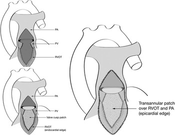
In patients with coexisting tetralogy of Fallot, the parietal extension of the muscular infundibular septum is excised before septation, as in repair of patients with isolated tetralogy of Fallot (Fig 5).Reference Nunn7 The ventriculo-pulmonary junction is usually hypoplastic, requiring transjunctional enlargement with a patch. To preserve pulmonary valvar competence, we employ the technique based on enlargement of the anterior leaflet of the pulmonary valve,Reference Karl, Sano, Pornvilawan and Mee8 creating a new sinus of Valsalva, which is then roofed with a transjunctional patch (Fig 6). A monocuspid polytetrafluorethylene patch can also be used or, in patients with pulmonary atresia, a xenograft conduit can be implanted between the right ventricle and the confluence of the pulmonary arteries.
The cartoons show excision of the parietal extension of the infundibular septum when repairing tetralogy of Fallot with atrioventricular septal defect (modified from Karl TR and Brizard CPR. In: Gardner TJ, Spray TL. Operative Cardiac Surgery. 5th Ed. New York: Oxford University Press 2004; 689–704).

The technique used to reconstruct the right ventricular outflow tract in tetralogy of Fallot as adapted for patients with associated atrioventricular septal defect (Anagnostopoulos et alReference Anagnostopoulos, Nolke and Alphonso9).

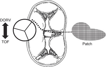
It is sometimes possible to septate hearts having a common atrioventricular valvar orifice in combination with tetralogy of Fallot using the Nunn technique. In this setting, of necessity, the superior bridging leaflet will show extensive bridging, fulfilling the definition for the Rastelli C variant. The best technique will depend to some extent on the degree of right ventricular origin of the aorta, and the size and shape of the superior rim of the ventricular septal defect in relationship to the atrioventricular valvar tissue. Despite obstruction of the outflow tract from the left ventricle being a theoretical concern, it has been rare in practice, either in patients with the isolated defect or in association with tetralogy of Fallot.Reference Nicholson, Nunn and Sholler6 As an alternative for those with coexisting tetralogy, a comma-shaped patch can be used to septate the ventricles, or the subaortic extension of the interventricular communication can be closed with a patch, the residual communication then being closed by using the Nunn technique (Fig 7).Reference Nicholson, Nunn and Sholler6, Reference Anagnostopoulos, Nolke and Alphonso9, Reference Malm, Karl and Mee10
Options for siting the patch when repairing tetralogy complicated by atrioventricular septal defect. The location of the aorta has been superimposed on Rastelli’s conceptual illustrations, showing the crest of the atrioventricular septum (dotted line), the approximate shape of the patch, and the proposed suture lie for the patch. This type of anatomy can also be corrected using a modification of the Nunn technique for isolated atrioventricular septal defect. The arrow indicates that a further rightward position of aorta would define a double outlet right ventriculo-arterial connection (modified from Idriss RF in: Mavroudis C and Backer CL, eds. Pediatric Cardiac Surgery. Philadelphia: Mosby, 2003).

Outcomes
Mortality has fallen significantly over the past three decades for patients undergoing repair of isolated atrioventricular septal defect, although post-operative abnormalities of conduction, atrioventricular valvar dysfunction, and obstruction to the left ventricular outflow tract remain problematic.Reference Karl11–Reference Meskishvili, Ishino and Dahnert15 Analysis of multi-institutional data reported for over 250 patients undergoing repair of atrioventricular septal defect with a common atrioventricular valvar orifice, where the patients had a mean comprehensive score within the Aristotle system for complexity of 10.7, revealed operative mortality to be 1.58%, with 95% confidence limits from 0% to 3%.Reference Najm, Williams and Chuaratanaphong16 For repair of the atrioventricular septal defect in isolation, the major risk factors for mortality included a left ventricular volume index of less than 20 cubic millimeters per metre squared, and a non-apex forming left ventricle, irrespective of the karyotype (D. R. Clarke, Personal communication, 2010). The congenital cardiac surgery database of the society of thoracic surgeons includes the outcome data for 1917 patients undergoing surgical repair of atrioventricular septal defect with common valvar orifice between 2005 and 2008.Reference Formigari, Di Donato and Gargiulo17 Mortality at discharge from hospital was 2.8%.
Results for the Australian repair in a cohort of over 100 patients, the majority having Down’s syndrome and unrestrictive ventricular septal defects, with repair being carried out at around 3 months of age, revealed operative mortality again to be 1.6%, with 95% confidence limits from 0.5% to 5%. Over a mean follow-up of 7.1 years, with a standard deviation of 3.5 years, only three re-operations have been required, with none of these involving re-intervention to relieve obstruction of the left ventricular outflow tract. Subsequent to the establishment of our new unit in Brisbane, we have carried out repair in 18 patients with common atrioventricular valvar orifice and unobstructed pulmonary outflow tracts, their mean comprehensive score with the Aristotle system being 12.4. All had repair with the Nunn technique, and all survived the procedure. Thus, experience with the Nunn technique compares favourably with most historical series employing alternate techniques for surgical repair.
Repair for patients having atrioventricular septal defect in combination with tetralogy of Fallot was first reported in 1969.Reference Jacobs18 By making a meta-analysis of almost 350 patients undergoing repair in 39 institutions, we calculate a mortality of 13% for this combination, with 95% confidence limits from 10% to 17%. Of note, in 16 of these series, with operations performed before 1990, there was zero mortality. For this period in Melbourne, there were 26 patients undergoing surgical correction of tetralogy combined with atrioventricular septal defect, this cohort accounting for one-twentieth of the patients with tetralogy of Fallot, and almost one-tenth of those with atrioventricular septal defect seen during the same interval.Reference Anagnostopoulos, Nolke and Alphonso9, Reference Malm, Karl and Mee10 Of the cohort, nine had double outlet right ventricle as assessed using the 50% criterion. Hospital mortality was 3.9%, with 95% confidence limits from 1.0% to 19%. Late death occurred in two patients over 1650 patient months of follow-up. Actuarial freedom from re-operation for residual septal defects or valvar dysfunction was significantly worse than that of either lesion in isolation, although the actuarial data relative to survival was similar.
Discussion
We can make some comparisons of the role of one as opposed to two patches in classical repairs, setting aside for the moment the Australian approach promoted by Nunn. The technique of using one patch, and dividing the bridging leaflets, probably does produce better exposure of the rim of the ventricular septum when using a transatrial approach. There is some flexibility in placing the line used to divide the superior bridging leaflet, and the surgeon is able, if necessary, to assign more valvar tissue to the left atrioventricular valve, although this is also possible, to a lesser extent, with other techniques. It is always necessary to sacrifice some valvar tissue when attaching segments of divided leaflets to the single patch. However, when using a classical two-patch repair, the superior bridging leaflet will remain anatomically intact, and the problems discussed above should not be encountered. This operation is probably easier to learn, and easier to teach, than other strategies. There may also be a lower potential for a dehiscence of the leaflets than with the classical single patch technique.
The Nunn operation combines some of the best features of the classical one and two patch repairs and eliminates some of the worst features. The technique as described has nearly universal applicability in patients with atrioventricular septal defect and common atrioventricular valvar orifice, and is quite reproducible. Furthermore, it is easy to teach to trainees, as there are fewer decisions to be made regarding geometry, size, and configuration of patches. The results obtained to date compare favourably with those achieved with other techniques, both in the short and medium term. Early concerns regarding the potential for creating an obstruction of the left ventricular outflow tract have thus far proved to be unwarranted. In this respect, it might be thought that suturing the superior bridging leaflet to the crest of the ventricular septum so as to obliterate the superior part of the ventricular component of the septal defect would, of necessity, narrow that subaortic outflow tract. Indeed, this was the presumption initially made by the anatomical member of our team. During the discussions we held last year in Brisbane, however, it became clear that this assumption is unwarranted. In case the superior leaflet is merely turned over, as it is attached to the septal crest, this process in no way narrows further the pre-existing dimensions of the outflow tract from the left ventricle.Reference D’Allaines, Colvez, Fevre, Levasseur, Facquet and Dubost19
Conclusion
We have presented a standardised technique for repair of patients with atrioventricular septal defects and common atrioventricular valvar orifice, which can be modified in the presence of tetralogy of Fallot. Despite the availability of other techniques, the Australian strategy as promoted by Nunn is very satisfactory, and to date has provided good clinical outcome in a large series of consecutive and unselected patients.
Acknowledgement
We thank our colleagues in intensive care, cardiology, anesthesia, perfusion, and nursing for their roles in the care of the patients reported herein.