Rheumatic heart disease, with progressive left ventricular dilation and dysfunction, is the most common form of acquired cardiac disease in young adults in the developing world.Reference Eisenberg 1 A number of factors may contribute to myocardial damage and detrimentally affect left ventricular myocardial contractility during rheumatic fever.Reference Narula, Chopra and Talwar 2 , Reference Taranta and Markowitz 3 The dimensions of the left ventricular cavity, and the ejection fraction, however, are indirect measurements of intrinsic myocardial function, and remain within normal limits, or are higher than normal, during the compensated phase of rheumatic heart disease.Reference Vasan, Shrivastava, Vijayakumar, Narang, Lister and Narula 4 , Reference Essop, Wisenbaugh and Sareli 5
Tissue Doppler imaging is able to measure the peak velocity of myocytic shortening in the longitudinal direction, and is believed to give an estimation of global left ventricular systolic function with high feasibility and reproducibility. The velocities derived from the annulus or the base of the left ventricle primarily reflect longitudinal motion, due to the longitudinally directed myocytes which are found in the subendocardium. This may explain why these measurements are particularly sensitive for the assessment of myocardial dysfunction in patients with volume overload.Reference Vinereanu, Ionescu and Fraser 6 , Reference Nagueh, Middleton, Kopelen, Zoghbi and Quinones 7 As the subendocardial myocytes are aligned longitudinally, we hypothesised that left ventricular long axis function may be altered in patients with rheumatic carditis. This has yet to be elucidated. We prospectively evaluated, therefore, the ventricular performance of patients after a primary episode of rheumatic carditis, using indexes of tissue Doppler imaging as potential echocardiographic indicators of subnormal myocardial dysfunction.
Materials and methods
Patients
We studied 82 patients presenting with primary episodes of rheumatic carditis to our hospital over a period of 24 months, basing the diagnosis of acute rheumatic fever on the modified Jones criterions. 8 – Reference Feinstein and Spagnuolo 11 All had evidence of continuing rheumatic activity, including elevated acute phase reactants. Mean age at the time of acute rheumatic fever was 10.3 ± 2.6 years, with a range from 4.6 to 15.8 years, and 32 (39%) were male. We also recruited 30 healthy subjects from children ssen with an innocent murmur over the same period. The protocol was approved by the Local Research Ethics Committee, and all subjects gave informed consent.
All children underwent conventional and tissue Doppler imaging initially and at the time of the follow-up examinaton.
Conventional echocardiographic study
All ultrasound examinations were performed with a commercially available echocardiographic machine (Vivid 3 System, Vingmed–General Electric) equipped with 2.5–5 and 4–8 MHz phased-array transducers. All patients underwent echocardiographic examination within 24 to 48 hours of establishment of the diagnosis of acute rheumatic fever and before starting anti-inflammatory treatment. Valvar regurgitation was diagnosed when color Doppler flow mapping demonstrated reversed flow through the valve in the closed position. To differentiate abnormal from physiological mitral regurgitation,Reference Brand, Dollberg and Keren 12 we required the high-velocity turbulent jet to extend beyond the paravalvar region by more than 15 millimetres, confirmed this by colour-guided pulsed Doppler spectral analysis.Reference Sahn and Maciel 13
On-line computerized planimetry of the maximal regurgitant jet areas was performed, and the mean of three cardiac cycles was taken. The severity of mitral and aortic regurgitation was graded on the basis of the maximal distance of the regurgitant jet from the valvar orifice using the criterions of Helmcke et al.,Reference Helmcke, Nanda and Hsiung 14 and Perry et al.,Reference Perry, Helmcke, Nanda, Byard and Soto 15 respectively. The dimensions of the left atrium and ventricle were measured from M-mode traces, and by using the recommendations of the American Society of Echocardiography.Reference Sahn, DeMaria, Kisslo and Weyman 16 Left ventricular ejection fraction and fractional shortening were estimated from M-mode tracings using the Teichholz formulaReference Teichholz, Cohen, Sonnenblick and Gorlin 17 and the cubed formula,Reference Feigenbaum, Popp and Wolfe 18 respectively. Measurements were obtained during four end-expiratory cardiac cycles, taking the average for each parameter.
Tissue Doppler imaging
Tissue Doppler imaging was performed in apical four-chamber planes with the pulsed-wave Doppler sample volume of 4 millmetre length placed successively at the lateral mitral annulus. The incident angle between the interrogating Doppler beam and longitudinal motion of the ventricle was kept as small possible. We measured the myocardial systolic wave velocity, the myocardial precontraction time from the onset of the QRS complex to the beginning of the myocardial systolic wave, and the myocardial contraction time, from the beginning to the end of the myocardial systolic wave, permitting calculation of the ratio between these times. At least 10 cardiac cycles were recorded from each site on a strip-chart recorder at a speed of 100 mm/s.
Follow-up
Patients with rheumatic carditis and congestive heart failure were treated with steroids, along with dietary salt restriction, digoxin, diuretics, and inhibitors of angiotension converting enzyme, whereas patients with carditis but without congestive heart failure were treated with aspirin. Injectable benzathine penicillin G for secondary prophylaxis was initiated or regulated at the time of the index attack in all patients.Reference Massel, Fyler and Roy 19 Follow-up examinations were routinely scheduled in all patients at 3 months after the index attack to study the evolution of valvar lesions. At the time of the follow-up examination, none had clinical or laboratory evidence of ongoing or recurrent rheumatic fever. Repeat echocardiographic examinations were performed by the same operator with the same ultrasound machine and with similar gain settings.
Statistical analysis
Results are expressed as mean, and range. The data were analyzed with SPSS version 10.0 software (SPSS, Chicago, IL). The differences between the groups were explored with the Student’s t test. Incidences in the groups were tested for significance with the χ 2 test. For comparison of tissue Doppler imaging, variables obtained in the same individual initially and at the time of the follow-up examinaton, the paired, two-sided Student’s t-test was used. A probability value of p < 0.05 was considered significant.
Results
Of the 82 patients, 50 (61%) had mild mitral regurgitation, while 20 (24%) had moderate, and 12 (15%) had severe mitral regurgitation. According to the valvar involvement at the time of index attack, we identified 32 patients with mitral regurgitation of grade 2 or more, and 50 patients with mild mitral regurgitation in the index attack. Improper coaptation of the aortic valvar leaflets without prolapse or annular dilation was seen in 7, and all were associated with mild degrees of valvar regurgitation. Of these, 3 also had mild mitral regurgitation, while the remaining 4 were associated with moderate degree of mitral regurgitation.
Conventional echocardiographic findings
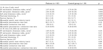
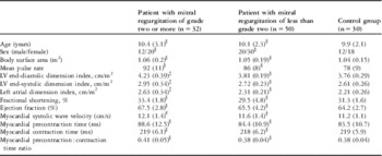
At the initial period, patients showed significantly larger left atrial and ventricle dimensions (Table 1). Furthermore, baseline left atrial and ventricle dimensions were significantly larger in patients with mitral regurgitation of grade 2 or more than control group, but no significant differences were noted between patients with mild mitral regurgitation and healthy subjects in the conventional echocardiographic parameters (Table 2). At follow-up examination, left atrial and ventricular dimensions significantly decreased (Table 1), albeit that ventricular dimensions were not significantly changed in patients with mild mitral regurtation without volume load (Table 3). Ejection fractions and fractional shortening values did not differ significantly between patients and their controls, neither at the initial nor the follow-up examinations (Tables 1, 2 and 3).
Conventional and tissue Doppler indexes of the patients and their controls.

Values are expressed as means with standard deviation shown in parentheses.
Demographic and baseline echocardiographic features of the groups.

Values are expressed as means with standard deviation shown in parentheses.
Patients’ groups compared to control group, §p > 0.05, €p = 0.007, ‡p < 0.001
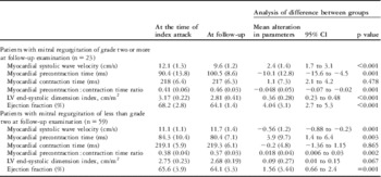
Comparison of conventional and tissue Doppler indexes of patients obtained initially and at the time of the follow-up examinaton (paired t test with confidence intervals).

Values are expressed as means with standard deviation shown in parentheses.
Tissue Doppler parameters
The myocardial systolic wave velocity of the mitral annulus was significantly higher than in control group (p = 0.038) at the time of index attack, but myocardial time intervals were not different between the patients and healthy subjects either at the time of index attack or during follow-up (Table 1). The myocardial systolic wave velocity of the mitral annulus was significantly higher in patients with mitral regurgitation of grade 2 or more when compared to control group (p = 0.007) but was not different between patients with mild mitral regurgitation and healthy subjects at the time of index attack. Myocardial time intervals of the mitral annulus were not significantly different between the two study groups and control group at the time of the index attack (Table 2).
According to the valvar involvement at the time of the follow-up examination, we identified 23 (28%) patients with mitral regurgitation of grade 2 or more, and 59 (72%) patients with mitral regurgitation of less than grade 2. Myocardial precontraction time, and the ratio between precontraction and contraction times, were significantly prolonged (p = 0.001), and the myocardial systolic wave velocity of the mitral annulus was significantly decreased (p < 0.001) in patients with mitral regurgitation of grade 2 or more at the time of the follow-up examination (Table 3). The myocardial systolic wave velocity of the mitral annulus was significantly lower (p < 0.001), and the myocardial precontraction awas longer, and the ration of contractions significantly greater (p < 0.001, p =0.038, respectively) between patients with mitral regurgitation of grade 2 or more and the control group, but myocardial systolic wave velocity and all myocardial time intervals of the mitral annulus were not significantly different between patients with mitral regurgitation of less than grade 2 and those making up the control group at follow-up examination (Table 4).
Tissue Doppler indexes of patients according to the valvar involvement at follow-up examination.

Values are expressed as means with standard deviation shown in parentheses.
Patients’ groups compared to control group, § p > 0.05; ¥ p = 0.038; ‡ p < 0.001
Discussion
Mitral regurgitation is the most common Doppler echocardiographic feature of both a first episode and recurrences of rheumatic carditis. Associated aortic valvar involvement is a less common feature.Reference Massel, Fyler and Roy 19 , Reference Sanyal, Thapar, Ahmed, Hooja and Tewari 20 Many efforts have been made to obtain an accurate estimation of the contractile performance of the ventricle in mitral regurgitation. Unfortunately, most indexes of ventricular function used in clinical practice, such as ejection fraction and stroke volume, do not solely reflect the contractile state of the ventricle. Ejection fraction, and left ventricular internal dimensions, are not reliable predictors of left ventricular systolic function after surgical correction of mitral regurgitation. The ejection fraction decreases significantly because of the increased afterload after surgical correction.Reference Enriquez-Sarano, Tajik, Schaff, Orszulak, Bailey and Frye 21 , Reference Enriquez-Sarano 22
The explanation for the uncertainty about left ventricular function is the considerable alteration in the loading conditions of the left ventricle in mitral regurgitation. The increased diastolic inflow volume in the left ventricle induces an increased preload. During systole, the left ventricle generally ejects the normal stroke volume through the aortic valve, but also produces a regurgitant flow to the left atrium, suggestive of a decreased impedance to ejection, producing doubt concerning the true contractile state of the myocardium.Reference Enriquez-Sarano, Tajik, Schaff, Orszulak, Bailey and Frye 21 , Reference Agricola, Galderisi and Oppizzi 23
The assessment of the systolic indexes of the mitral annulus with tissue Doppler imaging allows for an estimation of global left ventricular systolic function, providing information on the velocity of the descent of the base as an estimate of global systolic ventricular function.Reference Pai, Bodenheimer, Pai, Koss and Adamick 24 The onset of subendocardial shortening coincides hemodynamically with an early rapid build-up of intraventricular pressure during the isovolumic contraction period. Shortening of circumferential fibres occurs later, temporally coinciding with the onset of systolic ejection. The temporal lag between the onsets of subendocardial and subepicardial contraction activities correlate with the duration of isovolumic contraction. The differential placement of the longitudinal myocytes in the subendocardium, therefore, may render longitudinal velocity uniquely sensitive to mild or early impairments prior to changes being detectable in the short axis. Thus, tissue Doppler imaging may provide evidence of subclinical myocardial dysfunction in the early period before other conventional Doppler parameters.Reference Skoularigis, Sinovich, Joubert and Sareli 25
Furthermore, tissue Doppler imaging not only defines and measures mitral annular movement but, because of its high temporal resolution, also provides a useful estimate of myocardial time intervals, both systolic and diastolic.Reference Henry, Bonow, Rosing and Epstein 26 – Reference Wisenbaugh 28 Myocardial precontraction time is the myocardial systolic time corresponding to the isometric period before the onset of myocardial contraction, whereas myocardial contraction time corresponds to the period of contraction. The ratio between these times, corresponding to the ratio of the left ventricular pre-ejection period to the left ventricular ejection time obtained by standard Doppler assessment of the left ventricular outflow tract, may be considered to be an index inversely correlated with myocardial contractility.Reference Starling, Kirsh, Montgomery and Gross 27 In a previous study,Reference Agricola, Galderisi and Oppizzi 23 the ratio, along with and myocardial systolic wave velocity of the mitral annulus velocity, were the best discriminant cut off values for a postoperative reduction in ejection fraction of less than 10% obtained by receiver operating characteristic curves in patients with asymptomatic severe mitral regurgitation and normal ejection fraction. Moreover, a postoperative ejection fraction of less than 50% was observed in 8% of patients, and all the preoperative systolic tissue Doppler imaging parameters had a significant correlation with changes in ejection fraction.
In our study, tissue Doppler imaging indexes of patients appeared normal in those with little valvar involvement, while we found that for an increase of preload and decrease of afterload in those with significant valvar lesions, there was a concomitant significant increase of myocardial systolic wave velocity of the mitral annulus. This phenomenon can be simply justified by the length-dependent activation seen in the Frank-Starling mechanism, which endows the ventricle with performance characteristics such that the heart ejects whatever volume it receives during diastole. Thus, an increase of the length of the myocytes caused by an increase of end-diastolic volume and decrease of ventricular tension caused by decrease of afterload will lead to an increase of stroke volume, the velocity of shortening, and and also an increase of myocardial systolic wave velocity of the mitral annulus. The sudden increase of preload, and decrease of afterload, has a complex impact on the haemodynamic behavior of the ventricle that is presently still not fully comprehended. As such, this parameter cannot be recommended as an index of ventricular contractility, where significant changes in ventricular filling occur at the time of index attack.
In patients with persistent significant valvar lesions, the systolic myocardial velocity and systolic myocardial time intervals of the mitral annulus deteriorated during follow-up.
Abnormal values of tissue Doppler imaging systolic indexes observed in patients with persistent valvar involvement may be explained by persistent left ventricular volume overload that increases wall stress, causing subendocardial ischaemia and fibrosis.Reference Villari, Campbell and Hess 29 In support of these prognostications, the tissue Doppler imaging indexes were observed to be normal in patients with little valvar involvement during follow-up. These observations concur with recent reportsReference Vinereanu, Ionescu and Fraser 6 , Reference Agricola, Galderisi and Oppizzi 23 , Reference Pai, Bodenheimer, Pai, Koss and Adamick 24 that the preservation of cardiac functions was paralleled by the disappearance of volume overload.
Some limitations of our study should be noted. Delay of surgery for mitral insufficiency often results in irreversible left ventricular dysfunction in adults. This has prompted a trend toward earlier surgery in adult patients with chronic mitral regurgitation based on echocardiographic assessment.Reference Ross 30 In this respect, our findings may have important clinical implications. In contrast, as do many paediatric centres, we usually continue to delay surgery for children until severe symptoms are present. This approach is a result of the limitations of the size of the patients for valvar replacement, the need for anticoagulation management, and the expectation for early reoperation with ongoing growth.Reference Sanyal 31 – Reference Schaffer, Clarke, Campbell, Madigan, Wiggins and Wolfe 34 Thus, all our current patients were asymptomatic, and had only subclinic impairment of left ventricular systolic functions assessed by tissue Doppler imaging, so we did not recommend surgery. In this respect, it is questionable whether the abnormal indexes may predict a significant reduction in postoperative ejection fraction in asymptomatic children with severe mitral regurgitation and normal ejection fraction, which need to be tested in further studies.
We conclude that, in patients with rheumatic carditis, assessment using tissue Doppler imaging may be useful in providing evidence of subclinical myocardial dysfunction, suggesting contractile disturbance due to ongoing left ventricular volume overload before conventional Doppler parameters. These findings may offer new diagnostic and therapeutic opportunities in the screening and management of these patients, and should be considered in these settings.