Introduction
Between 1980 and 2007, 474,273 cases of AIDS were notified in Brazil and 710,000 adults were living with the human immunodeficiency virus (HIV) (UNAIDS/WHO, 2008). In a transversal study in Brazil, Pottes et al. (Reference Pottes, de Brito, Gouveia, Araújo and Carneiro2007) found that 10.8% of notified AIDS cases affected older adults. In Goiás State, the incidence coefficient of AIDS in the elderly population rose from 1.67 cases/100,000 inhabitants in 2000 to 5.75 cases in 2006 (IBGE, 2006).
Advanced age alone is an independent predictor of neuropsychiatric syndromes. HIV patients aged over 50 years are at higher risk of developing HIV-associated neurocognitive disorders (Valcour and Paul, Reference Valcour and Paul2006). Aging and HIV infection are independent determinants of progressive brain atrophy (Stout et al., Reference Stout1998; Ohnishi et al., Reference Ohnishi, Matsuda, Tabira, Asada and Uno2001). Becker et al. (Reference Becker, Lopez, Dew and Aizenstein2004) conducted a longitudinal study involving 290 HIV-positive subjects and 124 controls and found a 22% prevalence of HIV-1 associated dementia (HIVD) in individuals aged over 50 years versus 9% in seropositive younger adults. In a transversal study of 202 seropositive patients, Valcour et al. (Reference Valcour2004) reported a 25.2% frequency of HIVD in individuals aged over 50 years, a statistically higher rate than in younger subjects (13.7%).
HIVD was first described in 1986 as a rapid onset disease which evolved from motor slowing to dementia, acinetic mutism and coma (Navia et al., Reference Navia, Jordan and Price1986). In 1987, the Center for Disease Control recognized its clinical relevance and classified HIVD as a defining disease of AIDS (Center for Disease Control, 1987). The sites most affected by HIV at disease onset include the hippocampus, frontal lobes, periventricular white matter and basal ganglia, where this constitutes the anatomic substrate which categorizes HIVD as subcortical dementia (Dubé et al., Reference Dubé, Benton, Cruess and Evans2005).
HIV-1 associated dementia is characterized by cognitive decline, behavioral disturbances and motor symptoms (McArthur et al., Reference McArthur, Brew and Nath2005). The introduction of highly active anti-retroviral therapy (HAART) has changed the natural history of HIVD, presenting insidious evolution, milder cases, oscillating symptoms, longer survival, manifestation in patients with higher CD4, and cortical as well as subcortical symptoms (Valcour and Paul, Reference Valcour and Paul2006). In a retrospective study performed by Welch and Morse (Reference Welch and Morse2002) involving 669 seropositive outpatients in Louisiana, U.S.A., HIVD ranked among the six most frequent AIDS defining diseases in the final year of life (91.8% frequency), suggesting that HIVD continues to have an impact on mortality in AIDS.
Recent studies suggest that the prevalence of HIVD is rising in the post-HAART era. No published papers on HIVD in Brazilian older adults are available. The aim of this study was to describe the clinical and tomographic characteristics of elderly HIV-positive patients with HIVD.
Methods
The present study describes a series of eight HIV-positive patients seen consecutively at the Hospital of Tropical Diseases (HTD) in Goiás State between November 2007 and September 2008. The inclusion criteria were: (i) confirmed HIV positive diagnosis on the Elisa and Western blot test, (ii) age 60 years or over, and (iii) diagnosed with HIVD based on 2007 updated nosology for HIV-associated neurocognitive disorders (Antinori et al., Reference Antinori2007). Subjects with dementia prior to HIV diagnosis were excluded. Subjects with pre-existent disease, such as mental retardation and psychotic disorders, or those in current delirium were also excluded.
This study was approved by the Research Ethics Committee of the Hospital of Tropical Diseases of Goiás State. Free and informed consent was signed by all study participants or their caregivers.
Patients were assessed for medical history and life habits and answered a questionnaire on neuropsychic symptoms which investigated the following variables: difficulty concentrating, forgetfulness, social isolation, difficulty learning, sluggish reasoning, slowed use of hands or gait, loss of initiative, spatial and temporal disorientation, crying without reason, loss of interest in favorite activities, and verbal and physical aggressiveness. Participants were submitted to a neuropsychological test battery and neurological physical examination. Laboratory exam data were collected from medical charts. The etiological investigation comprised the following exams: serology for syphilis, vitamin B12 and folic acid levels, TSH and free T4 levels, liver and kidney function.
Seven tests were used in the cognitive assessment: Trail Making A and B Tests (TMT), Digit Span Test (forward and backward), Victoria Stroop Test (cards 1 and 3), Rey Auditory-Verbal Learning Test, Verbal Fluency Test, Mini-mental State Examination (MMSE) and Grooved Pegboard (Brucki and Rocha, Reference Brucki and Rocha2004; Strauss et al., Reference Strauss, Sherman, Spreen, Strauss, Sherman and Spreen2006; Malloy-Diniz et al., Reference Malloy-Diniz, Lasmar, Gazinelli, Fuentes and Salgado2007; Diniz et al., Reference Diniz, Volpe and Tavares2007). These tests were chosen based on their ease of application and their high frequency in international HIV studies, except for MMSE, which was chosen because it is largely studied in Brazil and commonly used in geriatric practice. All tests were transformed to whole number z-scores using appropriate normative sets.
Lawton's nine-item version of the Instrumental Activities of Daily Living (IADL) scale was used to assess functional status (Strauss et al., Reference Strauss, Sherman, Spreen, Strauss, Sherman and Spreen2006). The 15-item Geriatric Depression Scale was applied to measure depressive symptoms. A cut-off score of greater than five was considered indicative of possible depression (Paradela et al., Reference Paradela, Lourenço and Veras2005).
Patients were submitted to head tomography using contrast on a Siemens tomograph device, somaton model AR SP/STAR 40/80.
Results
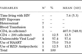
A total of eight confirmed HIVD patients with a mean age of 71 years, and mean schooling of 6.5 years, were assessed. Seven were men and the majority were Caucasian, in whom the main route of infection was through heterosexual activity. Six patients were using highly active anti-retroviral therapy (HAART), four of whom presented with undetectable viral load, while only one individual had a CD4 count lower than 200cells/mm3 (Table 1).
Number and percentage of patients studied by laboratorial and clinical characteristics – Hospital of Tropical Diseases (HTD), 2007/2008

*Viral Load not obtained in one patient.
HAART = highly active anti-retroviral therapy; BZD = benzodiazepines.
The majority of patients presented with mild dementia (87.5%). One case was classified as moderate dementia based on 2007 updated nosology for HIV-associated neurocognitive disorders (Antinori et al., Reference Antinori2007). Exams investigating etiology ruled out other causes of dementia. No patients had a history of cranial-encephalic trauma with loss of consciousness, epilepsy, meningitis or Parkinson's disease. All participants presented functional deficit on Lawton's IADL scale.
The predominant clinical presentation of HIVD in the elderly in the present study was subcortical dementia with psychiatric alterations including social isolation, aggressiveness, cognitive and motor symptoms such as psychomotor slowing and poorer fine motor control, attentional deficit, impaired working memory, compromised immediate and delayed memory, while recognition memory remained preserved. Cortical symptoms were also frequent, including dyscalculia, executive dysfunction and visuo-spatial alterations, and language deficits.
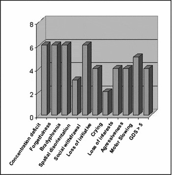
The most frequent neurocognitive symptoms in descending order were: concentration difficulties, forgetfulness, bradyphrenia, social isolation, motor slowing, loss of interests, loss of initiative and aggressiveness. Depressive symptoms with a GDS score above the 5-point cut-off were detected in four cases, none of which met criteria for major depression. None of the patients was using antidepressants (Figure 1).
Frequency of neuropsychiatric and motor symptoms, HTD, 2007/2008.

The neuropsychiatric assessment found poor performance, with a mean z-score of less than -2, for all the cases on the forward and reverse digit span test, referring to attention and working memory deficits. The Rey Auditory Verbal Learning test, items A1 to A5 and item 7 (immediate and delayed recall, respectively), the Trailmaking test and Stroop test (mental agility and selective attention), presented low scores in 87.5% of the subjects. Poor performance was also found in 75% of cases on the Grooved Pegboard test (motor skills) and MMSE. The most frequently affected sub-items of the MMSE in descending order were as follows: calculations (100% of cases), memory retrieval (87.5% of cases), temporal orientation (75% of cases), praxia (62.5% of cases) and language (57.14% of cases). In the MMSE analysis, one subject was disregarded due to poor eyesight.
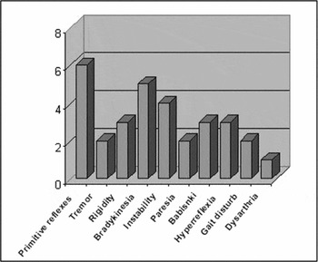
The disturbances most frequently found on neurological examination were, in descending order: presence of primitive reflexes, bradykinesia and postural instability. Three patients presented Babinski's sign, hyperreflexia and rigidity (Figure 2).
Frequency of neurological alterations on physical exam, HTD 2007/2008.

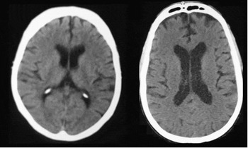
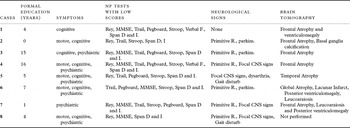
Cortical atrophy was observed on tomography in all patients (Figure 3). Atrophy was limited to the frontal region in five patients (71.4%) and ventriculomegaly in three cases (42.8%). Atrophy of the temporal lobes was found in one case (case 5), a pattern not expected for HIVD. However, the neuropsychological profile of this patient presented a pattern of subcortical dementia with altered immediate and delayed recall, compromised executive function (Trailmaking B and Digit Span tests), associated with preserved recognition memory, temporo-spatial orientation and verbal fluency (Table 2).
Tomographic findings for cases 1 and 3, respectively. HTD, 2007/2008: mild frontal atrophy and ventriculomegaly.

Distribution of clinical and tomographic findings by case, HTD 2007/2008

*Case 8 did not perform Stroop, Pegboard or Trailmaking tests due to poor eyesight.
Case 2 did not perform Pegboard test due to hand trembling.
Rey = A1 to A5 of the Rey Auditory Verbal Test; Verbal F = Verbal Fluency; Span = Forward and Reverse Digit Span Test; Primitive R = Primitive reflexes; CNS = Central Nervous System; parkins = parkinsonism.
Case 2 presented another unusual finding with frontal lobe atrophy and bilateral calcification in basal ganglia. Clinical assessment revealed a patient with a 13-year history of HIV who presented gross tremors in both hands and feet, and paresthesia in lower limbs for five years prior to manifestation of cognitive symptoms, rigidity, depressive symptoms and tactile hallucinations, as Ekbom's syndrome.
Analysis of medical history revealed that case 7 had had a stroke six years prior to the study, with mental confusion and gait disturbances at the time of the episode, which reached full remission. The imaging exams of this patient evidenced no cortical lesion. Two other cases presented episodes of self-limiting delirium during previous clinical complications, although they were not in delirium at the time of dementia diagnosis. No diagnoses of HIVD or cognitive impairment were recorded in their medical files.
Discussion
In contrast to degenerative dementias, 87.5% of presented HIVD cases occurred in males, a higher-than-expected percentage in view of the ratio of AIDS distribution by gender of 2:1 (men: women). In a transversal study in Brazil, Pottes et al. (Reference Pottes, de Brito, Gouveia, Araújo and Carneiro2007) found that over 75% of HIV older adults were men. These data suggest a higher-than-expected predominance of HIVD in men given the distribution of AIDS by gender in advanced age (Chiesi et al., Reference Chiesi1996).
The majority of our patients (seven cases) described the route of HIV infection as being heterosexual, currently the most common form of HIV exposure in Brazil (IBGE, 2006; Pottes et al., Reference Pottes, de Brito, Gouveia, Araújo and Carneiro2007). The average schooling among our sample was relatively low (6.5 years). National studies have confirmed that AIDS has spread over the last decade to afflict less socially privileged strata of the population (IBGE, 2006). Groups with lower schooling are at higher risk of exposure to HIV and to late diagnosis of the disease as a result of misinformation.
With the rising number of AIDS cases among the elderly, albeit due to an aging population or longer life expectancy in HIV-infected individuals, HIVD has become an increasingly significant differential diagnosis for cognitive loss in old age.
The clinical presentation of HIVD in the elderly in the present study was a pattern of cortical and subcortical compromise, also described in studies carried out in the U.S.A. (Brew, Reference Brew2004; Dubé et al., Reference Dubé, Benton, Cruess and Evans2005). Low fine-motor ability and parkinsonian signs were frequent in our sample (75% and 37.5% of cases, respectively; see Figure 3). Similar to our findings, Valcour et al. (Reference Valcour, Watters, Williams, Sacktor, McMurtray and Shikuma2008) described an association among age, HIV and presence of bradykinesia and tremors in a study involving 433 participants. The study found three or more parkinsonian signs in 28% of HIV positive individuals with normal cognition versus 68% of HIVD participants (p<0.0001) (Valcour et al., Reference Valcour, Watters, Williams, Sacktor, McMurtray and Shikuma2008).
In 1994, Maj et al. carried out a multi-center transversal study, with one arm run in São Paulo, Brazil. A neuropsychological test battery was used to analyze 178 HIV-infected people (aged 29 to 34 years), who were split into symptomatic and asymptomatic infection groups. The authors found a significant association between cognitive complaints and neuropsychological deficits in symptomatic HIV-seropositive individuals (p<0.001). Akin to our study, the HIV-seropositive group presented worse scores on Digit Span, TMT-A and Verbal Fluency Tests while no difference was observed across groups on the Grooved Pegboard Test.
A study conducted in Uganda assessed the neuropsychological patterns of 110 HIV positive patients (Robertson et al., Reference Robertson2007). Compared to seronegative individuals, HIV patients performed worse on the Digit Span forward and backward, Stroop and WHO-UCLA Auditory Verbal Learning Test. As in our study, executive function and memory were affected in the HIV positive patients. In contrast to our findings, however, Robertson et al. (Reference Robertson2007) found no significant difference between the groups on the Grooved Pegboard or Timed Gait tests (motor skills). The absence of motor symptoms could be explained by the lower mean age (36.7 years) of the particpants, or the fact that the study did not select individuals with cognitive impairment, or by differences in HIV subtypes. In Africa, clade D and A of HIV-1 predominate, whereas in Brazil clade B, of greater neurovirulence, is more common (Stefani et al., Reference Stefani2007).
Half of our sample presented depressive symptoms on the GDS, although none of the patients presented with symptoms of major depression. Depressive symptoms are common in subcortical disturbances (Casanova-Sotolongo et al., Reference Casanova-Sotolongo, Casanova-Carrillo and Casanova-Carrillo2002) and akin to cognitive symptoms, can resolve with anti-retroviral treatment. In cognitively normal seropositive individuals, depressive and motor symptoms have proved independent predictors of global neurological deficits (Robinson-Papp et al., Reference Robinson-Papp, Byrd, Mindt, Oden, Simpson and Morgello2008). Major depression frequently occurs in seropositive individuals, ranging from 4% to 37% of cases, (Asch et al., Reference Asch2003) and may constitute a cause of cognitive impairment. However, pseudodementia mimics subcortical dementias, presenting with attentional compromise, slowed mental processing and working memory dysfunction, all signs that are reversible by treatment with antidepressants (Turner et al., Reference Turner, Moran and Kopelman2002). Depression, alcoholism and hepatitis C can be cofactors for cognitive loss and must be treated (Janssen et al., Reference Jansen1991).
In our case load, 71.4% were found to have frontal cortical atrophy and 42.8% ventriculomegaly. No changes were found on brain images to suggest any association with other neurological diseases, with the exception of case 5 who presented bitemporal atrophy, possibly related to comorbidity with AD.
The hallmark pattern on HIVD imaging exams are periventricular hypersignal in white matter, ventriculomegaly, and cortical and subcortical atrophy (McArthur et al., Reference McArthur, Brew and Nath2005; Garcia-Moncó et al., Reference Garcia-Moncó, Brochado and Cortina2007). Cortical atrophy is also described in non-demented HIV-positive individuals. Thompson et al. (Reference Thompson2005) studied 26 non-demented HIV-positive individuals, and described atrophy in pre-motor and motor sensitive gyri, in addition to degenerative changes in white matter. The same authors, in a study on 30 seropositive patients, reported atrophy of the corpus collosum and frontal ventriculomegaly (Chiang et al., Reference Chiang2007). The brain of seronegative elderly may also present atrophy as a function of the aging process where loss in brain weight of up to 3% per decade occurs after the age of 50. Moreover, elderly subjects experience a loss of up to 12% in frontal lobe mass compared to younger individuals (Drachman, Reference Drachman2006). Our findings may reflect the aging brain or HIV infection and are limited by the absence of a control group and by the lower sensitivity of head tomography compared to magnetic resonance imaging (MRI) techniques.
Another limitation of the present study is that internationally standardized versions of some neuropsychological tests (Stroop, Digit Span, Trailmaking and Grooved Pegboard) were used due to a lack of Brazilian normative data for these tests. Ascertaining the prevalent HIV subtype in our series would be useful but access to this exam was not available. In Goiânia, there is a predominance of subtype B (prevalence of 84–100%), the most frequently found subtype in America (Stefani et al., Reference Stefani2007).
Cognitive loss in HIV-positive elderly persons is a diagnostic challenge. They are at higher risk of presenting with comorbidities such as thyroid dysfunction, vitamin B12 deficiency and cardiovascular diseases, which can lead to cognitive impairment (Valcour et al., Reference Valcour2004). In the elderly, primary degenerative dementia such as AD and frontotemporal dementia (FTD) constitute comorbidities that can contribute to cognitive deficits in HIVD. Moreover, clinical and neuroimaging data for our sample seemed to mimic, to some degree, several FTD features (personality change of apathetic type, focal frontal atrophy on CT). However, our patients did not fulfill diagnostic criteria for AD (McKhann et al., Reference McKhann, Drachman, Folstein, Katzman, Price and Stadlan1984) or FTD (Neary et al., Reference Neary1998). HIVD patients present with a dementia syndrome with a relatively fast onset, focal neurologic signs, and gait disturbance early in the course of the illness, features that preclude AD and FTD diagnosis. In any event, even considering the possibility of comorbidity, our patients did not present with recognition memory deficits (an important feature of AD), or insight deficits (a central feature to FTD diagnosis) in a presenile dementia form (supportive FTD criteria). Moreover, the co-occurrence of these conditions does not exclude HIVD diagnosis (Antinori et al., Reference Antinori2007). We observed no reports of dementia in these patients’ medical records. This may possibly be due to underdiagnosis of the disease.
The most common form of dementia in the elderly population is AD. Some studies point to a possible association between HIVD and AD in seropositive individuals using HAART (Alisky, Reference Alisky2007; Xu and Ikezu, Reference Xu and Ikezu2009). HIV induces chronic inflammation and β-amyloid deposition in the brain. HAART, particularly protease inhibitors, may exacerbate the accumulation of this peptide (Xu and Ikezu, Reference Xu and Ikezu2009). HIVD usually responds to HAART but there is a lack of studies proving the efficacy of cholinesterase inhibitors or memantine in anti-retroviral treatment-refractory HIVD cases (Alisky, Reference Alisky2005).
Half of the patients in the present study presented undetectable viral loads and high CD4, yet despite the infection being under laboratory control, these subjects still evolved to HIVD. It is possible that the chronic brain inflammation caused by the HIV remains after the virus has been eradicated to undetectable levels in serum (Xu and Ikezu, Reference Xu and Ikezu2009). The presence of HIVD in patients under laboratory control raises several questions. It may be the case that the central nervous system was not being adequately treated or there exists a physiopathological association of HIVD with other neurodegenerative diseases in HIV-positive older adults. Further, it may be possible that cholinesterase inhibitors improve cognitive functioning in HIVD.
Conclusion
This study presented a series of HIV-positive cases, mostly men; half of them presented undetectable viral loads and high CD4, with mild HIVD. Deficits on neuropsychological tests showed results similar to multi-center transversal studies on HIVD. The classic HIVD triad observed in younger individuals was also seen in this elderly population, namely motor impairment together with cognitive and psychiatric deficits. Cortical alterations were also detected including dyscalculia, visuo-spatial alterations and language changes. Tomographic findings revealed cortical atrophy to be similar to that found in older adults with mild dementia and on imaging of younger subjects with HIV-associated neurocognitive disorders. HIVD in elderly individuals is a challenge and becomes an increasingly significant differential diagnosis for cognitive loss in old age. This dementia must be clinically recognized and image exams are useful in excluding other central disorders. Prospective studies in HIV-positive elderly persons are warranted to better understand HIVD.
Conflict of interest
None.
Description of authors' roles
E.T.B. Silva participated in designing the study, collecting data, analyzing the data and writing the paper. L.F. Caixeta designed the study, helped with analyzing the data and assisted with writing the paper. V.L.D. Soares supervised the data collection and analysis. G.R.F Sagawa collected the data and assisted with writing the paper.
Acknowledgments
We sincerely thank the patients who agreed to be part of this research and our network of colleagues and HTD professionals who have helped us.