


Introduction
Newborn infants face numerous physiological challenges, including gastrointestinal tract immaturity, impaired nutrient absorption, delayed motility, and an underdeveloped immune system.(Reference Hackam and Sodhi1) Consequently, early-life nutrition strategies must be designed not only to meet the metabolic demands of rapid growth but also to support intestinal development, microbial colonization, and mucosal immunity.(Reference Lindquist and Hernell2) Lipids in human milk, mainly present as milk fat globules in the form of medium-chain triglycerides (MCTs) and long-chain triglycerides (LCTs), are a vital energy source, supplying around 50% of an infant’s total energy needs as well as a source for milk polar lipids and essential vitamins and fatty acids.(Reference Lindquist and Hernell2) MCTs, which consist of fatty acids with chain lengths of 6 to 12 carbons (notably caprylic and capric acids), present a promising avenue in neonatal nutrition due to their efficient absorption and rapid metabolism.(Reference Brink and Lonnerdal3) Unlike LCTs, MCTs do not require bile salt micelle formation and are absorbed directly through the portal vein, making them an attractive lipid source for infants with fat digestion impairments.(Reference Bach and Babayan4) Historically used to address malabsorption syndromes and hepatobiliary disease, MCTs are now being reevaluated as part of a broader ‘BioLipids’ strategy – one that incorporates bioactive lipids with structural and functional roles in epithelial maturation, barrier function, and inflammation modulation.(Reference Burge, Vieira and Eckert5) Several clinical studies have assessed the benefits of MCT-enriched formulas in promoting weight gain, improving feeding tolerance, and enhancing fat absorption in newborn infants.(Reference Brink and Lonnerdal3,Reference Burge, Vieira and Eckert5–Reference Nilsson, Duan and Ohlsson7) A Cochrane review by Henderson et al. evaluated randomized trials comparing high versus low MCT content in newborn formulas and found no consistent evidence for differences in growth velocity.(Reference Perretta, Ouldibbat and Hagadorn8) The present BioLipids formulation was developed to evaluate whether a composite lipid strategy incorporating MCTs, mono- and diglycerides (MDGs), and milk phospholipids (MPLs) could collectively enhance intestinal resilience during neonatal inflammatory injury. Importantly, the goal of the current study was not to isolate the mechanistic contribution of individual lipid species, but rather to assess the translational efficacy and tolerability of the integrated formulation in clinically relevant neonatal injury models. It emphasizes bioactive lipids – MCTs, phospholipids, and omega-3s – that promote gut development, reduce inflammation, and shape the neonatal microbiome. MCTs have demonstrated anti-inflammatory and gut-protective benefits in animal models, highlighting their importance in disease prevention and nutrition.(Reference Perretta, Ouldibbat and Hagadorn8) A recent systematic review and meta-analysis indicate that medium-chain fatty acids may help prevent or treat Alzheimer’s disease.(Reference Castro, Dias and Hillebrandt9) Murine studies demonstrate that MCT supplementation significantly influences intestinal maturation and host defense. MCTs improve villus architecture, promote goblet cell differentiation, and upregulate antimicrobial peptides in neonatal mice.(Reference Kono, Fujii and Asakawa10) In necrotizing enterocolitis (NEC), an inflammatory bowel disease affecting premature infants, hydrolysed fat-based diets lessen intestinal injury and lower proinflammatory cytokines such as IL-6 and TNF-α.(Reference Sodhi, Gonzalez Salazar and Kovler11,Reference Sodhi, Fulton and Good12) Mechanistic insights from murine models have also shown that MCTs can promote beneficial changes in gut microbial composition, favouring genera such as Lactobacillus and Bifidobacterium while suppressing pathobionts associated with dysbiosis and inflammation.(Reference Yuan, Cheng and Shen13)
MPLs – essential components of the milk fat globule membrane (MFGM) – are increasingly recognized for their diverse roles in infant digestion, nutrient absorption, and gut–brain–immune development.(Reference Ortega-Anaya and Jimenez-Flores14) These BioLipids support digestion by facilitating fat emulsification, enhancing nutrient uptake, influencing gut microbiota, and providing crucial compounds like choline and sphingomyelin for neurodevelopment and immune modulation. Their significance is especially pronounced in synthetic formula-fed infants, where mimicking the bio-functionality of human milk is vital.(Reference Brink and Lonnerdal3,Reference Nilsson, Duan and Ohlsson7,Reference Ortega-Anaya and Jimenez-Flores14) As dietary triglycerides undergo enzymatic digestion in the gastrointestinal tract, they are sequentially broken down into diacylglycerols and monoacylglycerols – the key intermediates in lipid absorption and metabolism.(Reference Zhiguang Huanga, Zhaoa and Guana15) Previous studies have demonstrated through both preclinical and clinical studies that a blend of specific MDGs enhances the absorption of lipophilic compounds such as lutein.(Reference Marriage, Williams and Choe16,Reference Tso, Vurma and Ko17) Despite promising preclinical data, the translational value of MCT and vital phospholipids fortification in routine neonatal care continues to develop. Variations in gestational age, clinical acuity, and baseline nutritional status may affect outcomes and responsiveness to lipid interventions. Additionally, long-term follow-up studies assessing neurocognitive and metabolic outcomes of infants fed MCT and vital phospholipids-enriched formulas are limited.
We have developed a BioLipids-fortified liquid diet that incorporates MCT, Whey Protein Phospholipid Concentrate (WPPC) as a MPL source, and MDG as functional lipids (Figure 1B, C) to enhance gut development and resilience in neonatal mice. We tested these BioLipids-fortified diets in a murine model of LPS-induced endotoxemia(Reference Sodhi, Wipf and Yamaguchi18) and the recently developed DSS-induced colonic injury in neonatal mice.(Reference Klerk, Moore and Scheese19) Our data show that BioLipids-fortified liquid diets are safe and provide multifactorial protection against inflammatory, oxidative, and neuromuscular gut injury in neonatal mice. Our study presents a preclinical framework for using BioLipids-fortified diets as a therapeutic approach to enhance neonatal gut development and resilience. Our findings support further exploration of BioLipids in clinical contexts, particularly concerning enteral nutrition for infants with significant inflammatory burdens or those at risk of gut–brain axis disruption. Although previous studies have investigated isolated MCT supplementation or hydrolysed lipid formulations in neonatal intestinal injury models, comparatively little is known regarding the combined effects of composite bioactive lipid formulations containing MCTs, MDGs, and MPLs on intestinal inflammation, oxidative stress, motility, and enteric nervous system (ENS) preservation in neonatal mice. Accordingly, the present study was designed to evaluate the safety, tolerability, and protective efficacy of BioLipids-fortified diets using complementary neonatal models of LPS-induced endotoxemia and DSS-induced intestinal injury.
BioLipid-fortified liquid diets for neonatal mice. (A) An illustration of a feeding strategy. (B, C) Composition of the study diets. (D) Body weight of neonatal mice raised on standard fat and BioLipid-fortified liquid diets. Statistical significance was determined using Nested 1-way ANOVA using GraphPad Prism 10 software. Each dot in the dot graphs represents data from an individual mouse. n = 10 or more mice per group.

Figure 1. Long description
Panel A: An illustration of the experimental treatment strategy. It shows a timeline starting with 7-day-old mice being fed a BioLipid-fortified liquid diet for four consecutive days until they are 11 days old, followed by tissue harvest and data collection. Panel B: A table detailing the composition of study diets. The table has 25 rows and 4 columns, comparing the amounts of various oils, fats, and other nutrients in Standard lipid, Bio-Lipid 1, and Bio-Lipid 2 diets. Panel C: A table showing the typical milk phospholipid composition of Bio-Lipid 1 and BAL2 diet. The table has 6 rows and 2 columns, listing different phospholipids and their concentrations in mg/L. Panel D: A dot graph showing the body weight of neonatal mice raised on standard fat and BioLipid-fortified liquid diets. The x-axis represents the duration of milk replacer feeding in days, and the y-axis represents body weight in grams. The graph compares three groups: Std Lipid, BioLipid 1, and BioLipid 2, with each dot representing data from an individual mouse.
Methods
Animal study approval
All mice experiments described in this study were carried out following the recommendations in the Guide for the Care and Use of Laboratory Animals of the National Institutes of Health and approved by Johns Hopkins University protocol (M023M234), according to the ‘NC3R’S The ARRIVE Guidelines.(Reference Kilkenny, Browne and Cuthill20) C57BL/6J mice were purchased from the Jackson Laboratory and bred in the pathogen-free facility at Johns Hopkins University for multiple generations to stabilize intestinal microbiota. All mice were given ad libitum access to water, food, and housed in a temperature-controlled room (22°C) with 12 hours’ light and dark cycles. All mice used in the study were euthanized humanely using isoflurane anesthesia by inhalation (∼3–4% isoflurane) followed by cervical dislocation.
BioLipids-fortified liquid diets
The study diets – StdLipid, BioLipids1, and BioLipids2 – were manufactured by Abbott Nutrition (Columbus, OH) and utilized in this preclinical study (Figure 1). These three rodent milk replacers were formulated with the same base ingredients and processed in the same manner, except for the experimental variables in BioLipids 1 and BioLipids 2, which were enriched with different types of functional lipids, including MCT oil, MDG oils, and milk lipid-enriched whey protein concentrate (WPPC). The StdLipid liquid diet consisted entirely of triglyceride oils, including a mixture of high oleic safflower oil (HOSO), soy oil, and coconut oil. The experimental BioLipids-fortified formulation diets enriched with the BioLipid system were created using HOSO, soy oil, coconut oil, MCT oil, and MDG oils, along with MPLs provided through a WPPC ingredient. The BioLipids 2 experimental formulation was created with double the MCT oil content compared to BioLipids 1. This enhancement was achieved by rebalancing the overall lipid matrix, where the increased MCT fraction was counterbalanced by a proportional reduction in LCT oils – specifically HOSO, soy oil, and coconut oil – to maintain consistent total lipid content and caloric density across formulations. All BioLipids-fortified formulation diets were subjected to identical manufacturing conditions, including equal emulsification, homogenization, and final thermal treatment.
BioLipids-fortified liquid diet feeding in neonatal mice
Neonatal mouse pups (7–8 days old) of both sexes from multiple litters were separated from their dams and randomly allocated into experimental treatment groups using litter-balanced assignment procedures to minimize litter-specific bias. Investigators performing image quantification, histologic analyses, and motility assessments were blinded to treatment allocation during data acquisition and analysis. The experimental treatment groups include (1) StdLipid, (2) BioLipids1, and (3) BioLipids2. They were housed in humidified warm incubators at 32°C. All mice were gavage fed (40 µL/g) five times a day (from 7 am to 7 pm) with their respective liquid diets containing either StdLipid, BioLipids1, or BioLipids 2 for 3 to 5 consecutive days, using a 24-gauge angiocatheter inserted into the oesophagus. The well-being tolerance of milk-replacer feeds was carefully evaluated for all mice, and they were weighed daily to monitor growth and development.
LPS-induced endotoxemia to induce small intestinal mucosal injury
To induce injury to the small intestine mucosa, mice of both sexes reared on StdLipid, BioLipids1, and BioLipids2 were divided into two groups. They received intraperitoneal injections of either saline or LPS from Escherichia coli 0127:B8 (Sigma; catalog no. L3129, 5mg/kg, once, 6 hours model) to stimulate a pro-inflammatory response that led to damage of the intestinal mucosa. The selected LPS dosing regimens were based on previously validated neonatal inflammatory injury models.(Reference Sodhi, Gonzalez Salazar and Kovler11,Reference Sodhi, Wipf and Yamaguchi18,Reference Sodhi, Scheese and Tragesser21) The intestinal mucosal damage was evaluated using a combination of molecular, immunohistochemistry (IHC), and intestinal transit time (motility) assays.
DSS model for inducing colonic mucosal injury
To induce colonic mucosal injury, StdLipid, BioLipids1, and BioLipids2 liquid diets were supplemented with 0.25–0.5% dextran sodium sulfate (DSS; molecular weight 40–50 kDa, Thermo Fisher Scientific Catalog #J14489-22). All mice were sacrificed on days 2 to 4 of the model to assess the DSS-induced colonic mucosal injury and gastrointestinal motility. The selected DSS dosing regimens were based on previously validated neonatal inflammatory injury models and optimized to induce reproducible inflammatory injury while minimizing neonatal mortality.(Reference Klerk, Moore and Scheese19)
Gastrointestinal motility assay
To measure gastrointestinal motility, saline or LPS- or DSS-treated mice were gavage-fed a 50 μl solution containing 70-kDa fluorescein-dextran (Sigma; catalog no. 90718, 10 mg/ml FITC-dextran) in their respective liquid diets, 30 minutes before the termination of the model as described previously.(Reference Klerk, Moore and Scheese19,Reference Kovler, Gonzalez Salazar and Fulton22) Each pup was euthanized exactly 30 minutes after FITC-dextran gavage feeding. The entire small intestine, from the stomach to the cecum, was cut into 1-centimeter segments. Tissue segments were placed in bead homogenizing tubes containing 200 μl of Dulbecco’s PBS (DPBS) and homogenized with a bead homogenizer (Fisher brand Bead Mill 24 Homogenizer, Thermo Fisher Scientific), followed by centrifugation at 12,000 g for 3 minutes. One hundred microliters of supernatant were used to measure FITC fluorescence (λ excitation, 492 nm; emission, 518 nm) with a SpectraMax M3 spectrophotometer (Molecular Devices).(Reference Klerk, Moore and Scheese19) The small intestinal transit time was then calculated based on the position of the geometric (Geom) center of FITC-dextran.(Reference Miller, Galligan and Burks23)
Quantitative real-time PCR (qRT-PCR)
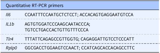
Total RNA was isolated from the snap-frozen full-thickness small intestine (ileum) and distal colon using the RNeasy® kit (Cat # 74106, Qiagen, Germantown, MD). RNA purity and concentration were checked on a SpectraMax® microplate reader (Molecular Devices, San Jose, CA). A total of 0.5 µg of RNA was reverse transcribed for cDNA synthesis using the QuantiTect® Reverse Transcription kit (Cat # 205313, Qiagen, Germantown, MD). Quantitative real-time PCR (qRT-PCR) was then performed on a Bio-Rad CFX96 Real-Time System (Bio-Rad Labs, Hercules, CA) using SYBR Green mix (Bio-Rad Labs, Hercules, CA), along with forward and reverse primers designed with the NCBI Primer-BLAST online programme and ordered from Integrated DNA Technologies, Coralville, Iowa (Table 1). The mRNA expression relative to the housekeeping gene ribosomal protein large P0 (Rplp0) was calculated using the 2-△△CT method as described previously.(Reference Livak and Schmittgen24)
Primer sequences

Table 1. Long description
A table titled ‘Quantitative RT-PCR primers’ with five rows and two columns. The columns are labeled ‘Gene’ and ‘Primer Sequences’. The rows list the following data: Row 1: Gene, Il6; Primer Sequences, CCAATTTCCAATGCTCTCCT; ACCACAGTGAGGAATGTCCA. Row 2: Gene, IL1b; Primer Sequences, AGTGTGGATCCCAAGCAATACCA; TGTCCTGACCACTGTTGTTTCCCA. Row 3: Gene, Tlr4; Primer Sequences, TTTATTCAGAGCCGTTGGTG; CAGAGGATTGTCCTCCCATT. Row 4: Gene, Rplp0; Primer Sequences, GGCGACCTGGAAGTCCAACT; CCATCAGCACCACAGCCTTC.
Immunohistochemistry (IHC) and Haematoxylin and Eosin (H&E) staining
Immunofluorescent staining of gut tissues was performed on 4% paraformaldehyde (PFA)-fixed 5 μm-thick paraffin sections. The sections were first warmed to 56°C in a vacuum incubator (Isotemp Vacuum oven, Fisher Scientific), immediately washed in xylene, gradually rehydrated through graded ethanol (100%, 95%, 70%) to water, and then processed for antigen retrieval in citrate buffer (10 mM pH 6.0) using a microwave (1000 W, 6 minutes). Samples were washed with PBS, blocked with 1% BSA/5% donkey serum (1 hour, room temperature), then incubated overnight at 4°C with primary antibodies (1:200 dilutions in 0.5% BSA), washed three times with PBS, and incubated with appropriate fluorescent-labelled secondary antibodies (1:1000 dilution in 0.5% BSA, Life Technologies Inc.) and the nuclear marker 4’,6-diamidino-2-phenylindole, dihydrochloride (DAPI, catalog no. D9542, Sigma, St. Louis, MO). The primary antibodies used were anti-E-cadherin (Ecad, catalog no. AF748, R&D Systems), anti-glial fibrillary acidic protein (GFAP, catalog no. PA110004, ThermoFisher), anti-3′-nitrotyrosine (3-NT, catalog no. ab61392, Abcam), anti-4-hydroxy-2-nonenal (4-HNE, catalog no. ab48506, Abcam), and anti-Tubulin beta 3 (Tubb3, catalog no. ab14545, Abcam). All IHC and H&E-stained slides were mounted using Gelvatol solution before imaging with a Nikon confocal microscope (Nikon, Inc.) and a Leica wide-field (DMi8) microscope.
Image quantification
Confocal images were analysed for fluorescent intensity using ImageJ software (ImageJ2 version: 2.14.0/1.54f). The region of interest (ROI) was delineated using a polygon icon tool and cleared for the area outside the ROI using the edit clear outside function. The ROI intensity was measured using the measure function. The means of the respective stains were computed relative to the nuclear-DAPI stain to normalize the ROI cellular densities. Finally, the averages of 3–4 areas of ROI per section per mouse were used to create graphs.
Statistical analysis
All data were analysed using GraphPad Prism 10 (GraphPad Software). Statistical significance was assessed using ordinary one-way ANOVA followed by Tukey’s multiple comparison test, or two-way ANOVA for grouped analyses where appropriate. Data are presented as mean ± SEM, and a P-value < 0.05 was considered statistically significant. Both male and female neonatal pups were included in all experiments. Group sizes were selected based on prior neonatal intestinal injury studies from our laboratory and related published models. Formal prospective power calculations were not performed because of the exploratory preclinical nature of the study. All experiments included at least three biological replicates with 2–3 mice per group per experiment.
Results
BioLipids-fortified liquid diets are well tolerated in newborn mice
The key difference in the lipid content of these composite BioLipids-fortified liquid diets – StdLipid, BioLipids1, and BioLipids2 – lies in the concentration of MCT oil: StdLipid (0 g/L), BioLipids1 (6.2 g/L), and BioLipids2 (12.5 g/L) (Figure 1B).
MCT oil is associated with various potential health benefits, such as enhanced cognitive function, weight loss, improved gut health, lower blood sugar levels, and increased energy levels.(Reference Kapoor, Malviya and Soll6,Reference St-Onge and Bosarge25,Reference Kanta, Lundsgaard and Havelund26) We anticipate that increasing the dosage of MCT oil in BioLipids composite formulations will be beneficial for these neonatal mice and crucial for combating pro-inflammatory agents. Besides MCTs, phospholipids, including sphingomyelin, phosphatidylcholine, phosphatidylserine, phosphatidyl ethanolamine, and phosphatidyl inositol, were additionally incorporated into the BioLipids1 and BioLipids2 liquid diets to replicate the natural composition of milk (Figure 1C). These MPLs are essential components of the milk fat globule membrane (MFGM) and play crucial roles in infant digestion, nutrient absorption, and gut–brain–immune development. They promote fat emulsification and enhance nutrient uptake, which is especially important for formula-fed infants to replicate the bio-functionality of human milk.(Reference Brink and Lonnerdal3,Reference Nilsson, Duan and Ohlsson7,Reference Ortega-Anaya and Jimenez-Flores14)
We first assessed whether the BioLipids-fortified liquid diets were safe and well tolerated in neonatal mice. To do so, we randomly allocated approximately 7–8 days- old mice from multiple litters into three groups. We reared them on either StdLipid or BioLipids-fortified liquid diets for four consecutive days while carefully monitoring for any signs of feed intolerance (e.g. stomach distention, emesis), dehydration, diarrhea, mobility issues, body weight loss, and mortality. We did not observe any signs of distress related to feed intolerance or overall well-being; all mice tolerated the feeds well. The body weights of the mice are presented in Figure 1D, indicating a trend toward improved body weight gain with BioLipids-fortified liquid diets compared to StdLipid groups. However, the differences were not statistically significant.
BioLipids-fortified liquid diets do not induce pro-inflammatory responses and protect against LPS-induced cytokine induction
Next, we evaluated the expression of pro-inflammatory cytokines by qRT-PCR analysis to determine whether the BioLipids-fortified diet feedings trigger any inflammatory response in the intestines. To achieve this, the mice were sacrificed on day 5 of the model to collect samples from the distal small intestine (ileum) and distal colon. These samples facilitated the assessment of gene expression related to pro-inflammatory cytokines, as well as the performance of IHC on critical oxidative, intestinal epithelial, and enteric neuron proteins. A targeted qRT-PCR approach was used to assess inflammatory responses based on prior neonatal intestinal injury studies. Tlr4 was analysed because of its established role in inflammatory signalling and neonatal intestinal injury,(Reference Hackam and Sodhi1,Reference Kovler, Gonzalez Salazar and Fulton22,Reference Takeuchi, Hoshino and Kawai27) whereas Il6 and Il1b were selected as canonical pro-inflammatory cytokines downstream of TLR4 activation.(Reference Bruno, Woller and Miller28) The data presented in Figure 2 clearly illustrate the baseline expression and the LPS-induced induction of Tlr4, Il6, and Il1b in the ileum (Figure 2B–D) and colon (Figure 2E–G). Data indicate no significant difference in Tlr4 expression at the baseline ileum between BioLipids-fortified and StdLipid-fed mice. Tlr4 expression was significantly lower in the colons of mice on BioLipids-fortified diets compared to those on StdLipid, suggesting that BioLipids differentially regulate TLR4, a key gene involved in gram-negative microbial recognition.(Reference Takeuchi, Hoshino and Kawai27) Pro-inflammatory cytokines Il6 and Il1b gene expression in the ileum and colon tissues was consistent across all StdLipid, BioLipids1, and BioLipids2 groups. As anticipated, LPS, the potent inducer of Tlr4-mediated pro-inflammatory responses across all biological systems,(Reference Hackam and Sodhi1) elicits a significant inflammatory response in both the ileum (Figure 2B–D) and the colon (Figure 2E–G) of StdLipid-fed mice. This response is relatively diminished in both BioLipids1 and BioLipids2 fortified groups, suggesting that BioLipids diets are indeed anti-inflammatory and protective. This finding holds clinical significance in this neonatal model and may have implications for humans.
BioLipid-fortified liquid diets are well-tolerated, non-inflammatory, and protective against LPS-induced intestinal pro-inflammatory responses. (A) An illustration of feeding and LPS treatment strategies. (B–G) Quantitative real-time PCR (qRT-PCR) analysis of pro-inflammatory cytokine gene expression in the ileum (B–D) and colon (E–G) of neonatal mice reared on StdLipid and BioLipid-fortified liquid diets for four consecutive days and subsequently injected with saline or LPS (5 mg/kg LPS, i.p. once, 6 hr treatment). Statistical significance was determined using a two-way ANOVA conducted with GraphPad Prism 10 software. ns = non-significant, *P < 0.05, **P < 0.01, ***P < 0.001. Each dot in the dot graphs represents data from a different mouse, n = 6 or more mice per group.

BioLipids-fortified liquid diets augment small intestine motility
Based on the observations in Figure 2 that BioLipids-fortified liquid diets have no adverse effects on gut health and an anti-inflammatory profile against LPS-induced pro-inflammatory responses, we next investigated whether BioLipids fortification is beneficial to the gut’s ENS. The ENS, comprising enteric neurons and glia, is essential for gut homeostasis, managing functions from digestion to immune response by coordinating with hormone-producing enterochromaffin cells.(Reference Gonzales and Gulbransen29) In our recent key publication, we have demonstrated using preclinical models that ENS in premature infants is critical for protection against NEC and is positively affected by human milk oligosaccharides derived from breast milk.(Reference Sodhi, Scheese and Tragesser21) Data presented in Figure 3 show that intestinal motility, as a function of ENS homeostasis,(Reference Kovler, Gonzalez Salazar and Fulton22) is enhanced with BioLipids-fortified diets compared to the StdLipid diet. The difference is most apparent in the Geom center calculations at the baseline (Figure 3C, D) and in relation to LPS-induced hypomotility (Figure 3F, G). At baseline levels, BioLipids-fortified diets show significantly higher Geom center as a function of intestinal motility (BioLipids1 P = 0.003 versus StdLipid; BioLipids2 P = 0.002 versus StdLipid). Furthermore, both BioLipids-fortified diets demonstrate protection against LPS-induced hypomotility (Figure 3F, G), suggesting that BioLipids-fortified diets may be clinically relevant, as hypomotility is a significant clinical issue after surgery, medicine usage, infections, and neurological disorders.(Reference Bielefeldt, Tuteja and Nusrat30)
BioLipid-fortified liquid diets enhance small intestine motility. (A) An illustration of the experimental treatment strategy. (B, G) Small intestinal motility, measured by the FITC-dextran transit time, is shown as the peak fluorescent intensity (mean and standard error of the mean) for each centimeter of the small intestine (B, E) and quantification of the geometric (Geom) center (C–D, F–G) of neonatal mice reared on StdLipid and BioLipid-fortified liquid diets for four consecutive days and subsequently injected with saline or LPS (5 mg/kg LPS, i.p. once, 6 hr treatment). Statistical significance was determined using one-way analysis of variance followed by the Tukey multiple comparisons test with GraphPad Prism 10 software. Each dot in the dot graphs represents data from individual mice. n = 6 or more mice per group.

Figure 3. Long description
Panel A: An illustration of the experimental treatment strategy. Panel B: A line graph showing small intestinal motility measured by FITC-dextran transit time, with the peak fluorescent intensity for each centimeter of the small intestine. The x-axis represents the segments of the small intestine (Sto, Duo, Jejunum, Ileum), and the y-axis represents FITC-fluorescence (λex 492/λem 518 nm). The graph includes three lines representing Std Lipid, BioLipid 1, and BioLipid 2. Panel C: A bar graph quantifying the geometric center of FITC-motility for Std Lipid and BioLipid 1, with the y-axis representing FITC-motility (Geom center). Panel D: A bar graph quantifying the geometric center of FITC-motility for Std Lipid and BioLipid 2. Panel E: A line graph showing small intestinal motility measured by FITC-dextran transit time after LPS injection, with the peak fluorescent intensity for each centimeter of the small intestine. The x-axis represents the segments of the small intestine (Sto, Duo, Jejunum, Ileum), and the y-axis represents FITC-fluorescence (λex 492/λem 518 nm). The graph includes three lines representing Std Lipid+LPS, BioLipid 1+LPS, and BioLipid 2+LPS. Panel F: A bar graph quantifying the geometric center of FITC-motility for Std Lipid LPS and BioLipid 1 LPS, with the y-axis representing FITC-motility (Geom center). Panel G: A bar graph quantifying the geometric center of FITC-motility for Std Lipid LPS and BioLipid 2 LPS.
BioLipids-fortified liquid diets protect against LPS-induced enteric neuron dysfunction
We next wanted to investigate whether intestinal hypomotility is caused by ENS insufficiency due to LPS treatment. The representative confocal images shown in Figure 4 illustrate immunostaining for the enteric neuron marker Tubulin, beta 3 class III (Tubb3, A–B), as well as the enteric glial marker Glial Fibrillary Acidic Protein (GFAP, C–D), which was quantified for fluorescence intensity using Fiji ImageJ software (Figure 4E, F). At baseline, robust Tubb3 immunostaining was observed in PFA-fixed, paraffin-embedded sections of the small intestine in the muscular layer from both StdLipid and BioLipids-fortified mice. However, the staining shows less colonization of neurons in the lamina propria of StdLipid sections compared to the remarkable fenestration of neurons in BioLipids-fortified mice intestinal sections (Figure 4Ai–Aiii). A similar pattern was observed in LPS-treated specimens from mice fed liquid diets fortified with StdLipid and BioLipids (Figure 4Bi–Biii). Furthermore, the quantification of data shows a significantly higher mean Tubb3 fluorescence when normalized to the nuclear DAPI staining (Figure 4E, BioLipids 1 & 2 + LPS vs. StdLipid + LPS, P = 0.0031). The GFAP immunostaining, on the other hand, is less robust and shows similar staining patterns in all mice (Figure 4C–D) and does not show any significant difference at the baseline levels but a significant increase was observed in StdLipid LPS-treated sections, which was preserved to the baseline levels in BioLipid1 and BioLipid2 LPS-treated mice (Figure 4F). The increase in GFAP reactivity in StdLipid LPS mice may indicate inflammation-induced gliosis leading to intestinal dysmotility, as shown in Figure 3. The LPS-induced enteric glial cell gliosis has recently been reported by Robertson et al.(Reference Robertson, Hahn and Robinson31) Overall, these data demonstrate preservation of enteric neuronal and glial marker expression in BioLipids-fed mice during inflammatory injury and support a protective association between BioLipids diets and maintenance of ENS integrity, which appears to correlate with the intestinal motility data presented in Figure 3.
BioLipid-fortified liquid diets protect against LPS-associated loss of enteric neuronal and glial marker expression in the small intestine of neonatal mice. (A–D) Representative confocal images of PFA-fixed, paraffin-embedded, 5 μm sections of the distal small intestine (ileum) stained with the enteric neuron marker Tubb3 (Ai–iii, Bi–iii) and the enteric glia marker GFAP (Ci–iii, Di–iii), along with quantification of mean fluorescent intensities (E, F) using Fiji ImageJ software (the area within the dotted lines quantified), from neonatal mice reared on StdLipid (Ai–Ci) and BioLipid-fortified liquid diets (Aii–Dii, Aiii–Diii), treated with saline (A, C) or LPS (5 mg/kg LPS, i.p. once, 6 hours before sacrifice) (B, D). Scale bar = 25 μm. Statistical significance was determined by a 2-way ANOVA using GraphPad Prism 10 software. **P < 0.01, ***P < 0.001. Each dot in the dot graphs represents data from a different mouse. n = 6 or more mice per group. Tubb3, tubulin, beta 3 class III; GFAP, glial fibrillary acidic protein.

BioLipids-fortified liquid diets protect against DSS-induced colonic injury
As shown above, BioLipids-fortified liquid diets are beneficial against LPS-induced injury. We next explored whether these diets are protective against DSS-induced colonic injury. To do so, we developed a mild to moderate colonic injury model by supplementing with low doses (0.25–0.5% of DSS) in both the StdLipid and BioLipids-fortified liquid diets, feeding every time (Figure 5A). Neonatal mice subjected to a higher dosage exceeding 0.5% experienced high mortality (Data not shown). We have recently published a low-dose DSS-induced colonic injury model that causes both colonic and ileal NEC injury in neonatal mice.(Reference Klerk, Moore and Scheese19) Our 0.5% DSS-colonic injury model did not reveal remarkable histological injury based on H&E staining (Figure 5B, C). However, the sections indicated excessive sloughing, as evident from its presence in the luminal faecal content, along with a reduction in the height and depth of colonic crypts (Figure 5Ci). No noticeable differences in the gross histological architecture were observed at the baseline among StdLipid, BioLipids1, and BioLipids2 fortified diet groups (Figure 5Bi–Biii). We also compared the colonic epithelial architecture by immunostaining with the epithelial marker protein E-Cadherin (Ecad). The representative confocal images of Ecad immunostained colonic tissue from StdLipid, BioLipids1, and BioLipids2 fortified diets, with or without DSS exposure, show a significant loss of Ecad in the StdLipid DSS mice and preserved Ecad staining in the BioLipid1 and BioLipid2 DSS mice (Figure 5D–F), indicating the protection offered by BioLipid fortification against DSS-induced injury. Interestingly, the gene expression levels of the pro-inflammatory cytokine Il6 show significant protection against DSS-induced increases in BioLipids-fortified liquid diets fed to mice compared to those fed with StdLipid (Figure 5E, P < 0.001). These data indicate that BioLipids diets protect against colonic injury caused by DSS, a toxicant widely used to study colon injury in mice.(Reference Klerk, Moore and Scheese19,Reference Raouf, Steinway and Scheese32)
BioLipid-fortified liquid diets protect against DSS-induced colonic injury in neonatal mice. (A) An illustration of an experimental treatment strategy. (B–E) Representative H&E-stained histological images (Bi-Ciii) and Ecad-stained confocal images (Di–Eiii) of PFA-fixed, paraffin-embedded, 5 µm sections of colon tissues. (F) qRT-PCR analysis of the pro-inflammatory cytokine Il6, and (G) quantification of mean fluorescent intensities of Ecad images using Fiji ImageJ software from neonatal mice reared on StdLipid (Bi–Ei) and BioLipid-fortified liquid diets (Bii-Eii, Biii-Eiii) exposed to 0.5% DSS. Scale bar = 50 µm (Bi–Ciii), 25 µm (Di–Eiii). Statistical significance was determined using a two-way ANOVA conducted with GraphPad Prism 10 software. *P < 0.05, **P < 0.01, ***P < 0.001. Each dot in the dot graphs represents data from a different mouse. n = 6 or more mice per group. Ecad, E-cadherin.

Figure 5. Long description
Panel A: The image is a flowchart illustrating the experimental treatment strategy. It starts with 7-day-old mice and includes stages such as feeding with BioLipid milk replacer, DSS treatment, and data collection at 11 days old. Panel B-E: The images are histological and confocal images of colon tissues. Panels Bi-Ciii show H&E-stained sections, while Panels Di-Eiii show E-cadherin-stained sections. The tissues are from mice fed with Standard Lipid, BioLipid 1, and BioLipid 2, both with and without DSS treatment. Panel F: The image is a bar graph showing qRT-PCR analysis of the pro-inflammatory cytokine Il6. The x-axis represents different diet groups (StdLipid, BioLipid1, BioLipid2) and the y-axis represents Il6 expression levels. Data points are shown for both no DSS and DSS conditions. Panel G: The image is a bar graph quantifying mean fluorescent intensities of E-cadherin images. The x-axis represents different diet groups and the y-axis represents mean fluorescence (MeanDens/Dapi). Data points are shown for both no DSS and DSS conditions. Statistical significance is indicated with asterisks (*P < 0.05, **P < 0.01, ***P < 0.001).
BioLipids-fortified liquid diets protect against DSS-induced oxidative-colonic injury
To further verify the protection afforded by BioLipids-fortified diets against DSS-induced colonic injury, we immunostained the PFA-fixed, paraffin-embedded colonic sections from StdLipid, BioLipids1, and BioLipids2-fed mice with or without DSS exposure using the protein peroxidation markers 3’-Nitrotyrosine (3’-NT)(Reference Sodhi, Gonzalez Salazar and Kovler11,Reference Sodhi, Wipf and Yamaguchi18,Reference Klerk, Moore and Scheese19) and 4-Hydroxy-2-Nonenal (4-HNE),(Reference Ma, Zhang and Li33) which are presented as representative confocal images in Figure 6. At the baseline level – without DSS exposure – both 3’-NT (Figure 6 Ai–iii, E) and 4-HNE (Figure 6Ci–iii, F) displayed a weak signal with no discernible differences in immunofluorescence staining across all three liquid diet groups. In contrast, significant epithelial immunofluorescence was observed in DSS-exposed images, which was more pronounced in StdLipid DSS-exposed colon sections (Figure 6Bi, Di) than in BioLipids-fortified DSS colon sections (Figure 6Bii–iii, Dii–iii), demonstrating a statistically significant difference when quantified for the mean fluorescent intensities (Figure 6E, F). Overall, the data shown in both Figures 5 and 6 clearly demonstrate that DSS-induced colonic epithelial injury is mitigated by feeding BioLipids liquid diets.
BioLipid-fortified liquid diets protect against DSS-induced oxidative colonic injury in neonatal mice. (Ai–Diii) Representative confocal images of PFA-fixed, paraffin-embedded, 5 µm sections of the colon stained with oxidative stress markers, 3’-NT (protein oxidation marker) (A–B) and 4-HNE (lipid peroxidation marker)(C–D). (E, F) Quantification of mean fluorescent intensities using Fiji ImageJ software, for neonatal mice reared on standard lipid or BioLipid-fortified liquid diets and treated with saline (A, C) or exposed to 0.5% DSS (B, D). Scale bar = 25 µm. Statistical significance was determined using a two-way ANOVA conducted with GraphPad Prism 10 software. *P < 0.05, **P < 0.01, ***P < 0.001. Each dot in the dot graphs represents data from a different mouse. n = 6 or more mice per group. 3’-NT, 3-Nitrotyrosine; 4-HNE, 4-Hydroxy-2-nonenal.

BioLipid-fortified liquid diets protect against DSS-induced small intestine hypomotility. (A) An illustration of the experimental treatment strategy. (B, C) Small intestinal motility, measured by the FITC-dextran transit time, is represented as the peak fluorescent intensity (mean and standard error of the mean) for each centimeter of the small intestine (B) and the quantification of the geometric (Geom) center (C) for neonatal mice reared on standard lipid or BioLipid-fortified liquid diets and treated with saline or exposed to 0.5% DSS. Statistical significance was determined using one-way analysis of variance followed by the Tukey multiple comparisons test with GraphPad Prism 10 software. ns = non-significant, ***P < 0·001. Each dot on the dot graphs represents data from individual mice. n = 6 or more mice per group.

Figure 7. Long description
Panel A: The illustration shows the experimental treatment strategy. It starts with 7-day-old mice, followed by feeding with bioactive lipid milk replacer (BAL) for one day, then BAL with 0.5% DSS for three days. On day 5, mice receive an oral gavage of FITC-dextran, and their intestines are harvested to measure FITC-fluorescence per centimeter. Panel B: The line graph displays FITC-dextran distance migration along the small intestine, measured in fluorescence units (λex 492/λem 518 nm). The x-axis represents sections of the small intestine (Sto, Duo, Jejunum, Ileum) divided into 1-cm sections, while the y-axis shows FITC-fluorescence. Three groups are compared: Std Lipid+DSS, BioLipid1+DSS, and BioLipid2+DSS. The graph shows a decline in fluorescence intensity from the stomach to the ileum, with BioLipid2+DSS maintaining higher fluorescence levels. Panel C: The bar graph quantifies FITC-motility using the geometric center of the small intestine. The x-axis lists the groups (Std Lipid+DSS, BioLipid1+DSS, BioLipid2+DSS), and the y-axis shows the geometric center values. BioLipid2+DSS shows significantly higher motility compared to the other groups, indicated by ***P < 0.001.
BioLipids-fortified liquid diets protect against DSS-induced small intestine hypomotility
Building on the observation from our recent publication that indicates DSS-induced colitis in mice causes small intestinal hypomotility.(Reference Raouf, Steinway and Scheese32) Next, we studied small intestinal motility to determine whether BioLipids-fortified diets provide protection against DSS-induced hypomotility. To study the small intestine motility, experimental mice reared on StdLipid and BioLipids liquid diets and unexposed or exposed to DSS were given 70kd FITC-dextran 30-min before the termination of models and assessed for the FITC-dextran as a tracer for the motility (Figure 7A). Mice raised on a StdLipid diet and exposed to DSS exhibit a significant reduction in motility, as indicated by both motility curves (Figure 7B) and Geom center (Figure 7C), a phenomenon not observed in DSS-exposed mice raised on BioLipids diets, indicating that the BioLipids diet protects against DSS-induced small intestine hypomotility.
BioLipids-fortified liquid diets protect against DSS-induced enteric neuronal loss
To confirm whether DSS-induced hypomotility arises from changes in the ENS of the bowel, we assessed both enteric neurons and enteric glia using immunostaining with Tubb3 (a neuronal marker) and GFAP (a glial marker) on PFA-fixed, paraffin-embedded 5 µm-thick ileal and colonic sections from StdLipid and BioLipids-fortified liquid diets exposed to DSS. The representative confocal images and quantification of fluorescent intensities are presented in Figures 8 and 9. The data indicate that DSS exposure results in a significant reduction in Tubb3 fluorescence in the ileum (Figure 8A, B, E), but not in the colon (Figure 9A, B, E) sections of mice reared on the StdLipid diet, with no notable loss observed in mice reared on BioLipids-fortified liquid diets. DSS exposure did not cause a significant change in GFAP fluorescence in the colon sections of mice reared on the StdLipid diet, but a significant reduction was observed in the BioLipids-fortified liquid diets exposed to DSS (Figure 9C, D, F). It is worth mentioning that the BioLipid1 and BioLipid2-fortified mice showed a significant increase in GFAP reactivity at baseline. The observed increase in baseline GFAP expression (Figures 8 and 9) in BioLipids-fed mice may reflect altered enteric glial activity; however, GFAP upregulation is context-dependent and may represent adaptive or reactive glial responses rather than a direct marker of improved ENS function. Overall, the loss of enteric neurons may be responsible for the hypomotility observed in StdLipid DSS mice. In contrast, the absence of enteric neuron loss in BioLipids DSS mice aligns with the preserved motility (Figure 7).
BioLipid-fortified liquid diets preserve enteric neuronal and glial marker expression during DSS-induced intestinal injury in the small intestine of neonatal mice. (A–D) Representative confocal images of PFA-fixed, paraffin-embedded, 5 µm sections of the distal small intestine (ileum) stained with the enteric neuron marker Tubb3 (Ai-iii, bi-iii) and the enteric glia marker GFAP (Ci–iii, Di–iii). (E, F) Quantification of mean fluorescent intensities was performed using Fiji ImageJ software (with the area within the dotted lines quantified) for neonatal mice reared on Standard lipid and BioLipid-fortified liquid diets and treated with saline (A, C) or exposed to 0.5% DSS (B, D). Scale bar = 25 µm. Statistical significance was determined using a two-way ANOVA conducted with GraphPad Prism 10 software. ns = non-significant, *P < 0.05, ***P < 0.001. Each dot in the dot graphs represents data from a different mouse. n = 6 or more mice per group. Tubb3, tubulin, beta 3 class III; GFAP, glial fibrillary acidic protein.

BioLipid-fortified liquid diets protect against DSS-induced loss of enteric neurons and enteric glia in the colonic tissues of neonatal mice. (A–D) Representative confocal images of PFA-fixed, paraffin-embedded, 5 μm sections of the colon stained with the enteric neuron marker Tubb3 (Ai–iii, Bi–iii) and the enteric glia marker GFAP (Ci–iii, Di–iii). (E, F) Quantification of mean fluorescent intensities using Fiji ImageJ software (area within the dotted lines quantified) of neonatal mice reared on standard lipid and BioLipid-fortified liquid diets and treated with saline (A, C) or exposed to 0.5% DSS (B, D). Scale bar = 25 µm. Statistical significance was determined using a two-way ANOVA conducted with GraphPad Prism 10 software. ns=non-significant, *P < 0.05, ***P < 0.001. Each dot in the dot graphs represents data from a different mouse. n = 6 or more mice per group. Tubb3, tubulin, beta 3 class III; GFAP, Glial fibrillary acidic protein.

Discussion
The present study offers compelling evidence that BioLipids-fortified liquid diets containing functional oils, namely MCTs, MPLs, and MDG oil, are safe, well-tolerated, and protective against intestinal inflammation and neuromuscular injury in neonatal mice. These findings signify a notable advancement in neonatal nutrition science and present a promising therapeutic pathway for mitigating gastrointestinal complications frequently seen in newborn infants. Our data show that BioLipids diets do not induce baseline inflammation in the gut, as indicated by the stable expression of Tlr4, Il6, and Il1b in both ileal and colonic tissues (Figure 2). More importantly, BioLipids diets significantly reduce the inflammatory response to lipopolysaccharide (LPS), a potent inducer of Toll-like receptor 4 (TLR4)-mediated signalling pathways.(Reference Takeuchi, Hoshino and Kawai27) These observations support the possibility that the composite BioLipids formulation containing MCTs, MDGs, and MPLs may collectively influence innate immune signalling and mucosal homeostasis during intestinal injury.(Reference Brink and Lonnerdal3,Reference Nilsson, Duan and Ohlsson7,Reference Sodhi, Gonzalez Salazar and Kovler11,Reference Ortega-Anaya and Jimenez-Flores14) Given TLR4’s central role in the pathogenesis of NEC, the anti-inflammatory effect observed in preclinical studies may have significant implications for managing newborn infants at risk of inflammatory bowel injury.(Reference Hackam and Sodhi1) An important consideration in interpreting the present findings is that the experimental BioLipids formulations differed simultaneously in multiple lipid-related variables, including MCT content, MDGs, and MPLs. Consequently, the current study design does not permit mechanistic assignment of the observed protective effects to any individual lipid component. Rather, the present data support the protective efficacy of the integrated BioLipids formulation as a whole. Future reductionist studies will be required to determine the relative contribution of specific lipid subclasses to intestinal protection and ENS preservation.
Newborn infants are particularly vulnerable to lipid malabsorption, with up to 30% of dietary fat excreted due to immature pancreatic enzymes and bile salt production. Fat absorption in this population is further compromised by shorter intestinal transit times and underdeveloped chylomicron assembly pathways.(Reference Lindquist and Hernell2,Reference Kovler, Gonzalez Salazar and Fulton22) In contrast to adults, neonatal lipid digestion relies heavily on bile salt-stimulated lipase (BSSL) from breast milk and pancreatic lipase-related protein 2 (PLRP2), which act synergistically to digest triglycerides in the absence of pancreatic triglyceride lipase (PTL). Importantly, pasteurization of human milk inactivates BSSL and has been shown to reduce fat absorption and growth in newborn infants, underscoring the need for enzymatically compatible lipid delivery strategies.(Reference Lindquist and Hernell2) Our use of MCT-rich composite BioLipids diets simulates the digestion-friendly profile of human milk lipids by circumventing bile salt dependence, thus offering a promising nutritional alternative. In addition to digestion and absorption, intracellular lipid handling is critical in enterocytes. Lipid droplets (LDs), once thought to be inert fat stores, are now recognized as dynamic organelles that regulate lipid storage, intracellular trafficking, and signalling.(Reference Beilstein, Carriere and Leturque34) LD-associated proteins like Plin2 and Plin3 regulate droplet formation; Plin3 is a key regulator following fat intake, while Plin2 aids long-term lipid retention. Enzymes for triglyceride synthesis, such as MGAT2 and DGAT2, are found on both ER and LD surfaces, linking storage and secretion. LDs act as buffers after meals and facilitate lipid delivery during fasting. Autophagic degradation (lipophagy) of LDs in enterocytes is crucial for HDL secretion and lipid clearance, and failure in these processes may lead to inflammation.(Reference Beilstein, Carriere and Leturque34) Murine studies from our lab and others have shown that undigested formula lipids can accumulate in the distal ileum, triggering epithelial sloughing and oxidative stress, which contribute to NEC pathology, effects that may be mitigated by predigested or MCT-enriched lipid formulations like BioLipids.(Reference Burge, Vieira and Eckert5,Reference Sodhi, Fulton and Good12)
Our findings are also supported by clinical data from parenterally-fed newborn infants, where lipid emulsions containing MCTs have been associated with improved tolerance, reduced inflammation, and lower risk of liver dysfunction.(Reference Kapoor, Malviya and Soll6) MCT-containing emulsions are metabolized more rapidly and exhibit a lower omega-6 profile, potentially reducing pro-inflammatory eicosanoid synthesis. Furthermore, structured emulsions such as SMOFlipid, which combine MCT, olive oil, and fish oil, are increasingly used to manage parenteral nutrition-associated liver disease and cholestasis, although high-quality evidence is still emerging. These clinical insights strengthen the rationale for incorporating MCTs into early-life enteral lipid strategies. Emerging evidence from neurodegenerative disease models reinforces the metabolic benefits of MCFAs. In Alzheimer’s disease, brain glucose utilization is compromised due to insulin resistance.(Reference Castro, Dias and Hillebrandt9,Reference Hadzic, Gregor and Kofler35) MCFAs rapidly generate ketone bodies that cross the blood–brain barrier and serve as alternative fuels. These ketone bodies improve cerebral energy metabolism without suppressing glucose uptake.(Reference Hadzic, Gregor and Kofler35) This mechanism may impact neonates, particularly those with glucose homeostasis issues due to inflammation, hypoxia, or immaturity.(Reference Burge, Vieira and Eckert5,Reference Nilsson, Duan and Ohlsson7,Reference Sodhi, Fulton and Good12) By supporting mitochondrial function and limiting oxidative stress, BioLipids formulations enriched with MCTs and phospholipids may aid energy-deficient tissues, including the developing brain and gut.(Reference Joshi, Richard and Gohil36,Reference Swiatkiewicz, Hanczakowska and Okon37) Recent work by Yuan et al. highlights the benefits of medium- and long-chain triacylglycerols (MLCTs) from human milk fat substitutes. MLCT-fed mice showed lower hepatic fat accumulation and enhanced liver function, along with favourable shifts in Firmicutes to Bacteroidetes ratios and reduced pro-inflammatory taxa like Desulfovibrionaceae. (Reference Yuan, Cheng and Shen13) These changes align with the protective effects observed in our neonatal inflammation models and support the relevance of BioLipids-MCT and vital phospholipid-based nutrition. We observed significant protection provided by BioLipids-fortified diets in a dextran sulfate sodium (DSS) model of neonatal colitis (Figures 5 and 6). Our neonatal mice fed BioLipids-fortified diets showed reduced epithelial damage, lower oxidative stress (as indicated by 3’-nitrotyrosine and 4-HNE staining) (Figure 6), and maintained small intestinal motility (Figure 7). These results support the hypothesis that the supplementation of MCTs and phospholipids in nutritional diets provides mucosal protection through both anti-inflammatory and anti-oxidative mechanisms.(Reference Zhiguang Huanga, Zhaoa and Guana15) The ability of BioLipids diets to mitigate DSS-induced enteric neuronal loss further reinforces their potential as neuroprotective agents during neonatal gut injury. Overall, our findings support using BioLipids-MCT and phospholipid-fortified formulas as a comprehensive strategy to enhance neonatal intestinal resilience. Our findings align with piglet studies, which prove that supplementation with MCTs, particularly caprylic acid (C8:0), results in enhanced gut structure, increased villus height, and greater duodenal and cecal tissue mass.(Reference Swiatkiewicz, Hanczakowska and Okon37) These studies demonstrated that maternal coconut oil diets reduced piglet mortality and improved daily weight gain. Postnatal supplementation with C8:0 acidified intestinal chyme supported immune maturation and increased serum IgG levels – benefits closely parallel the anti-inflammatory, motility-preserving, and ENS-protective effects observed in our neonatal mouse model. Similarly, in a murine model of DSS-induced ileitis, Hadžić et al(Reference Hadzic, Gregor and Kofler35) showed that ketogenic diets with LCTs more effectively suppressed inflammatory cytokines (TNF-α, IL-1β). These effects were linked to increased glutathione-related antioxidants and improved cecal microbiota. While MCTs promoted ketosis, the lipid composition, not just ketogenesis, determined the therapeutic outcome, underscoring the need to consider lipid type and structure in enteral nutrition formulation. Our findings with BioLipids-fortified liquid diets support this principle, as specific MCT and phospholipids-based blends were necessary to achieve motility rescue, ENS protection, and inflammation mitigation. Additionally, recent data from a murine DSS ileitis model underscore the integral role of TLR4 in regulating enteric neuroplasticity, gut motility, and neuromuscular integrity. Faggin et al.(Reference Faggin, Cerantola and Caputi38) demonstrated that TLR4 knockout mice were significantly protected from DSS-induced ileitis, maintaining ENS architecture, motility, and neurotransmission, including nitrergic and serotonergic pathways. These mice exhibited reduced neuronal loss, diminished reactive gliosis, and lower 5-HT3 and 5-HT2A receptor activity, further confirming that TLR4-driven inflammation impairs excitatory and inhibitory neuromuscular signalling.(Reference Faggin, Cerantola and Caputi38) Our observation that BioLipids diets mitigate TLR4-mediated dysfunction suggests that MCT-phospholipid-based lipid formulations might serve as nutritional modulators of TLR4 signalling and ENS plasticity.
In addition to their immunomodulatory properties, BioLipids diets significantly improved gastrointestinal motility (Figure 3B–D). This was most evident in mice challenged with LPS, where BioLipids-fed animals exhibited higher intestinal transit velocities and geometric centers of FITC-dextran distribution (Figure 3E–G). This effect coincided with the preservation of enteric neurons (Tubb3+) and glial cells (GFAP+), suggesting that BioLipids diets not only prevent inflammation but also preserve the integrity of the ENS (Figure 4). These findings are particularly intriguing, considering the well-established role of ENS dysfunction in gastrointestinal dysmotility, feeding intolerance, and postoperative ileus and NEC.(Reference Kovler, Gonzalez Salazar and Fulton22,Reference Gonzales and Gulbransen29) By targeting key aspects of gut physiology – immune activation, epithelial integrity, ENS preservation, and motility regulation – these diets offer a comprehensive solution for the nutritional support of newborn infants. Importantly, all ingredients used in these formulas are GRAS (Generally Recognized As Safe) and are found in ready-to-feed infant products, indicating high translational relevance.(39) This study also opens new avenues for mechanistic exploration. For example, medium-chain fatty acids (MCFAs) have been shown to signal through G protein-coupled receptors such as GPR84, which are expressed on immune and epithelial cells and may play a role in regulating inflammation and barrier function.(Reference Kimura, Ichimura and Ohue-Kitano40) Moreover, the microbiome is likely to mediate or modulate many of the observed effects, and future studies should examine changes in microbial composition and metabolite production in response to BioLipids diets.(Reference Hadzic, Gregor and Kofler35) The limitations of our study include the usage of a murine model that, although physiologically relevant, may not completely replicate human newborn intestinal development. Furthermore, while we concentrated on early outcomes (days 2–5), long-term follow-up studies will be necessary to evaluate the durability of these protective effects and their influence on neurodevelopmental and metabolic outcomes.
Several limitations should be acknowledged. First, the present study was designed primarily as a translational and exploratory preclinical investigation and did not include mechanistic dissection of individual lipid signalling pathways. Second, although complementary inflammatory and functional endpoints were evaluated, formal blinded histopathologic injury scoring was not performed in the neonatal DSS model because injury severity was intentionally maintained at mild-to-moderate levels to minimize mortality. Third, microbiome alterations, long-term neurodevelopmental outcomes, and lineage-tracing analyses of ENS populations were beyond the scope of the current study. Future studies addressing these mechanistic and longitudinal questions will further strengthen the translational relevance of BioLipids-based nutritional interventions.
In conclusion, BioLipids-fortified liquid diets provide a novel and effective strategy to protect against inflammatory, oxidative, and neuromuscular gut injuries in neonates. These findings establish a foundation for future translational studies and clinical trials aimed at optimizing lipid-based enteral nutrition for Newborns and high-risk infants.
Supplementary material
The supplementary material for this article can be found at https://doi.org/10.1017/jns.2026.10141.
Data availability statement
The data supporting the findings of this study are available within this article and its Supplementary Materials. Additional data generated and analysed during the current study are available from the corresponding author upon reasonable request.
Acknowledgements
None.
Author contributions
CPS and DJH conceived and designed the study. CPS, CT, DS, JWD, KT, MEB, SWM, JY, YA, SW, MW, TP, and HSJ performed experiments and analysed data. MV and TD contributed to formulation design and scientific interpretation. CPS and DJH drafted the manuscript. All authors reviewed and approved the final manuscript.
Financial support
DJH receives support from R35GM141956 from the National Institutes of Health, and this research was partly funded by a sponsored research grant from Abbott Nutrition.
Competing interests
This work was partly supported by a sponsored research grant from Abbott Nutrition to DJH. Mustafa Vurma and Tapas Das are employees of Abbott Nutrition. The remaining authors declare no competing financial interests related to this work.



 Open access

