Introduction
In spite of many improvements, surgery for stenosis of the cricotracheal region and the trachea itself remains a difficult and challenging task. The established concept of laryngotracheal reconstructionReference Cotton1,Reference Rethi2 is based on augmenting the stenotic lumen by interposition of autologous rib grafts. However, more recent studies suggest that resection of laryngotracheal stenoses together with primary anastomosisReference Grillo, Mathisen and Wain3,Reference Pearson4 may be a superior approach in many cases, even in the paediatric population.Reference Monnier, Lang and Savary5 Although not always necessary, appropriate techniques of laryngeal and tracheal release may be performed to allow for reanastomosis after resection of significant tracheal length.Reference Merati, Rieder, Patel, Park and Girod6 No matter what the limits of excision and reconstruction, the driving principle of segmental resection is to preserve as much healthy trachea as possible above and below the stenotic segment.
The precise localisation and characterisation of the extent of the stenotic tracheal segment can be assessed by fibreoptic examination or direct laryngoscopy combined with 0° telescopy. However, these data are insufficient to exactly localise the stenotic segment after surgical exposure of the trachea, since the level of the vocal folds can only be estimated, and since other landmarks (such as the cricoid arch and the first tracheal ring) may be significantly altered due to scarring in the area. Opening the trachea too far from the diseased area may result in a longer defect, making the reconstructive anastomosis technically more difficult and prone to post-operative complications.
With this in mind, a technique of marking the site of tracheal stenosisReference Lichtenberger7 was developed. This report describes an easy, quick and reliable technique of projecting the extent of endoscopically characterised stenotic segments of the subglottic and tracheal airway onto its external aspect.
Methods
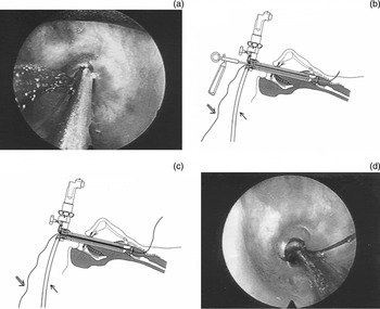
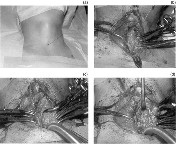
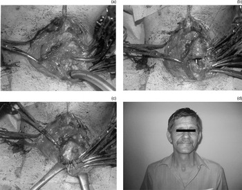
The first step of the procedure is to secure the airway by jet ventilation. It is important to note that the proposed marking depends on the use of jet technique, so that the airway is not obscured by an endotracheal tube. Microlaryngoscopy is performed using standard equipment. The proximal end of the stenosis is visualised directly through the laryngoscope, aided by a 0° telescope. The distal end of the jet catheter is positioned 3 mm above the stenosis. Through the telescope, the marking thread is seen on the left side and the tip of the needle carrier on the right (Figure 1a). At this point, the endo-extratracheal suture technique and needle carrier (Richard Wolf, Knittlingen, Germany) is used to drive a 2-0 Prolene suture directly above the proximal end of the stenosis and through the trachea, subcutaneous tissue and skin (Figure 1b–c). The needle carrier is withdrawn and the jet catheter and the endoscopic part of the marking thread are left in place (Figure 1d, Figure 2a). After skin incision, the external end of the marking thread can be used as a guide, taking the surgeon directly to the anterior tracheal wall at the exact level of the upper margin of the stenotic segment (Figure 2b). Opening of the airway may be performed with greater confidence due to precise localisation and marking of the diseased area. Only the stenotic segment is resected, thus minimising the gap between the proximal and distal end of the trachea (Figure 2c). After the resection, the posterior wall of the trachea is anastomosed (Figure 2d, Figure 3a). Finally, the anterior wall of the trachea is repaired (Figure 3b–c). Figure 3(d) shows a patient one year after tracheal resection.
(a) Endoscopic view of placement of endo-extratracheal suture. The tip of the jet catheter is positioned 3 mm above the stenosis on the left, the marking thread is in the middle and the tip of the needle carrier is on the right. (b) The Lichtenberger endo-extratracheal suture technique. The needle with thread is pushed through the trachea from inside out. (c) The marking thread lying in the stitching canal. (d) Endoscopic view of the stenosis, with the marking thread at top right and the jet catheter tip at bottom right.

(a) The marking thread, as seen before skin incision. (b) The marking thread, as seen before opening the trachea. (c) After resection of the stenotic tracheal segment. (d) Anastomosis of the back wall of the trachea.

(a) Suturing of the back wall of the trachea. (b) Anastomosis of the anterior wall of the trachea. (c) Suture of the anterior wall of the trachea. (d) The patient one year after surgery.

Results
The authors have used this technique to mark the upper margin of stenosis in 16 cricotracheal or tracheal resections, without complications or limitations. The technique is also feasible in children weighing more than 15 kg. In children of this weight, the use of smaller adult laryngoscopes allows sufficient room for the endo-extratracheal suture technique.
Discussion
The Lichtenberger needle carrier is an instrument used to pass a suture from the endoluminal airway to external structures.Reference Lichtenberger and Toohill8 It was originally developed for the endoscopic management of bilateral vocal fold immobility.Reference Lichtenberger9 The Lichtenberger technique of lateral fixation of one vocal fold offers both permanent and temporary solutions for almost every stage of presentation of glottic obstruction due to neurogenic vocal fold immobility. This technique also results in excellent voice preservation. It can also be used for the positioning of endoscopic keels to reconfigure the anterior commissure after dissection of anterior webs,Reference Lichtenberger and Toohill10 for the performance of bilateral temporary lateralisation, and for securing endoscopic stents in the larynx after the division of posterior commissure scarring.Reference Lichtenberger11 The endo-extratracheal suture technique described in this paper proved to be advantageous in marking the site of stenosis, within three different institutions and countries. The approach offers the following advantages. Firstly, the upper site of the stenosis is marked with a precision not possible via the external approach. Secondly, the surgeon is much less likely to unnecessarily sacrifice healthy tissue below and above the stenosis. Thirdly, the surgeon does not underestimate the amount of trachea to be resected, avoiding potential post-operative airway and healing problems due to inadequate stenosis resection.