





Introduction
Ascomycetes of the Histoplasma capsulatum complex are the etiological agents of histoplasmosis, a primarily respiratory disease that is frequently reported in tropical and subtropical regions worldwide [Reference Tewari, Ajello and Hay1], with frequent outbreaks in Latin American countries [Reference Queiroz-Telles2].
The H. capsulatum complex comprises cryptic species that exhibit high genetic diversity, which is associated with their geographical distribution [Reference Taylor3]. The related disease has been reported on five continents [Reference Kasuga4]. This fungal complex is globally distributed, evidenced by the detection of autochthonous outbreaks at extreme latitudes, such as 54°N in Canada [Reference Anderson5] and 38°S in Argentinean Patagonia [Reference Calanni6]. Lately, a specimen of this complex was detected in soil samples and penguin excreta from the sixth continent, Antarctica, using a nested-PCR approach with a specific molecular marker for H. capsulatum [Reference Machado-Moreira7].
In nature, H. capsulatum species are found in natural habitats as geophilic microorganisms, forming heterothallic mycelium with thin-hyaline hyphae and containing macroconidia and microconidia in their infective morphotype (M-phase). Fungal growth occurs in environments with relative humidity >60%, temperatures between 18°C and 28°C, darkness, and high concentrations of micronutrients, particularly nitrogen and phosphorus [Reference Taylor3]. These sceneries are usually found in environments with accumulated bird and/or bat guano, which contribute to the maintenance of H. capsulatum microfocus [Reference Taylor3, Reference MacMurray and Russell8]. In the infected host and in special culture media at 37°C, H. capsulatum develops as unicellular yeast-budding cells (Y-phase), representing its parasitic and virulent morphotype [Reference Taylor3].
Host respiratory infection is an accidental event that occurs after inhaling infective-aerosolized M-phase propagules, mainly hyphal fragments and conidia, present in infectious sites [Reference Tewari, Ajello and Hay1–Reference Taylor3]. These sites are commonly found in caves, abandoned buildings and mines, poultry farms, among other places. After inhalation, M-phase infectious propagules convert to the Y-phase in the upper and lower respiratory tract [Reference Taylor3, Reference Suárez-Álvarez9], leading to a primary pulmonary infection. Depending on the host immune response as well as the genotype and virulence of the H. capsulatum strains, the infection can cause acute or chronic inflammation, which characterizes the disease in its localized or disseminated clinical forms [Reference Tewari, Ajello and Hay1, Reference Taylor3, Reference Sahaza10]. The disease is not contagious, and transmission through nasal secretions has never been reported [Reference Taylor3].
H. capsulatum species have a wide range of mammalian hosts, including humans and wild, captive, and domestic animals [Reference Vargas-Yáñez11–Reference Shacklette, Diercks and Gale15]. A broad compilation data on bat species infected with H. capsulatum was published by Kunz [Reference Kunz16], which has increased over time, particularly in the Americas. Bats are considered the main reservoirs and dispersers of H. capsulatum in nature, based on substantial numbers of fungal isolations from randomly captured infected bats and recurrent evidence of these pathogens in bat guano [Reference Taylor3, Reference Hoff and Bigler17–Reference Gómez22].
In Mexico, the disease is listed in section 130 of the ‘Ley Federal del Trabajo de los Estados Unidos Mexicanos’ (Federal Labour Law of the United Mexican States, 2024, Website: https://www.dof.gob.mx/, accessible at 01 May 2024), which mentions several occupational activities with possible risk of histoplasmosis infection, such as mining, guano collection, and farming work. Other professional and recreational activities should be considered when exposing people to environments containing fungal infective propagules, such as speleology, archaeology, biology, ecotourism in caves, chicken farming, and work involving reformation and cleaning of abandoned buildings and spaces. Although histoplasmosis notification in Mexico is not mandatory, outbreaks have been reported in different states for years.
The Mexican state of Nayarit combines environmental and occupational factors with the presence of many abandoned buildings used by bats as shelters, creating favourable niches for the growth of H. capsulatum. Although human histoplasmosis has been reported in Nayarit, isolation or molecular detection of the H. capsulatum species complex from environmental or bat-associated samples was never documented in the state; therefore, this study aimed to detect the presence of these pathogens in guano samples collected in different sites selected by the occurrence of bat colonies, as well as in surrounding spaces that tourists or local people frequently visited. Fungal presence was also monitored in organ samples from bats donated freely by communities in the municipalities of Nayarit.
Materials and methods
Guano and bat samples
Guano samples were randomly collected in different municipalities of the Mexican state of Nayarit: Tepic, Santa María del Oro (SAMAO), and La Yesca. Sampling sites were selected based on the presence of bats, either in caves or urban buildings. Approximately 20–30 g of the superficial guano layer was collected using a disposable spatula per sample. Each sample was placed in a 10 × 15 cm plastic container. Afterwards, 5 mL of a cryoprotective agent (10% glycerol) that had been previously heat-sterilized was added to each container. Samples were labelled and transported under refrigerated conditions to the laboratory of the ‘Unidad Especializada en Biodiversidad y Agrobiología del Centro Nayarita de Innovación y Transferencia de Tecnología, UAN’, and stored at −80°C until processing. Safety measures were implemented during sampling, and all equipment utilized was carefully transported to the laboratory for sterilization.
Bats were collected by local populations, who spontaneously donate specimens to our team. They were identified according to the bats key published by Medellín et al. [Reference Medellín23]. Each specimen was externally disinfected with 10% ethanol before its dissection and subsequent organ extractions (lungs, liver, and intestines), which were performed under a laminar flow cabinet. Each organ sample was divided into two portions: one for molecular detection of H. capsulatum and the other for fungal isolation using appropriate culture media.
H. capsulatum detection by nested-PCR
DNA extractions from guano and bat organ samples were conducted using the DNeasy PowerSoil Pro Kit and the DNeasy Tissue Kit (Qiagen, Germany), respectively. All generated DNAs were processed by nested-PCR targeting the Hcp100 gene, which is considered a unique molecular marker for H. capsulatum. The first PCR used as external primers HcI (5′-GCG TTC CGA GCC TTC CAC CTC AAC-3′) and HcII (5′-ATG TTC CAT CGG GCG ATC TAG TCG CGG CCA GGT TCA-3′), which amplify a 391 bp fragment. This product was used as a template for the second PCR, using internal primers HcIII (5′-GAG ATC TAG TCG CGG CCA GGT TCA-3′) and HcIV (5′-AGG AGA GAA CTG TAT CGG TGG CTT G-3′), which amplify a final fungal-specific 210 bp fragment [Reference Gómez22, Reference Bialek24–Reference Taylor26]. As a positive control, DNA from an H. capsulatum human strain (EH-53), previously standardized at the ‘Laboratorio de Inmunología de Hongos, Unidad de Micología, Facultad de Medicina, UNAM’, was used. Milli-Q water was used as the negative control.
Nested-PCR for each sample was performed in a 25 μL reaction mixture containing 200 μM of each dNTP, 2 mM MgCl₂, 100 pmol of each primer, 1 U of Taq DNA polymerase, 1× reaction buffer, and 75 ng of DNA template. The PCR was performed using a thermocycler (Bio-Rad Laboratories Inc., Hercules, CA, USA) under the following conditions: initial denaturation at 94°C for 5 min, followed by 30 cycles of 1 min at 94°C, 1 min at 50°C, and 1 min at 72°C, with a final extension step at 72°C for 5 min.
All amplified products (amplicons) were analysed in individual lanes using electrophoresis on 1.5% agarose gels in TBE buffer (45 mM Tris-base, 45 mM boric acid, 1 mM EDTA, dissolved in Milli-Q water). Gels were stained with ethidium bromide (10 mg) in TBE buffer. To corroborate the size of the generated amplicons, a standard molecular weight marker ranging from 3 000 to 50 bp (Sigma Aldrich, Merck Darmstadt, Germany) was included into the gels. Gel visualization was conducted in a UV trans-illuminator (Analytik Jena, Thuringia, Germany).
Isolation of H. capsulatum from bat organs
The sampled bat organs were cultured to isolate H. capsulatum. Individual organs were washed in phosphate-buffered saline solution, pH 7.2, supplemented with gentamicin (50 μg/μL). Organs were plated on Mycobiotic-agar (Bioxón, CdMx, Mexico) and incubated at 28°C for 4–6 weeks, discarding contaminated cultures. After the sixth week, colonial morphologies like H. capsulatum were separated by plating on Brain Heart Infusion-Agar (Bioxón) for fungal macromorphology and micromorphology identification and, finally, on potato dextrose-agar (PDA) to stimulate conidia formation. Positive H. capsulatum culture isolation was confirmed by a nested-PCR reaction. Colonial macromorphology was documented with a Samsung digital camera ST150F 16MP (Samsung Electronics Co., Suwon, South Korea) and micromorphology was photographed with a BX50 Olympus microscope (Olympus Corporation of the Americas, Center Valley, PA), equipped with a digital camera and Infinity Analyse software, version 6.3.0.
Results
A total of 27 guano samples were collected from sites characterized by bat presence, located in both urban and rural areas across the municipalities of Tepic, SAMAO, and La Yesca, in the state of Nayarit (Table 1). Data from each collection site are referred to in Table 1, including an active government building (G1); abandoned mine (G2, G3); a hill in an agave farm (G4–G6); abandoned houses (G8–G10, G15, and G16); a ruin (G11); and caves (G7, G12, G13, G14, and G17–G27).
Bats’ guano sampling sites, in urban and rural areas

Table 1. Long description
The table consists of three columns: Guano samples, Collection sites, and Municipality.
* G 1: S E M A R N A T building, Tepic.
* G 2 and G 3: Abandoned mine at 3 meters and 20 meters respectively, SAMAO.
* G 4, G 5, and G 6: Hill collapsed cave, hill partially collapsed cave, and hill below a tree, all in SAMAO.
* G 7: Cave of hematophagous bats, SAMAO.
* G 8, G 9, and G 10: Abandoned houses, SAMAO.
* G 11: Jauja ruin, Tepic.
* G 12: La Herradura cave, Tepic.
* G 13 and G 14: Guadalupe Ocotan cave 1 and 2, La Yesca.
* G 15 and G 16: El Portezuelo and La Cieneguilla abandoned houses, La Yesca.
* G 17: Chinda cave, La Yesca.
* G 18: Rio Muerto cave, La Yesca.
* G 19, G 20, and G 21: Las Palomas cave at 3 meters, 10 meters, and 50 meters respectively, La Yesca.
* G 22, G 23, and G 24: Cueva Azul cave, La Yesca.
* G 25: El Guamuchil cave, La Yesca.
* G 26 and G 27: Los Tigres cave, La Yesca.
a Secretaría de Medio Ambiente y Recursos Naturales (Secretary of Environment and Natural Resources).
b Guano collected at different distances from the mine access.
c Hill of the Buckingham locality (SAMAO).
d Guano collected at different distances from the cave access.
Out of 27 guano samples processed, 21 amplified by nested-PCR the fungal-specific 210 bp fragment of the Hcp100 marker (Table 2 and Figure 1a,b), indicating the presence of H. capsulatum in several localities and municipalities of Nayarit.
Detection of Histoplasma capsulatum by nested-PCR in guano samples

Table 2. Long description
The table is organized into four columns representing two identical sets of data headers: Guano samples and H c p 100 amplification.
Left side results (Samples G 1 to G 14):
* Samples G 1 through G 14 all show Positive results for H c p 100 amplification.
Right side results (Samples G 15 to G 27):
* G 15, G 16: Positive.
* G 17: Negative.
* G 18, G 19: Positive.
* G 20, G 21: Negative.
* G 22: Positive.
* G 23: Negative.
* G 24: Positive.
* G 25: Negative.
* G 26: Positive.
* G 27: Negative.
The final row in the fourth column is empty.
Gel amplification of the Hcp100 marker by nested-PCR in the processed guano samples. (a) Lane 1: molecular weight marker; Lane 2: positive control; Lane 3: negative control; Lane 4–15: G1, G2, G3, G4, G5, G6, G7, G8, G9, G10, G11, and G12 guano samples. (b) Lane 1: molecular weight marker; Lane 2: positive control; Lane 3: negative control; Lanes 4–18: G13, G14, G15, G16, G17, G18, G19, G20, G21, G22, G23, G24, G25, G26, and G27 guano samples. The arrow indicates the specific 210 bp marker for each amplified guano sample. Details are described in the Materials and methods section.

Figure 1. Long description
Panel a contains 15 lanes. Lane 1 shows a molecular weight ladder with multiple horizontal bands. Lane 2 is the positive control showing a distinct band. Lane 3 is the negative control with no band. Lanes 4 through 15 contain guano samples G 1 through G 12. A horizontal arrow on the left points to a consistent row of bands across lanes 2 and 4 through 15, labeled as the 210 b p marker.
Panel b contains 18 lanes. Lane 1 shows the molecular weight ladder. Lane 2 is the positive control. Lane 3 is the negative control. Lanes 4 through 18 contain guano samples G 13 through G 27. A horizontal arrow points to the 210 b p marker level. In this panel, distinct bands are visible in lanes 2, 4, 5, 6, 7, 9, 10, 13, 15, and 17, while other lanes show faint or no amplification.
In all municipalities of the Nayarit state where bat guano was collected, the presence of H. capsulatum was detected by the nested-PCR (see Table 2). However, the present results documented the absence of this pathogen at a few collection sites in the ‘La Yesca’ municipality, where six guano samples (G17, G20, G21, G23, G25, and G27) showed negative Hcp100 amplifications (Table 2 and Figure 1b).
Out of 12 bats processed, 5 species were identified – Glossophaga mutica, Artibeus jamaicensis, Desmodus rotundus, Choeroniscus godmani, and Rhogeessa parvula – collected from various sites within the municipalities of Tepic and SAMAO (Table 3).
Data of the studied bats

Table 3. Long description
The table consists of three columns: Bat codes, Bat species, and Collection sites.
* B 0 1: Glossophaga mutica, Abandoned house in Tepic municipality.
* B 0 2: G. mutica, Abandoned house in Tepic municipality.
* B 0 3: Artibeus jamaicensis, Urban park in Tepic municipality.
* B 0 4: Desmodus rotundus, S A M A O lake in the crater lake of S A M A O municipality.
* B 0 5: D. rotundus, S A M A O lake in the crater lake of S A M A O municipality.
* B 0 6: D. rotundus, S A M A O lake in the crater lake of S A M A O municipality.
* B 0 7: Rhogessa parvula, Tepic-Vallarta highway in Tepic municipality.
* B 0 8: A. jamaicensis, Sports Center ‘Sta. Teresita’ in Tepic municipality.
* B 0 9: Choeroniscus godmani, Morelos park in Tepic municipality.
* B 0 10: G. mutica, Occupied house in Tepic municipality.
* B 0 11: C. godmani, University building in Tepic municipality.
* B 0 12: A. jamaicensis, Street near houses in Tepic municipality.
a Tepic municipality.
b Crater lake of SAMAO municipality.
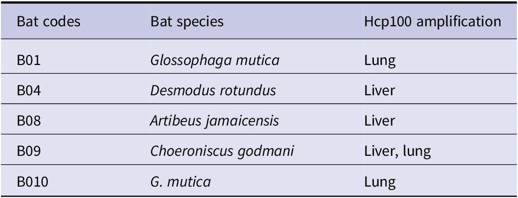
After looking for H. capsulatum infection in all the bats collected, 5 specimens showed amplification of the Hcp100 marker, representing a 41.6% infection rate. Table 4 enlisted positive reactions in at least one of the analysed organs of the following bat specimens: B01 (G. mutica); B04 (D. rotundus); B08 (A. jamaicensis); B09 (C. godmani); and B010 (G. mutica).
Detection of Histoplasma capsulatum by nested-PCR in bat organ samples

Table 4. Long description
The table consists of three columns: Bat codes, Bat species, and Hcp 100 amplification.
* Row 1: Bat code B 0 1, species Glossophaga mutica, amplification in Lung.
* Row 2: Bat code B 0 4, species Desmodus rotundus, amplification in Liver.
* Row 3: Bat code B 0 8, species Artibeus jamaicensis, amplification in Liver.
* Row 4: Bat code B 0 9, species Choeroniscus godmani, amplification in Liver and lung.
* Row 5: Bat code B 0 1 0, species G dot mutica, amplification in Lung.
Histoplasma capsulatum was isolated from a lung sample of a G. mutica female bat (B010), which had previously tested positive for H. capsulatum infection by nested-PCR (see Table 4). The fungal isolate exhibited slightly brown cotton-like mycelial culture (Figure 2a). Microscopic observations revealed the presence of hyaline branching septate hyphae, microconidia, and thick-walled tuberculate macroconidia (Figure 2b–d). This fungal isolation was confirmed by nested-PCR.
Histoplasma capsulatum cultured from a bat tissue sample. The figures show macro and microscopic morphologies of a fungal isolate from a Glossophaga mutica lung sample (B010). (a) Colonial morphology on PDA-culture at 27°C, at 15 days-culture; (b) mycelium with numerous macroconidia; (c,d) microscopies underlining tuberculate thick-walled macroconidia. Scale bars = 20 μm. Macrophotography was obtained with a digital camera, and microphotography was obtained at 40×, in an equipped Olympus microscope (see details in Materials and methods section).

Figure 2. Long description
A multi-panel display consisting of one vertical photograph on the left and three square micrographs on the right.
* Panel a, on the left, shows a macroscopic photo of a fungal isolate cultured on P D A in a glass test tube. Two distinct, white, fuzzy, cotton-like spherical colonies are visible growing on the surface of the light-tan agar medium.
* Panel b, at the top right, is a microphotography showing a blue-stained mycelium network. Numerous small, circular macroconidia are scattered across the fibrous fungal tissue. A scale bar at the bottom right indicates 50 micrometers.
* Panel c, at the bottom middle, provides a closer view of the macroconidia. The structures are thick-walled and tuberculate, appearing as dark blue circles with spiked or bumpy outer edges. A scale bar at the bottom right indicates 20 micrometers.
* Panel d, at the bottom right, is a high-magnification micrograph of a single macroconidium. It clearly shows a central circular body surrounded by prominent, rounded finger-like projections or tubercles. A scale bar at the bottom right indicates 20 micrometers.
Discussion
The present study was designed to cover three municipalities of Nayarit, where sampling sites were selected according to their accessibility, logistical feasibility, safety, availability of guano deposits, and ongoing community collaborations. The findings provide robust evidence of H. capsulatum in specific environments of the Nayarit state and cannot be extrapolated to the all-state scenario.
The isolation of H. capsulatum from soil samples is often unsuccessful due to contamination with other microorganisms that share the same habitat and can mask their growth in culture media [Reference Taylor19,Reference González-González25,Reference Taylor26]. Fortunately, molecular approaches, such as nested-PCR with the specific Hcp100 marker, were highly effective for detecting H. capsulatum, in both clinical and environmental samples, with detection limits as low as 3–5 fungal cells [Reference Gómez22, Reference Bialek24].
All guano samples analysed in the present study were collected from sites with confirmed bat activity and signs of human access. The sampling sites studied here were diverse, ranging from urban environments such as the SEMARNAT government building in Tepic-downtown, to rural and mountainous areas of the Nayarit state (Table 1).
The detection of H. capsulatum in 21 guano samples indicates a potential risk of infection not only for humans but also for different animals inhabiting contaminated environments. Data of Table 2 and Figure 1a,b, display the presence of H. capsulatum in bat guano samples either in urban (G1) or rural (G2–G27) areas, which carry a risk of infection to their populations.
It is important to emphasize that in some collection sites, such as a hill from the SAMAO municipality, predominantly in G4–G6 bat guano samples (details in Table 1), their soils were modified by anthropogenic activities. This inference was based on the fact that these sites are adjacent to an agave farm, which is irrigated with agrochemicals that drag contaminant residues to them. These circumstances could produce unknown effects on the surrounding soil, modifying their microbiota and affecting, particularly, the H. capsulatum growth conditions. Thus, several agricultural practices underscore the need to investigate feasible changes on soil microbiota, particularly, the generation of pernicious alterations in their pathogenicity.
At an abandoned house (G8–G10) on a rural site near a crater lake, in SAMAO, a shelter of frugivorous bats (Phyllostomidae) was identified with substantial accumulated guano. The presence of this guano, up to 15 cm deep, together with optimal humidity and temperature levels, would facilitate H. capsulatum growth.
The importance of the G12 sample collected in ‘La Herradura’ cave lies in the use of bat guano as agricultural fertilizer by local farmers, which constitutes a risk of infection. In this context, environmental conditions are closely intertwined with occupational and agrarian health of the resident population, reinforcing the need for an integrated surveillance approach under the ‘One Health framework’ [27].
Most of the samples collected in the municipality of La Yesca were obtained from caves (G13, G14, and G17–G27) with difficult access and are rarely visited by people (see Table 1). However, local community members have reported guano extraction. In addition, two samples from La Yesca were obtained from abandoned houses (G15, G16), which were used as resting refuges by rural workers and as shelters for insectivorous bats, creating favourable conditions for H. capsulatum infection.
The use of guano as a natural fertilizer has become very common in the state of Nayarit. Thus, it is fundamental to implement sanitary measures to protect the health of individuals involved in guano exploitation. For this reason, regional producers should be advised to implement sanitary measures at sites where H. capsulatum is detected [Reference Gómez22].
The detection of H. capsulatum in bat guano collected from diverse natural and human-modified environments underscores the need for sanitary measures to prevent histoplasmosis infection.
In the Nayarit state, 67 bat species have been documented, including 21 migratory species [Reference Ceballos and Ceballos28–Reference Ramírez-Silva and Hernández-Cadena30]. Bats are known to use a wide range of roosting sites, including caves, tree hollows, and rocky gaps, among others. However, due to accelerated land-use changes, bat colonies are also found in structures constructed by humans, mainly in abandoned buildings within urban or rural areas, where guano can accumulate over time.
Bats are frequently infected with H. capsulatum [Reference Taylor19, Reference González-González25, Reference Taylor26, Reference Canteros31–Reference Ruiz-Muñoz36]. Out of 12 bats collected, H. capsulatum was detected by nested-PCR in 6 organs from 5 individuals (see Table 4). The detection of H. capsulatum in the liver of tree bat specimens suggests possible fungal dissemination in these infected hosts.
Bat specimens collected in the present study come from urban and rural areas. They were obtained through spontaneous donations and do not represent the whole bat populations of Nayarit. Therefore, the present results should not be interpreted as a prevalence estimation or state-wide distribution data. The collected specimens were found dead in peridomestic or anthropogenic environments, such as abandoned houses or in areas with frequent human transit. All bats that showed Histoplasma-positive infection belonged to non-migratory species, ruling out seasonal movements. As bats were randomly collected, it is unknown whether these individuals shared roosts with migratory bats. Thus, our data cannot distinguish native or introduced presence of the fungus. Considering that our findings provide direct confirmation of the Histoplasma existence in bat-associated biological material, exploring this question would require further population-genetic studies of the fungal isolates.
Conclusions
In the Mexican Nayarit state, environmental factors and the presence of bats in urban and rural structures favour the proliferation of H. capsulatum. Considering that we demonstrated the presence of Histoplasma in bats guano and their infected tissues, our findings suggest a potential health risk to exposed populations, primarily agricultural workers and visitors to bat roosts. Thus, among several sanitary protections for the population exposed to this fungal infection, it is recommended to avoid guano accumulation in inhabited houses and to restrict people’s access to bat refugees.
Data availability statement
The data used in this study are available from the corresponding author upon reasonable request.
Acknowledgements
G.R.P.S. acknowledge the ‘Honorable XXXIII Ayuntamiento de La Yesca, Nayarit’ for the facilities to interact with the Nayarit state communities.
Author contribution
G.R.P., J.R.N., and M.L.T. were involved in the design of this study. J.R.N.L., E.J.A.N., and A.G.G. collected guano samples. G.R.P.S. and G.R.A. extracted DNA from guano samples and performed all molecular assays. E.J.A.N. and A.G.G. extracted DNA from bat organ samples. G.R.P.S. and M.L.T. analysed the results and drafted the manuscript. All authors revised the final version of the manuscript.
Funding statement
This paper constitutes a bilateral academic collaboration between MLT from UNAM-Mexico and GRPS from UAN-Mexico. GRPS contributed to this research with two grants: (1) Project Ref: PRIMSO-017 of the ‘Consejo Estatal de Ciencia y Tecnología del Estado de Nayarit (COCyTEN)’; (2) Project Ref: RENAJEB-2023-2029 of the ‘Secretaría de Ciencia, Humanidades, Tecnología e Innovación (SECIHTI)’. MLT contributed to this research with her laboratorial and institutional supports.
Competing interests
The authors declare no conflicts of interest.




 Open access

