Introduction
Gynogenesis refers to the fertilization of eggs with inactivated sperm, and prevents any contribution of the paternal genome to the progeny. As a result, embryonic development proceeds with the inheritance of only the maternal chromosome set(s). In nature, the well studied examples of spontaneous gynogenetic fish species include the Ginbuna (Carassius auratus gibelio; Yamashita et al., Reference Yamashita, Jiang, Onozato, Nakanishi and Nagahama1993) and the Amazon molly (Poecilia formosa; Schartl et al., Reference Schartl, Wilde, Schlupp and Parzefall1995). In fish, due to pre-embryonic events such as insemination, the extrusion of the second polar body and the first mitotic cleavage can be manipulated; the techniques for artificially induced gynogenesis are well established and have been widely utilized. Induced mitogynogenesis is the most convenient and powerful approach to generate homozygous progeny at the first filial generation (Bertotto et al., Reference Bertotto, Cepollaro, Libertini, Barbaro, Francescon, Belvedere, Barbaro and Colombo2005). It can be achieved by blocking cell division with temperature, pressure, or chemical treatments after ultraviolet (UV) light-irradiated sperm inseminated eggs have completed their second meiosis, in which the maternal haploid chromosome set is doubled. In 1981, Streisinger et al. (Reference Streisinger, Walker, Dower, Knauber and Singer1981) published a key study about induced gynogenesis in zebrafish (Danio rerio) and described how to produce meiotic diploids, mitotic diploids, and homozygous and heterozygous clonal lines of zebrafish. The publication marked the rise of the zebrafish as the leading animal model for research on embryonic development in vertebrates. Mitogynogenesis has been successfully induced in a total of 19 fish species and doubled haploid animals have been produced, including 16 freshwater species and three marine species (Komen & Thorgaard, Reference Komen and Thorgaard2007).
There are two distinct hypotheses explaining the mechanism of mitogynogenesis and chromosome doubling. The prevalent hypothesis is that the treatments may disable or disorganize spindles, block the anaphase movement of chromosomes, and form a doubled nucleus without cell division, which is also called ‘suppression of the first cleavage’ (Streisinger et al., Reference Streisinger, Walker, Dower, Knauber and Singer1981; Chourrout, Reference Chourrout1984; Ihssen et al., Reference Ihssen, McKay, McMillan and Phillips1990; Kobayashi, Reference Kobayashi1997; Nam et al., Reference Nam, Choi, Park and Kim2001; Morelli & Aquacop, Reference Morelli2003; Sakao et al., Reference Sakao, Fujimoto, Tanaka, Yamaha and Arai2003). However, after careful study of the behaviour of spindles and nuclei in eggs treated with hydrostatic pressure or heat shock around the time of the metaphase of the first cell cycle in the rainbow trout (Oncorhynchus mykiss), Zhang & Onozato (Reference Zhang and Onozato2004) found that heat shock or hydrostatic pressure did not inhibit the first cleavage owing to the regeneration of the bipolar spindle but inhibited the second cleavage by forming a monopolar spindle during the second cell cycle, and thus, the chromosome set was doubled. They proposed a new hypothesis in which the destruction of the centrosome by hydrostatic pressure treatment leads to chromosome set doubling. Zhu et al. (Reference Zhu, You, Zhang, Xu and Xu2006, Reference Zhu, You, Zhang, Xu and Sun2007) adapted indirect immunofluorescence staining to detect cytological changes during mitosis of UV light-irradiated homosperm inseminated eggs from the Oliver flounder (another name of the Japanese flounder) treated with cold shock or hydrostatic pressure. The results of both studies supported the theory of Zhang & Onozato.
The objective of this paper was to detect the effects of hydrostatic pressure treatment on the induction of mitogynogenesis in Japanese flounder eggs inseminated using UV-irradiated red sea bream (Pagrus major) sperm by analyzing histological sections.
Material and methods
Animal and gamete collection
All experiments were conducted at Beidaihe Central Experiment Station, Chinese Academy of Fishery Sciences, Qinhuangdao, Heibei Province, China. The fish were reared at 14°C with a 18-h light/6-h dark photoperiod. Sperm from mature red sea bream (3 years old; total length: 35 cm; body weight: 1.4 kg) were collected using a 5-ml plastic syringe by gently pressing the abdomen and avoiding water and urine contamination. Eggs were collected from one fertile, mature Japanese flounder (4 years old; total length: 45 cm; body weight: 2.4 kg). The eggs were stripped from the female and collected in a 1000-mL glass beaker. Totally, 3 ml of sperm (1.01 × 10⁶ cells per ml) and 50 ml eggs (1000 eggs per ml) were collected, and were stored at 4°C before use.
UV irradiation of sperm, insemination, and hydrostatic pressure treatment
The UV irradiation of red sea bream sperm was performed according to the method of Yamamoto (Reference Yamamoto1995, Reference Yamamoto1999). Before irradiation, the sperm were diluted 50 times with Ringer's solution (0.1408 mol·L–1 NaCl, 0.0052 mol·L–1 KCl, 0.0049 mol·L–1 CaCl2· 2H2O, 0.0011 mol·L–1 MgCl2· 6H2O, 0.0008 mol·L–1 NaH2PO4·2H2O, 0.0024 mol·L–1NaHCO3, 0.0050 mol·L–1 C6H12O6·H2O; pH 6.5–7.2), and then irradiated under UV light at an intensity of 73 mJ/cm2 (VLX-3W, Cole Parmer Instrument Company, Illinois, USA). Artificial insemination was performed by mixing 1 ml UV-irradiated sperm and 40 ml eggs for 3 min in a 1000-mL glass beaker, and the mixture was diluted with 1000 ml filtered sea-water and incubated at 17°C. The mixture was subjected to a pressure of 63.7 MPa for 6 min after 60 min of insemination (Yamamoto, Reference Yamamoto1995). The eggs were then transferred to 17°C sea-water and incubated after the treatment.
Cytology
For the cytological observations, after hydrostatic pressure treatment, the eggs were fixed as follows: from insemination to 60 min after insemination (AI) (time just before hydrostatic pressure treatment), the eggs were fixed with the time interval of 3 min in 4.5 ml Bouin's solution. From 68 min AI to 3.5 h AI, the eggs were fixed with the time interval of 2 min. After a fixation of 14 h, the eggs were transferred to 70% ethanol until use.
The histological sections were made according to the method of Sun et al. (Reference Sun, You, Zhang and Xu2005). The membrane of each egg was pricked with a needle at the vegetal pole. Eight to 10 eggs at each stage were embedded in a hot agarose solution (1–3%), and dehydrated in an ethanol series and terpineol. The eggs were then embedded in paraffin, serially sectioned into 8-µm slices, and stained with Harris’ hematoxylin and eosin. All slides were observed under an Olympus BX 51 research microscope and photographs were taken with a DP71 CCD (Olympus Corporation, Tokyo, Japan).
Ploidy determination
The relative content of DNA in 1-day-old hatching larvae was measured to determine their ploidy status by using a flow cytometer (Ploidy Analyzer; Partec GmbH, Münster, Germany). Each larva was placed in a Petri dish, 0.5 ml CyStain DNA 1step, including 4′6-deamidino-2-phenylindole (DAPI; Partec GmbH, Münster, Germany) was added, and the larva was rubbed with two tweezers in order to release the cells. Subsequently, added an additional 1.5 ml of CyStain DNA 1step. The cells were then incubated at room temperature for 5 min, filtered through a 30 µm CellTrics disposable filter (Partec GmbH, Münster, Germany), and analyzed using flow cytometry. The normal diploid larvae hatched from normal fertilization of male and female Japanese flounder were used for the 2C diploid value, which were set as the standard, together with the larvae from UV-irradiated sperm inseminated eggs without pressure treatment and normal larvae from mitogynogenetic group. For each group, 10 larvae were analyzed.
Results
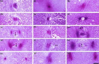
Before the application of hydrostatic pressure at 60 min AI, most of the eggs (8 of 10) were at the metaphase of the first mitosis. For each egg, the spindle was very conspicuous, the deeply stained chromosomes were arranged in an orderly manner at the centre of the metaphase plate, and the axis of the chromosomes was perpendicular to the spindle axis (Fig. 1 a). The spindle was disassembled by hydrostatic pressure immediately after the treatment at 68 min AI and the previously well-arranged chromosomes became disordered (Fig. 1 b). At 82 min AI, the spindle resembled in its former position and the chromosomes were rearranged. In this section, a dense chromatin body (DCB) was also observed (Fig. 1 c). The egg then entered the normal anaphase (Fig. 1 d) and telophase (Fig. 1 e) of the first mitosis, which was not blocked.
The developmental process of mitogynogenetic eggs of Japanese flounder (Paralichthys olivaceus) from the first mitosis to the third mitosis when incubated at 17°C. (a)–(e) First mitotic cycle, (f)–(j) second mitotic cycle, and (k)–(o) third mitotic cycle. (a) Metaphase of the first mitosis (60 min after insemination, AI); (b) 68 min AI, the spindle was disassembled by the hydrostatic pressure treatment; (c) 82 min AI, the spindle resembled in its former position, the arrow indicates the dense chromatin body (DCB); (d) anaphase, the arrow indicates the DCB (94 min AI); (e) telophase (100 min AI); (f) interphase of second mitosis (104 min AI); (g) prophase, one nucleus and one aster (118 min AI); (h) metaphase, monopolar spindle (152 min AI); (i) anaphase, the arrow indicates the dense chromatin body (154 min AI); (j) telophase (156 min AI); (k) interphase of the third mitosis (160 min AI); (l) prophase, one nucleus and two asters (162 min AI); (m) metaphase, bipolar spindle, the arrow indicate the dense chromatin body (174 min AI); (n) anaphase, the arrow indicates the DCB (182 min AI); and (o) telophase (190 min AI). Scale bar = 20 µm.

After the first mitosis, the egg entered the second mitosis. At 118 min AI, the egg was at the prophase, a nucleus was formed, and at only one side of the nucleus, there was an aster that was produced by the centrosome (Fig. 1 g). Thirty-four minutes later, the nuclear membrane was breakdown and a monopolar spindle was assembled instead of the normal bipolar spindle. The chromosomes were arranged in a line on the microtubules of the monopolar spindle, facing the first cleavage furrow, and the egg entered the metaphase of the second mitosis (Fig. 1 h). The monopolar spindle then became bigger and arranged chromosomes were randomly disturbed on the microtubules, this represents the anaphase and telophase of the second mitosis (Figs. 1 i, j). In the second mitosis, the chromosomes were replicated, but cell cleavage was blocked.
At 162 min AI, the egg entered the prophase of the third mitotic cycle, and two asters at opposite ends of the nucleus (Fig. 1 l). As the egg developed into metaphase, a bipolar spindle was assembled, the deeply stained chromosomes arranged orderly in the centre of metaphase plate, and the axis of the chromosomes was perpendicular to the spindle axis at 174 min AI (Fig. 1 m). The chromosomes were then separated and dragged to the two poles by the spindles, and the cell entered the anaphase and telophase of the third mitosis (Fig. 1 n, o).
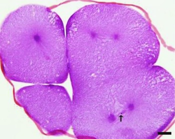
The deeply stained DCB, which was formed by the UV-irradiated red sea bream sperm, was observed in the sections (Fig. 1 c, d, i, m, n). In the first and third mitotic cycles, the DCB disturbed on the equatorial plate of the bipolar spindle. In the second cycle, the distance between the DCB and the monopolar spindle was half of that between the equatorial plate and one aster in the bipolar spindle (Fig. 1 i). The DCB randomly entered one blastomere. In the section shown in Fig. 2, only one DCB was observed in one of the four blastomeres.
Four-cell stage, the arrow indicates the only dense chromatin body disrupting the equatorial plate of one blastomere. Scale bar = 20 µm.

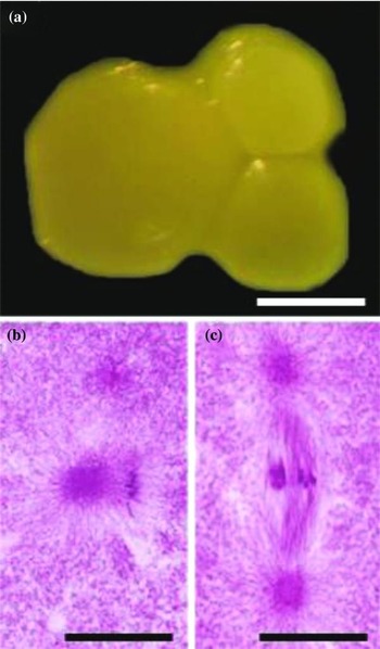
Seven of 150 eggs at 150 min AI were observed to have three blastomeres – one big blastomere and two small blastomeres (Fig. 3 a). In the histological section, the bigger blastomere contained a monopolar spindle (Fig. 3 b), but the two smaller blastomeres contained a bipolar spindle (Fig. 3 c). The chromosomes arranged on the monopolar spindle were parallel to the first cleavage furrow, while the bipolar spindle's chromosomes were perpendicular to it. The presence of three blastomeres in one egg is one example of the abnormal cell events that occur in eggs following hydrostatic treatment.
Histological and morphological observation of an egg with three blastomeres. (a) An embryo with three blastomeres at 150 min after insemination; (b) monopolar spindle in one blastomere; and (c) bipolar spindle in the other blastomere. Scale bar in (a) indicates 160 µm; scale bars in (b) and (c) indicates 20 µm.

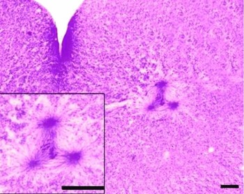
Some other abnormal cell events were also observed. At 126 min AI, a tripolar spindle was clearly assembled in one blastomere of two (Fig. 4). The areas of the three asters were 47.29 µm2, 98.24 µm2 and 144.56 µm2. The sum of the two small asters’ areas (145.53 µm2) was approximately equal to the area of one large aster. Another abnormal cell event was observed in an egg at 116 min AI wherein two nuclei were formed in one blastomere. These two nuclei were approximately the same size, and both had one aster each located beside it (Fig. 5).
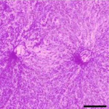
At 126 min after insemination, a tripolar spindle was well assembled in one blastomere of two. Scale bars = 20 µm.

At 116 min after insemination, two nuclei formed in only one blastomere. Scale bar = 20 µm.

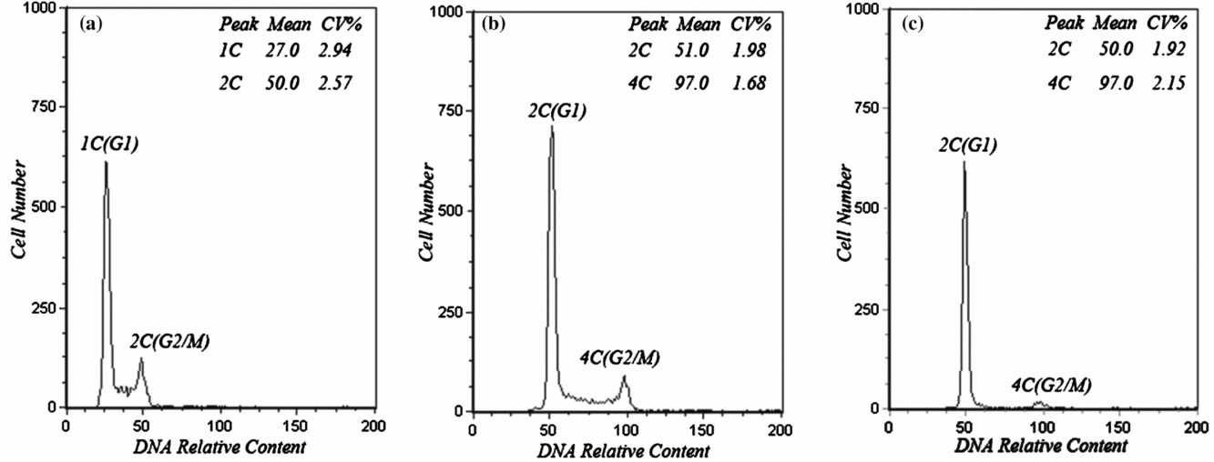
The results of flow cytometric analysis indicated that the relative DNA content of the diploid control was 51.0 ± 1.9 (mean ± SD), while for the haploid it was 27.0 ± 2.5, which was almost half that of the diploid control. The relative DNA content of the mitogynogenetic diploid was 50.0 ± 1.7, which was equal to that of the diploid control (Fig. 6).
Histograms of ploidy analysis. (a) Haploid; (b) diploid from normal fertilization; and (c) diploid from mitogynogenesis.

Discussion
Most of the eggs were treated by hydrostatic pressure during the metaphase of the first mitosis; the bipolar spindle of each egg was well assembled and then destroyed by the treatment. Fourteen minutes later, the bipolar spindle reassembled and the first mitosis normally continued. In the second mitotic cycle, a monopolar spindle assembled in each blastomere instead of a bipolar spindle. Mitosis proceeded with a monopolar spindle in each blastomere, and, as a result, the chromosomes were doubled but the cell cleavage was blocked. Subsequently, the third cycle proceeded normally. Flow cytometric analysis indicated that the relative DNA content of mitogynogenetic larva was equal to that of the diploid control, and the relative DNA content of larvae developed from eggs inseminated with UV-irradiated red sea bream sperm without hydrostatic pressure treatment was almost half of the diploid control. These observations demonstrated the effectiveness of UV irradiation and hydrostatic pressure treatment.
In the studies of rainbow trout (Zhang & Onozato, Reference Zhang and Onozato2004), and olive flounder (Zhu et al., Reference Zhu, You, Zhang, Xu and Xu2006, Reference Zhu, You, Zhang, Xu and Sun2007), pooled eggs of several females were used. Although eggs from only one female were used in this study, the results we obtained are similar to those observed by Zhang & Onozato (Reference Zhang and Onozato2004), and Zhu et al. (Reference Zhu, You, Zhang, Xu and Xu2006, Reference Zhu, You, Zhang, Xu and Sun2007). This indicated that the maternal effect to the embryo development process after hydrostatic pressure treatment could be eliminated. Zhang & Onozato (Reference Zhang and Onozato2004) proposed a new model for chromosome set doubling. The spindle and the daughter centrioles are dissembled by hydrostatic treatment. The remaining two single-mother centrioles and pericentriolar matrices (PCMs) regenerate a bipolar spindle and the first cleavage progresses normally. A centriole distributed to a daughter blastomere generates a whole centrosome with a newly formed procentriole and, as a result, a monopolar spindle is formed, which leads to the failure of the anaphase disjunction of duplicated chromosomes of the second cleavage, eventually resulting in chromosome set doubling (Zhang & Onozato, Reference Zhang and Onozato2004).
The animal cell's centrosome contains a pair of centrioles surrounded by a PCM. At the time of DNA synthesis, each centriole duplicates to produce a mother and daughter centriole pair, which forms two centrosomes to organize the mitotic spindle poles (Loncarek et al., Reference Loncarek, Sluder and Khodjakov2007). The number of centriole pairs determines the number of spindle poles. In sea urchin zygotes, when mitosis is prolonged by artificial methods, the two spindle poles do not further subdivide even when mitosis is prolonged to 20 times its normal duration. Ultrastructural analysis of such tetrapolar spindles reveals that each pole contains only one centriole. After the cell divides into four, each of the half centrosomes assembles a daughter centriole, and then becomes a normal centrosome with full reproductive capacity. However, they do not undergo centriole splitting or centrosome disjunction, and each cell assembles a monopolar spindle at the next mitosis. In effect, the two mitotic centrosomes with normal reproductive capacity subdivide into four centrosomes with half the normal reproductive capacity (Mazia et al., Reference Mazia, Harris and Bibring1960; Sluder & Begg, Reference Sluder and Begg1985; Hinchcliffe et al., Reference Hinchcliffe, Cassels, Rieder and Sluder1998; Hinchcliffe & Sluder, Reference Hinchcliffe and Sluder2001).
The formation of three blastomeres in one egg can also be interpreted using the model of Zhang & Onozato (Reference Zhang and Onozato2004). As they develop asynchronously, the daughter centrioles in one egg may be mature, immature, or in a transition state from the immature to the mature state when hydrostatic pressure is applied. After the application of hydrostatic pressure, the mature centriole is not destroyed but the immature centriole is, resulting in one of the centrosomes containing one centriole and the other forming a pair. After the first mitosis, the blastomere that receives the centrosome containing only one centriole assembles a monopolar spindle instead of a bipolar spindle, resulting in a diploid blastomere. Another blastomere that has a complete centrosome undergoes normal mitosis. Those embryos may stop developing at some stage or develop into haploid–diploid mosaics.
The probable cause of the tripolar spindle is that the centrosomes are not destroyed by hydrostatic pressure and the first mitosis proceeds normally. At the second mitotic cycle, the centrosome in one blastomere is duplicated at the interphase and assembles into two integrated centrosomes. An unexpected event can result in the separation of two centrioles from one centrosome. The one centrosome and two separated centrioles then assemble a tripolar spindle (Fig. 4). A tripolar spindle is rarely observed in the finfish and it is only observed in spontaneous gynogenetic fish species such as the Ginbuna (Carassius auratus gibelio; Yamashita et al., Reference Yamashita, Jiang, Onozato, Nakanishi and Nagahama1993). A tripolar spindle is formed at the first meiosis and the three sets of chromosomes present in triploid Ginbuna oocytes are separated in three directions; this ensures the retention of the original ploidy during gynogenesis. In our study the tripolar spindle was formed after hydrostatic pressure treatment, but it was not spontaneously formed. Chromosomes were not uniformly distributed on the tripolar spindle and resulted in chromosomal malsegregation.
Financial support
This study was financially supported by the earmarked fund for the State 863 High-technology R&D Project (2012AA10A408-5), and Modern Agro-industry Technology Research System (CARS-50-G02, CARS-50-Z03), China.
Statement of interest
None