Introduction
Trichinella spiralis is a parasite that causes the zoonotic disease trichinosis. It is considered a health problem, as it affects humans in many countries worldwide (Thawornkuno et al. Reference Thawornkuno, Nogrado, Adisakwattana, Thiangtrongjit and Reamtong2022). This viviparous nematode completes its life cycle in a single host. Most of the adult worm’s life is spent in the intestines, where it releases thousands of larvae that invade muscles of the host. The larval forms become encapsulated as small cystic structures within muscle cells of the infected host (Thawornkuno et al. Reference Thawornkuno, Nogrado, Adisakwattana, Thiangtrongjit and Reamtong2022). Human infections occur by eating inappropriately cooked pork infected by the encysted larvae (Mohammed et al. Reference Mohammed, Youseef, Mubarak, Mawas, Khalifa and Felefel2022).
Trichinosis symptoms can be categorised into three clinical phases: enteral, migratory, and parenteral. The signs and symptoms could appear starting from 12 hours to 2 days after eating infected meat. Inflammation that is brought on by the adult worms migrating through the intestinal epithelium manifests as nausea, vomiting, sweating, and diarrhoea. Edema of the face and fever could develop later (Mohib et al. Reference Mohib, Clevenbergh, Truyens, Morissens and Castro Rodriguez2022). Ten days after infection, severe muscle pain, breathing difficulties, weakened pulse and blood pressure, heart damage, and different mental diseases may appear. Eventually, death due to heart failure, lung problems, or renal dysfunction may result (Ribicich et al. Reference Ribicich, Fariña, Aronowicz, Ercole, Bessi, Winter and Pasqualetti2020). The existence of matching symptoms and signs, as well as the presence of Trichinella larvae in biopsy muscle tissue or particular antibodies in serum, all contribute to the diagnosis of trichinosis (Zolfaghari Emameh et al. Reference Zolfaghari Emameh, Kuuslahti, Näreaho, Sukura and Parkkila2016).
Benzimidazole derivatives such as mebendazole and albendazole (ABZ) have been used for decades in the treatment of trichinosis. Despite being efficient killers of adult worms, they showed limited efficacy against encapsulated and new-born larvae (Chai et al. Reference Chai, Jung and Hong2021; Rong-Yu and Feng-qi Reference Rong-Yu and Feng-qi2015). Therefore, scientific research aims to discover novel, secure, and effective anthelmintic agents. Recently, drug repurposing has recently emerged as a tool for developing new indications for existing, failed, or abandoned drugs (El-Sayad et al. Reference El-Sayad, El-Wakil, Moharam, Abd El-Latif, Ghareeb and Elhadad2023). Metformin (MET) is an orally administered biguanide that is commonly prescribed for the treatment of type II diabetes mellitus and gestational diabetes. Other uses include the protection against diabetes development in high-risk populations and the treatment of antipsychotic-induced weight gain and oligomenorrhea due to polycystic ovarian syndrome. MET is usually prescribed at daily doses ranging from 500 to 2550 mg. This anti-hyperglycaemic and potentially anti-cancer drug may inhibit cell proliferation by inducing energy stress (Lv and Guo Reference Lv and Guo2020). Recently, it has been tried as an anti-parasitic agent against Echinoccocus granulosus, Plasmodium spp., and T. spiralis with promising results (Loos et al. Reference Loos, Dávila, Rodrígues, Petrigh, Zoppi, Crocenzi and Cumino2017; Miyakoda et al. Reference Miyakoda, Bayarsaikhan, Kimura, Akbari, Udono and Yui2018; Othman et al. Reference Othman, Abou Rayia, Ashour, Saied, Zineldeen and El-Ebiary2016). Several parasitic infections are now being treated with nanotechnology (Hatam-Nahavandi Reference Hatam-Nahavandi2019; Volpedo et al. Reference Volpedo, Costa, Ryan, Halsey, Satoskar and Oghumu2019). Chitosan is one of the polymeric nanoparticles of natural sources. It is characterized by being a biocompatible, biodegradable, and nontoxic material that can be used safely in different applications (Mohammed et al. Reference Mohammed, Syeda, Wasan and Wasan2017). The aim of this study was to compare the effectiveness of albendazole and the anti-diabetic medication metformin loaded on chitosan nanoparticles in treating mice with various stages of T. spiralis infection.
Materials and methods
Ethical consideration
All procedures were performed on mice in accordance with the Institutional Animal Care and Use committee (IACUC), Alexandria University, Egypt (Protocol approval number: AU0122092011). The study also followed ARRIVE guidelines and complied with the National Research Council’s guide for the care and use of laboratory animals.
Preparation of drugs
Albendazole was purchased as a pure powder (Pharco Company), while metformin tablets (Merck Sante S.A.S Company) were crushed using a pestle and mortar. The active ingredient of each drug was calculated separately, and the dose needed was calculated for all mice and dissolved in 100 ml of distilled water for oral administration using an esophageal syringe (0.5 ml/each mouse) (Nassef et al. Reference Nassef, Moharm, Atia, Brakat, Abou Hussien and Mohamed2018).
Preparation of blank chitosan nanoparticles
Chitosan powder was weighed (40 mg) using an electrical balance, added to 40 ml of 1% acetic acid in a glass flask, and allowed to dissolve using magnetic stirrer at 300 rpm at room temperature (22–26°C). Sodium tripolyphosphate (TPP) was weighed (40 mg) and dissolved in 40 ml of distilled water using magnetic stirring at 300 rpm. In order to prepare the Cs nanoparticles (NPs), the sodium TPP aqueous solution was added dropwise to the above-prepared Cs solution with stirring at 300 rpm for half an hour. Then Tween 80 (50 ml) was added to the solution. The prepared solution was left overnight at room temperature (22–26°C) for proper ionic gelation. Nanoparticles were formed by centrifugation at 15,000 rpm for 30 minutes. The supernatant was carefully separated from the sediment. The sediment containing Cs NPs was collected in tubes (Kandav et al. Reference Kandav, Bhatt and Jindal2019).
Preparation of ABZ and/or MET/ Cs NPs
The desired concentrations of albendazole (ABZ) dissolved in ethanol (96%) and metformin (MET) dissolved in acetic acid solution were added to the chitosan solution and mixed using magnetic stirrer for 30 min at 25°C. Sodium TPP solution was mixed with the chitosan solution containing either ABZ or MET using a magnetic stirrer (300 rpm) for 30 min at 40°C. Tween 80 (50 ml) was added to solution. Finally, nanoparticles were purified by centrifugation for 30 min at 15,000 rpm and then washed twice with distilled water. The resulting precipitates were lyophilized (Kandav et al. Reference Kandav, Bhatt and Jindal2019).
Physio-chemical characterization of Cs NPs and drugs loaded Cs NPs
Transmission electron microscope (TEM) examination was used to assess the shape and morphology of Cs NPs and drugs loaded Cs NPs (Michen et al. Reference Michen, Geers, Vanhecke, Endes, Rothen-Rutishauser, Balog and Petri-Fink2015).
Zeta potential was measured using a laser light-scattering spectrophotometer. Zeta potential is related to the magnitude of the electrical charges on the particle surface. It indicates the degree of repulsion between adjacent similarly charged particles in dispersion. The significance of zeta potential is that its value can be related to the stability of NPs.
Fourier transform infrared (FTIR) spectroscopy measured the absorption of infrared radiation of materials at the atoms vibrating about their bonds using Shimadzu FTIR-8400S (Tokyo, Japan). It is primarily used to identify bonds types, structures, and functional groups in organic and inorganic compounds (Bhargava Reference Bhargava2012).
Drugs loading efficiency (LE) was calculated using the following formula:
 $$ \mathrm{LE}=\frac{\mathbf{Ct}-\mathbf{Cu}}{\mathbf{Ct}}\times \mathbf{100} $$
where Ct is the total concentration of drug added to the system and Cu is the concentration of free drug in the supernatant (Figure 1).
Calibration standard curves of ABZ/ Cs NPs- MET/ Cs NPs using spectrophotometer at 342, 256 nm.

Experimental animals
The present study was carried out on 160 apparently healthy laboratory-bred male Swiss albino mice, weighing about 20–25 gms and aged five to six weeks. They were purchased from the animal house in Theodor Bilharz Research Institute (TBRI), Giza, Egypt. The mice were fed a standard commercial pellet diet with free accessible water all through the time of the study. Mice were housed in plastic cages with wood shaving bedding and nesting materials under controlled adequate conditions (Temperature 22–26°C, humidity 30–70%, 12 hrs light and 12 hrs dark cycle) as per the ethical consideration of institutional and national guidelines.
Parasite and dose of infection
The T. spiralis strain was kindly provided from experimentally infected Swiss albino mice in the Parasitology Department, TBRI, Giza, Egypt. The strain of Trichinella spiralis (Istituto Superiore di Sanita` code: ISS6158) obtained in this work was kindly provided by the Medical Parasitology Department, Tanta Faculty of Medicine, Tanta university, Egypt. Then it was kept in the Parasitology Department at Theodor Bilharz Research Institute (TBRI) (Giza, Egypt) by consecutive passages on mice according to the method of Gamble (Reference Gamble1996). Briefly, the muscles of infected mice were minced and digested in 1% pepsin-hydrochloride. After incubation overnight at 37°C, larvae were collected by using the sedimentation technique and washed several times with saline, and the larvae number per ml was counted. The standard experimental infective dose for each mouse was 200–300 orally inoculated larvae (El-Sayad et al. Reference El-Sayad, El-Wakil, Moharam, Abd El-Latif, Ghareeb and Elhadad2023).
Mice grouping and experimental design
Mice groups included in the present work were designed to represent different treatment options for T. spiralis infection. They were divided into control and experimental groups. The experimental group was further subdivided into six groups depending on the administrated drug (Figure 2). Each experimental group was divided into 2 subgroups (a and b) according to the time of sacrifice (10 mice/subgroup). The control groups were subdivided into two groups (infected non-treated and non-infected groups with 4 subgroups).
Experimental design.

All drugs were administered according to the corresponding design of groups and subgroups as follows:
-
• ABZ was used as the gold standard drug for treatment in a dose of 50 mg/kg orally for 5 successive days starting from the 1st day of infection for the enteral phase and from the 30th d.p.i for the parenteral phase (Kang et al. Reference Kang, Lee, Ng, Kim and Park2015).
-
• ABZ/ Cs NPs were orally administrated in a dose of 50 mg/kg daily for 5 successive days starting from the 1st day of infection for the enteral phase and from the 30th d.p.i for the parenteral phase.
-
• MET/ Cs NPs were administrated in a dose of 50 mg/kg daily orally for 5 successive days starting from the 1st day of infection for the enteral phase and from the 30th d.p.i for the parenteral phase.
-
• ABZ & MET/ Cs NPs were orally administered in a dose of 50 mg/kg daily of each prepared drug orally for 5 successive days starting from the 1st day of infection for the enteral phase and from the 30th d.p.i for the parenteral phase.
-
• ABZ & MET were used in administered in a dose of 50 mg/kg daily orally for 5 successive days starting from the 1st day of infection for the enteral phase and from the 30th d.p.i for the parenteral phase.
The dose was adjusted in each drug so that each mouse received the calculated dose of the chosen drug according to its body weight in ~0.5 ml by esophageal syringe. Eating and drinking were allowed after one hour of drug administration.
Parasitological assessment
In subgroups (a), the ten mice were sacrificed 6 d.p.i. Infected mice were starved the night before the sacrifice. Mice were sacrificed under light anesthesia by isoflurane inhalation (Forane®, UK). The abdominal cavity was dissected, and the whole intestine was taken off and washed rapidly under warm tap water to remove debris. Small portions (1 cm from the middle region of the small intestines) were taken and immediately fixed in 10% formol-saline for histopathological studies. The remaining parts of the small intestines were split longitudinally along its entire length. Finally, the intestine was cut into two-centimeter pieces and placed in a beaker full of normal saline at 37°C for three to four hours. After that, the intestine was shaken well in the saline, rinsed in a similar amount of saline, and finally removed and discarded. Sediment of the adult worms was collected by standing in the container for half an hour. Then after removal of the supernatant fluid, adult worms were counted under the dissecting microscope at 10× power.
In subgroups (b), mice were sacrificed 36 d.p.i, each mouse was dissected, and its muscles were digested in 1% pepsin and 1% concentrated HCl in 200 ml distilled water. A section was immediately fixed in 10% formal saline for histopathological studies. The mixture was incubated at 37°C for two hours under continuous agitation using an electric stirrer. The digested product was passed through a 50-mesh/inch sieve to remove the coarse particles. Encysted larvae were collected on a 200-mesh/inch sieve, washed twice with tap water, and then suspended in 150 ml of tap water in a conical flask. The supernatant fluid was then discarded and the muscle larvae in the sediment were counted microscopically using a McMaster counting chamber.
Histopathological assessment
Tissue samples from small intestine biopsies and skeletal muscle specimens from the hind legs of the studied groups were fixed in 10% neutral buffered formalin for 24 h, dehydrated in ascending grades of alcohol, cleared in xylene, and embedded in paraffin blocks. Hematoxylin & eosin (H&E) were used to stain 4-micron thick sections (Nassef et al. Reference Nassef, Moharm, Atia, Brakat, Abou Hussien and Mohamed2018).
Scanning electron microscope examination (SEM)
T. spiralis adult worms and ML harvested from sacrificed infected mice treated with drugs loaded on Cs NPs and infected untreated mice were directly pipetted into a fresh fixation solution of 2.5% glutaraldehyde (w/v) in 0.1 M sodium cacodylate at pH 7.2 and left overnight at 37°C. Trichinella stages were washed in 0.1 M sodium cacodylate buffer at pH 7.2 for 5 min and post-fixed in a 2% (w/v) osmium tetroxide in sodium cacodylate buffer for one hr. The specimens were dehydrated in an ethanol series and dried using liquid carbon dioxide. The dried parasite stages were sprinkled onto and mounted on stub bearing adhesive. After sputter coating with gold, they were examined by SEM (Jeol Corp., Mitaka, Japan) at 5 to 20 KV in SEM laboratory in TBRI, Giza, Egypt (Bughdadi Reference Bughdadi2010).
Statistical analysis
The data were analyzed using Microsoft Excel 2016 and statistical package for social science ‘IBM SPSS Statistics for Windows’, version 26 (IBM Corp., Armonk, NY, USA). Continuous normally distributed variables were represented as mean±SD with 95% confidence interval; a p value < 0.05 will be considered statistically significant. To compare the means of normally distributed variables between groups, the student’s t test was performed. The ANOVA followed by Tukey HSD as a post-hoc test in multigroups was performed.
Results
Physio-chemical characterization of Cs NPs and drugs loaded Cs NPs
The TEM morphological characteristics of Cs NPs and drugs-loaded Cs NPs are shown in Table 1 and Figure 3. Cs NPs appeared as crystallites with rutile structure, with a size of 21.81–41.61μm. Other drugs-loaded Cs NPs were spherical with increased size. This increase in size pointed to successful drug encapsulation.
Sizes and shapes of Cs NPs and drugs loaded Cs NPs by TEM

TEM pictures of Cs NPs (a,b), ABZ/ Cs NPs (c,d), MET/ Cs NPs (e,f), ABZ & MET/ Cs NPs (g,h).

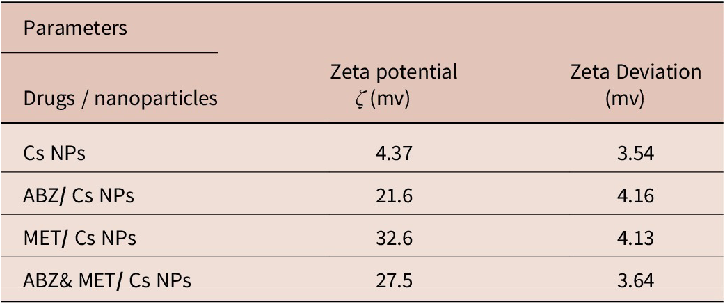
The ζ potential (mV) of prepared Cs NPs, ABZ/ Cs NPs, MET/ Cs NPs, and ABZ & MET/ Cs NPs were 4.37, 21.6, 32.6, and 27.5, respectively (Table 2, Figure 4).
Zeta potential and Zeta deviation measurements of the studied drugs nanoparticles

Zeta potential of Cs NPs (a), ABZ/ Cs NPs (b), MET/ Cs NPs (c), ABZ & MET/ Cs NPs (d).

FTIR spectra corresponding to the Cs NPs are shown in Figure (5a) .The basic characteristic peaks are at 3436.41 cm−1 (O-H stretch and N-H, overlap), 2079.19 cm−1 (C-H stretch), 1642.97 cm−1, 1633.87 cm−1 (NH2 deformation, C-C), 1268.52 cm−1 (C-O stretch), 699.19 cm−1 (C-H stretch), and 666.28 cm−1 (C-O-H twist broad). The basic characteristic peaks of ABZ/Cs NPs are at 3436.03 cm−1 (O-H stretch and N-H, overlap), 2079.63 cm−1 (C-H stretch), 1640.23 cm−1, 1633.89 cm−1 (C=C stretching), 696.27 cm−1 (C-H stretch), and 666.27 cm−1 (C-O-H twist broad) (Figure 5b). The basic characteristic peaks of MET/ Cs NPs are at 3910.01 cm−1 (O-H stretch), 3436.90 cm−1 (O-H stretch and N-H, overlap), 2076.10 cm−1 (C-H stretch), 1633.87 cm−1 (NH2 deformation, C-C), 1276.90 cm−1 (C-O stretch), and 698.88 cm−1 (C-H stretch) (Figure 5c). The basic characteristic peaks of ABZ & MET/ Cs NPs are at 3435.95 cm−1 (O-H stretch and N-H, overlap), 2076.47 cm−1 (C-H stretch), 1634.23 cm−1 (NH2 deformation, C-C), 1273.28 cm−1 (C-O stretch), 1017.11 cm−(C=C bending), and 695.46 cm−1 (C-H stretch) (Figure 5d).
FTIR spectroscopy of Cs NPs (a), ABZ/ Cs NPs (b), MET/ Cs NPs (c), ABZ & MET/ Cs NPs (d).

Effect of treatment regimens on the intestinal phase of T. spiralis
Table 3 shows the recovered number of adult worms in each subgroup and the reduction (LR %) percentage compared to the infected untreated control. All subgroups (1–6a) showed a statistically significant reduction of the recovered adult worms compared to non-treated control subgroup (7a). The highest reduction was achieved by ABZ & MET/ Cs NPs (7.3±1.6), followed by ABZ/ Cs NPs (14.3±3.4), combined ABZ and MET (23.3±4.6), ABZ alone (32.7±6.4), and MET/ Cs NPs (53.2±6.3). Finally, MET alone showed the lowest reduction of mean worm counts (77.2±8.4).
T. spiralis adult worm counts recovered from small intestines of the studied subgroups

T. spiralis worm count is represented as mean±SD.
The LR is represented as percent depending on the following law.
LR % = (Mean worm counts in infected untreated control subgroup (a) − Mean worm counts in experimental subgroup (a)
Mean worm counts in infected untreated control subgroup (a) x 100.
a p value is significantly different comparing between subgroups (a) depending on One Way ANOVA test.
b p value is significantly different comparing with control depending on student t test.
c p value is significantly different comparing between subgroups (a) depending on a post hoc test (Tukey HSD).
1 Initial p value <0.05 is significant;
2 Initial p value <0.01 is highly significant.
Effect of treatment regimens on the parenteral phase of T. spiralis
Table 4 shows the count of the recovered larvae per gram of muscles in the studied subgroups (b). All subgroups showed a statistically significant differences compared to the control non-treated subgroup (7b) (p< 0.001). The highest reduction of the muscle larvae count was achieved by ABZ & MET /Cs NPs (100.8±9.1), followed by ABZ /Cs NPs (156.8±7.4), combined ABZ & MET (243.0±44.3), ABZ alone (298.3±25.1), and MET (469.5±36.6). Finally, the group treated with MET alone showed the lowest reduction in count (703.8±17.2). Regarding the reduction percentage, it was noticed that the highest percentage was recorded among subgroup (b3) treated with ABZ & MET/ Cs NPs (93.1%) followed by subgroup (1b) treated with ABZ /Cs NPs (89.2%), subgroup (6b) treated with ABZ/ MET (83.3%), subgroup (4b) treated with ABZ (79.5%), and subgroup (3b) treated with MET/ Cs NPs (67.7%). The lowest reduction percentage was recorded in subgroup (5b) treated with MET (51.5%).
T. spiralis larvae count per gram of muscles recovered from the studied subgroups

T. spiralis larvae count is represented as mean±SD.
The LR is represented as percent depending on the following law.
LR % = (Mean worm counts in infected untreated control subgroup (b) − Mean worm counts in experimental subgroup (b)
Mean worm counts in infected untreated control subgroup (b) x 100.
a p value is significantly different comparing between subgroups (b) depending on One Way ANOVA test.
b p value is significantly different comparing with control depending on student t test.
c p value is significantly different comparing between subgroups (b) depending on a post hoc test (Tukey HSD).
1 Initial p value <0.05 is significant;
2 Initial p value <0.01 is highly significant.
Intestinal histopathological examination
Concerning the histopathological changes in the intestinal phase, examination of the intestines of infected positive control subgroup (7a) revealed severe inflammation, dense inflammatory infiltrate in lamina propria, reactive lymphoid follicles, oedema, destruction of intestinal villi, and ulceration. Marked improvement of histopathological changes was observed in infected subgroup treated with ABZ/ Cs NPs (1a) followed by infected subgroup treated with ABZ & MET/ Cs NPs (3a), infected subgroup treated with ABZ (4a), infected subgroup treated with MET (5a) and infected subgroup treated with ABZ/ MET (6a), as the intestines showed mild mucosal chronic mononuclear inflammatory infiltrate. The infected subgroup treated with MET/ Cs NPs subgroup (2a) showed multiple coiled adult worms in the lumen with moderate stromal inflammatory infiltrate with eosinophils (Figure 6).
Histological photomicrographs showing transverse sections of the small intestines. (a) ABZ/Cs NPs subgroup (1a) showing intact villi with mild stromal chronic inflammatory infiltrate (X 100). (b) MET/Cs NPs subgroup (2a) showing coiled T. spiralis worms in the lumen (arrows) (×400). (c) ABZ and MET/Cs NPs subgroup (3a) showing intestinal villi heavily infiltrated by chronic inflammatory cells as well as eosinophils (arrows) (×400). (d) ABZ subgroup (4a) showing eosinophilic rich inflammatory infiltrates (arrows) (×400). (e) MET subgroup (5a) showing the small intestinal lumen full of worms (red arrows) and the intestinal villi are infiltrated by chronic inflammatory cells (blue arrows) (×100). (f) ABZ and MET combination subgroup (6a) showing intestinal lumen entangling Trichinella worm with calcified embryos (red arrow) (×400). (g,h) Infected control subgroup (7a) showing an evident lymphoid follicle causing focal blunting of the villi (arrow) (g: ×100, h: ×400).

Muscular histopathological examination
Concerning the histopathological changes in the skeletal muscles, muscles of the positive control subgroup (7b) revealed multiple nurse cells entangling degenerating larvae surrounded by moderate chronic mononuclear inflammatory infiltrate with adjacent reactive lymph nodes. Marked improvement was observed in the group treated with ABZ/ Cs NPs (1b), as the muscles were free of infection. Similar results were observed in the infected mice that were treated with a combination of ABZ & MET/ Cs NPs (3b) and the infected mice treated with a combination ABZ & MET (6b). The infected mice that were treated with MET/ Cs NPs (2b) and those treated with MET (5b) showed several nurse cells entangling the encysted larvae with surrounding mild to moderate chronic mononuclear inflammatory infiltrate. Muscles of the infected mice that were treated with ABZ (4b) showed multiple foci of chronic mononuclear inflammatory infiltrate with no evident nurse cells (Figure 7).
Histological photomicrographs of skeletal muscles of the studied subgroups. (a) Muscle fibers of ABZ/Cs NPs subgroup (1b) showing free muscle fibers with minimal inflammatory infiltrate (×100). (b) MET/Cs NPs subgroup (2b) showing multiple nurse cells surrounded by a cuff of chronic inflammatory cells (arrows) (×100). (c) ABZ and MET/Cs NPs subgroup (3b) showing encysted larva surrounded and infiltrated by mixed acute and chronic inflammatory cells with degenerative changes manifested by fragmentation of the capsule and vacuolization (arrows) (×1,000). (d) ABZ subgroup (4b) showing skeletal muscle fibers entangling 2 nurse cells; one is completely digested (arrows) and the other shows degenerative changes and vacuolization (H&E ×400). (e) MET subgroup (5b) showing muscle fibers entangling a homogenized degenerated T. spiralis cyst in the center (arrows) surrounded by a viable cysts in lower and upper parts with moderate inflammatory infiltrate (×). (f) ABZ and MET combination subgroup (6b) showing homogenized degenerated larva in the nurse cell (star) within skeletal muscle fibers (arrows) (×1,000). (g) Positive control infected mouse subgroup (7b) showing several coiled larvae within skeletal muscle bundles with surrounding heavy mixed acute and chronic nonspecific inflammatory infiltrate (×400). (h) Negative control subgroup (8b) showing completely free muscle fibers from any larvae (×1,000).

SEM morphological examination
SEM examination of the tegument of the control untreated T. spiralis ML groups showed the typical coiling behaviour and the characteristic pattern of tegument. Also, T. spiralis adult worms of the control subgroup (8b) showed normal morphology with preserved tegument and normal appearance of the hypodermal gland opening. SEM examination of the tegument of T. spiralis adults (Figure 8) and muscle larvae (Figure 9) recovered from all treated subgroups treated with the studied drugs loaded on chitosan nanoparticles showed destruction with multiple degenerative changes. The destruction was more obvious in the subgroup treated combined ABZ & MET/ Cs NPs. The degenerative changes were in the form of the appearance of multiple masses and sloughing of the tegument in some areas.
SEM of T. spiralis adult worms. (a) Worms from infected control subgroup (7a) showing primary folds with large spacing, transverse creases and hypodermal glands opening. (b) Adult worm from ABZ/Cs NPs subgroup (1a) showing sloughing of the wall with large opening due to rupture of cuticle and multiple fissures with loss of normal annulations. (c) Adult worm from MET/ Cs NPs subgroup (2a) showing minimal changes in the cuticle. (d) Adult worm from the ABZ and MET/Cs NPs subgroup (3a) showing complete destruction of the cuticle, loss of normal annulations of cuticle with the appearance of cauliflower masses.

SEM of T. spiralis larvae. (a) Larvae recovered from the infected control subgroup (7b) showing normal cuticle with transverse creases and longitudinal ridge. (b) Larvae recovered from ABZ/Cs NPs treated mouse subgroup (1b) showing degenerative changes of cuticle with loss of striations. (c) Larvae recovered from MET/Cs NPs subgroup (2b) showing contracture and deformity of some areas of the cuticle and loss of the normal annulations of cuticle. (d) Larvae from ABZ and MET/Cs NPs subgroup (3b) showing severe degenerative changes of cuticle with loss of striations.

Discussion
Trichinellosis is a zoonotic illness that is still a major global public health concern with important clinical implications (Abou Rayia et al. Reference Abou Rayia, Saad, Ashour and Oreiby2017). It has been categorised as an emerging or re-emerging disease, especially in developing countries, because there are approximately 10,000 new cases of infection each year (Bai et al. Reference Bai, Hu, Liu, Tang and Liu2017). Chemotherapy using benzimidazole derivatives is most effective when given in the early stages of infection; however, the majority of cases are discovered several weeks after infection, when the larvae have already invaded the muscles (Gottstein et al. Reference Gottstein, Pozio and Nöckler2009). The current situation has aroused the need for the next generation of anthelmintics. Nanotechnology is a promising era for developing effective de novo drugs at the nano dimension (Hatam-Nahavandi Reference Hatam-Nahavandi2019). The current study assessed the efficacy of albendazole and metformin loaded on chitosan nanoparticles in treating mice infected with various stages of Trichinella infection.
Regarding the intestinal phase in the present study, a statistically significant difference in the recovered worm counts was found between albendazole treated and the infected non-treated control. In addition, the difference in the number of recovered ML from the parenteral phase was statistically significant. Similar results were previously reported by Chung et al. (Reference Chung, Joo, Quan, Kwon and Cho2001), who asserted that a single oral dose of ABZ given two hours or eight hours after the initial infection completely eradicated the infection as demonstrated by absence of adult worms in the gut on the seventh day following infection. They also claimed that albendazole was highly effective against immature worms; however, the susceptibility to ABZ decreased as the worms matured. Chung et al. (Reference Chung, Joo, Quan, Kwon and Cho2001) also studied the effect of albendazole on ML and reported a reduction rate of 80.8% as compared to the untreated controls. Many studies confirmed that ABZ is more effective against the intestinal stage of Trichinella infection than against the muscular stage, with a reduction percent that varied from 26% to 91% for muscle larvae and from 62% to 100% for adult worms (Attia et al. Reference Attia, Mahmoud, Farrag, Makboul, Mohamed and Ibraheim2015; Nada et al. Reference Nada, Mohammad, Moad, El-Shafey, Al-Ghandour and Ibrahim2018; Shalaby et al. Reference Shalaby, Moghazy, Shalaby and Nasr2010; Shoheib et al. Reference Shoheib, Shamloula, Abdin and El-Segai2006). It should be mentioned that benzimidazoles were found to progressively lose their anthelmintic efficacy in livestock in Brazil, which could be due to the emergence of drug-resistant helminths (Jaeger and Carvalho-Costa Reference Jaeger and Carvalho-Costa2017).
It has been documented that the bioavailability and intestinal absorption of albendazole is limited, which affects its therapeutic efficacy; hence, Eid et al. (Reference Eid, Ashour, Essa, El Maghraby and Arafa2020) studied chitosan-coated nanostructured lipid carriers for albendazole oral delivery in trichinellosis treatment and reported that ABZ nanoparticles decreased the number of worms. This was further supported by a decrease in the number of larvae and enhanced histological characteristics.
Metformin is a biguanide derivative that is often prescribed for type 2 diabetes to decrease hepatic glucose production and increase muscle sensitivity to insulin (Chinnaiyan et al. Reference Chinnaiyan, Deivasigamani and Gadela2019; Rena et al. Reference Rena, Hardie and Pearson2017). Several studies suggested that metformin influences immunological responses in mice models of cancer and autoimmune illness (Kunisada et al. Reference Kunisada, Eikawa, Tomonobu, Domae, Uehara, Hori, Furusawa, Hase, Sasaki and Udono2017; Pearce et al. Reference Pearce, Walsh, Cejas, Harms, Shen, Wang, Jones and Choi2009). The effects of metformin on the intestinal and muscular phases of T. spiralis were investigated in the current study. When compared to the infected untreated control, the counts of recovered worms were lower, and the difference was statistically significant (77.2±8.4 vs. 195.2±7.6). While in the muscular phase, the count of the recovered ML decreased to 703.8±17.2 vs. 1451±55.8 in the infected non-treated control, and the difference was statistically significant. This effect was much ameliorated when MET was loaded on chitosan nanoparticles, as the recovered number of worms decreased to 53.2±6.3 and the number of recovered larvae to 469.5±36.6.
In the present study, metformin was combined with albendazole. The effect of albendazole was greatly enhanced by this combination compared to albendazole alone, both in the intestinal and muscular phases. In the intestinal phase, the count of the recovered worms from the combination treated group decreased to 23.3±4.6 vs. 32.7±6.4 in ABZ only treated group. While in the muscular phase, the count of the recovered ML decreased to 243.0±44.3 vs. 298.3± 25.1 in ABZ-only treated group, though the difference was not statistically significant. The effect of this combination was much ameliorated when they were loaded on chitosan nanoparticles, as the recovered number of worms decreased to 7.3±1.6 worms and the number of recovered larvae to 100.8±9.1 larvae. Recent studies started to combine albendazole as the standard treatment of trichinellosis with other drugs in a trial to decrease the dosage and improve its therapeutic efficacy. Fahmy and Diab (Reference Fahmy and Diab2021) studied how well mefloquine (MQ) works against T. spiralis infection when used alone or in combination with albendazole at lower doses. The worm count was reduced by the greatest percentage in the ABZ & MQ-treated group (93.7%), then in the ABZ-treated group (88.7%), and finally in the MQ treated group (84.7%), while a drug efficacy of 86.2% was reported with this combination followed by 82.2% in MQ treated group and 79.6% in ABZ treated group.
Histological analysis of small intestines of the infected positive control demonstrated severe inflammation, dense inflammatory infiltration in the lamina propria, reactive lymphoid follicles, the loss of intestinal villi, and ulceration. Infected mice treated with ABZ/ Cs NPs showed the greatest improvement in histopathological alterations, followed by infected mice treated with MET/ Cs NPs, infected mice treated with ABZ, infected mice treated with MET, and infected mice treated with ABZ & MET. Concerning the histopathological changes the parenteral phase of the infection, the skeletal muscles of the infected positive control revealed multiple nurse cells entangling degenerating larvae surrounded by a moderate chronic mononuclear inflammatory infiltrate with nearby reactive LN. A noticeable improvement was seen in the group receiving ABZ/ Cs NP treatment. Similar results were observed in the combination of ABZ & MET/ Cs NPs and ABZ & MET subgroups. These histopathological changes correlated with previous studies of Shalaby et al. (Reference Shalaby, Moghazy, Shalaby and Nasr2010) and Abou Rayia et al. (Reference Abou Rayia, Saad, Ashour and Oreiby2017). They reported a marked improvement in the intestinal and muscular architecture in all treated groups compared to the non-treated control group. Also, Fahmy and Diab (Reference Fahmy and Diab2021) reported that the treated groups’ intestinal and skeletal muscles showed only little inflammatory infiltrates and cellular inflammation.
In the current study, SEM examination of the tegument of worms and larvae from the control untreated groups showed the typical characteristic pattern, while adult worms and muscle larvae recovered from all subgroups treated with drugs loaded on chitosan nanoparticles showed destruction with multiple degenerative changes. The destruction was more obvious in the subgroup treated combined ABZ & MET/ Cs NPs. These SEM findings agreed with Abou Rayia et al. (Reference Abou Rayia, Saad, Ashour and Oreiby2017) who found significant degeneration and destruction in adult teguments in mice infected with T. spiralis and treated with the artemisinin and mebendazole. They reported that changes in the parasite shape were secondary to changes due to interference of drugs with DNA synthesis of parasite or interference with folic acid cycle. Also, Nassef et al. (Reference Nassef, Moharm, Atia, Brakat, Abou Hussien and Shamseldeen2019) and Nassef et al. (Reference Nassef, Moharm, Atia, Brakat, Abou Hussien and Mohamed2018) reported changes in tegument of muscle larvae and adults of T. spiralis treated with albendazole-loaded chitosan nanoparticles. The degenerative changes were blebbing, vesicle formation and focal sloughing.
In conclusion, the present study provides evidence into the potential benefits of MET as a new treatment that could be combined with ABZ to treat T. spiralis in experimentally infected mice. Chitosan nanoparticles loaded with ABZ proved to be a promising drug against both muscle larvae and adults of T. spiralis in experimentally infected mice. Further studies about the evaluation of the immunological and anti-inflammatory effects of the studied treatments are highly recommended. Being an effective and safe drug, MET should be tried using different doses and duration of treatment to enhance its therapeutic effectiveness. Studies about the detailed interactions between ABZ and MET are strongly advised.