As it has been stated before, education is a fundamental tool to guide the multidisciplinary team in charge of the treatment of adolescents and adults with congenital cardiac disease.Reference Gatzoulis1 In the past, it was rare for the pathologist to face problems of adults having congenitally malformed hearts, the defects considered complex usually causing death within the first few months of life. These hearts present modifications in structure and function dependent not only on the natural history of the lesion, but also on the post-surgical changes.
The increase in life expectancy in these individuals occurred in parallel with advances in diagnostic, surgical, and therapeutic procedures. The condition, or conditions, that eventually cause the death of these patients is worth knowing, as well as the relation to the surgical state and other cardiovascular and clinically relevant conditions. Necropsy data in this population is scarce,Reference Oechslin, Harrison, Connelly, Webb and Siu2, Reference Gerlis, Ho and Somerville3 or else appears in the form of case reports.Reference Matsuura, Akizuki, Nakamura, Ishibashi-Ueda and Moriyama4 Post mortem assessment, nonetheless, can contribute new and unsuspected findings. This knowledge, and the pathological analysis of the main complications and sequels, may help to improve both clinical diagnosis and surgical management.
Patients and methods
Over a period of 19 years from 1989 through 2007, a total of 102 patients with congenital cardiovascular defects aged 15 years and above were submitted to autopsy at a tertiary Cardiology centre in São Paulo, Brazil, specifically the Heart Institute-InCor of the University of São Paulo Medical School. This study reviews retrospectively the autopsy records, analysing the primary morphological diagnosis or diagnoses of the cardiac anomaly, the terminal cause of death, the existence of gross and microscopical signs of heart failure, pulmonary hypertension, thrombosis, infective endocarditis either present or previous, concomitant rheumatic heart disease, relevant systemic findings, and mainly thromboembolic episodes, again present or previous. The state of being submitted to previous corrective or palliative surgery or to interventional catheterization was also searched for every patient, as well as the number of surgical procedures and the time elapsed between the surgical or interventional procedure and death, when available. Other clinical conditions, such as puerperal period or known genetic syndromes, were also reported. We sought to detect also a possible influence of the occasion when death occurred, trying to analyse if the quality of specialized care had any impact on the patient survival. Cases of bicuspid aortic valve operated over the 3rd decade of life because of stenotic calcification were excluded from the present study.
The morphological lesions were classified as complex or not based on the report of the British Cardiac Society Working Party, published in 2002.5
According to the local law, sudden deaths occurring outside the hospital must be submitted to necropsy by a forensic pathologist in a specialised centre, and for this reason such cases were also not included in this study.
Statistical tests were performed with the aid of “Sigma Stat version 3.5” software package (Systat Software, Inc., Richmond, California, USA).
Results
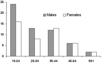
Of the patients, 57 were male (55.9%), and the ages ranged from 15 to 69 years, with a mean of 31.12 years, and a median of 28 years. No significant difference was detected regarding the age of death according to the gender (Fig. 1). Relative to the total number of necropsies in the period, numbering 6076, cases of adolescents or adults with congenital cardiac disease accounted for 1.7%.
Age distribution according to gender. Range values are in years.

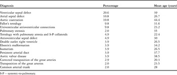
The types of cardiovascular defects and relative percentages are showed in the Table 1.
Main diagnoses, their relative percentages and mean ages of death for each group.

S-P – systemic-to-pulmonary.
Of the group, 66 individuals (64.7%) had been submitted to at least one or more cardiac surgeries during their lifespan, in order to correct or palliate their defects. Among the non-operated patients, the great majority comprised outpatients followed at the same institution, usually with contraindications for surgery, but in 3 cases the congenital defect was only discovered at necropsy. In 2 of these cases, with aortic coarctation, a misdiagnosis had been done elsewhere, 1 considered to have systemic hypertension and the other hypertrophic cardiomyopathy. In the third case, the patient had been followed with the diagnosis of idiopathic pulmonary hypertension, but the necropsy showed the presence of a large inlet ventricular septal defect occluded by the leaflets of the tricuspid valve.
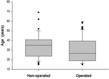
The mean age of death was significantly higher in the group of patients who had never undergone surgery, with median ages of 35 versus 25.5 years (p = 0.03, Fig. 2)
Box plots of the ages in the groups of operated and non-operated patients. The upper and lower ends of each box indicate the 75th and 25th percentiles, respectively, of the data, and the central line indicates the median. Outliers appear as individual points outside the box.

Dividing the patients in quarters according to the period of death, with the first period extending from 1989 to 1993, the second from 1994 to 1998, the third from 1999 to 2003, and the final quarter from 2004 until 2007, a significant difference was detected regarding the mean age of death, with lower values detected in the last quarter compared to the second and third ones (Kruskal-Wallis, p = 0.004). Using the same time division, no difference was found regarding the state of having or not having been submitted to surgery, according to the period when death occurred.
Among the patients who had undergone surgery, 34 of the 66 (51.5%) died within the first 30 days post-operatively. In 19 of them, the surgical procedure was the first one during their lives, 16 (84%) presenting with so-called complex defects as judged by the classification of the British Cardiac Society Working Party. In the group of patients not undergoing surgery, complex lesions were present in 24 of 36 (67%).
Main causes of death
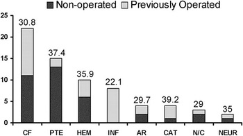
The most prevalent cause was death related to surgery, with 34 of 102 patients (33.3%) dying within the first 30 days after being submitted to corrective or palliative surgery. Of these, 14 of the 34 (42%) were reoperations. The distribution of the causes of death in the 68 patients either not undergoing surgery, or else not having recent surgery, is shown in Figure 3. Although no significant difference in age was detected according to the terminal cause of death, the lowest ages were found in the groups of individuals who died of infectious causes, these patients dying at a mean age of 22.1 years. Cardiac failure was the main terminal cause in this group, being found in one-third, and including chronic failure and cardiogenic shock.
Numbers of patients according to the cause of death among non-recently operated and never-operated patients, and mean age in each group (values above each column, in years). CF – cardiac failure, including cardiogenic shock; PTE – pulmonary thromboembolism; HEM – haemorrhage/haemorrhagic complications; INF – infection/septicaemia, including infective endocarditis; AR – no morphologic cause, with clinical data accounting for arrhythmias; CAT – death occurring in the immediate post-procedural (cardiac catheterization) period; NEUR – neurologic causes; N/C – morphologically undefined (non-conclusive).

Pulmonary thromboembolism was considered the cause of death in 15 of these 68 patients (22.1%), including two with recent occlusion of systemic-pulmonary collateral arteries. The mean age of death of theses patients was 37.4 years. There was a statistically significant association between pulmonary thromboembolism and histopathological signs of pulmonary hypertension (p = 0.011). In other 8 patients, pulmonary thromboembolism and/or pulmonary infarction appeared as a complication, although not extensive enough to be considered the cause of death.
Infection was detected as the main cause of death in 8 of 68 patients (11.8%), with 5 showing acute infective endocarditis, one septicaemia following acute appendicitis, one purulent pleuritis 37 days following a Fontan procedure, and the remaining one showing bronchopneumonia.
Acute infective endocarditis was the indication for surgery in 5 of the patients who died in the short post-operative period, and was detected as an incidental necropsy finding in a non-operated patient with hepatic cirrhosis who died due to digestive bleeding. Overall, acute infective endocarditis was present in 11 of 102 patients (10.8%). The affected sites of endocarditis were native valves in 6 cases, and prosthetic material in the remaining 5, involving a Blalock-Taussig shunt in 2, prosthetic valves in 2, and the patch used to close a ventricular septal defect in the other. In all cases, the infectious agents described inside the lesions were bacteria, specifically coccuses. The mean age of death for all the cases of acute endocarditis was 27.8 years.
There was statistically significant association between the occurrence of endocarditis and defects causing low pulmonary blood flow (p = 0.043). No difference was observed in the prevalence of endocarditis related to gender (p = 0.108). Gross features compatible with sequels of previous and treated endocarditis were detected in 6 of 102 patients (5.9%), and consisted of valvar perforations with no bacteria in the lesions.
Haemorrhagic episodes consequent to arterial rupture appeared as the main cause of death in 10 of the 68 patients (14.7%). In 5 of them, rupture or dissection of a great artery was present, with 3 dissections of the aorta, one of a common trunk,Reference Gutierrez, Binotto, Aiello and Mansur6 and the other of the pulmonary trunk. The dissections of the common and pulmonary trunks occurred in cases with severe histological signs of pulmonary hypertension, the patients being aged 35 and 39 years respectively, while 2 of the 3 aortic dissections occurred in individuals with aortic coarctation, aged 58 and 69 years, with 1 having undergone a previously operation. Interestingly, spontaneous aortic rupture occurred in a 17 year-old boy with non-operated transposition.
Death occurred shortly after diagnostic or interventional catheterization in 4 cases (5.9%). In 2 of them, cardiac tamponade was found, and in the remaining 2 no morphological cause was detected.
In 2 patients, a cerebral infarction was extensive enough to be assigned as the cause of death. In one case, it was a late complication of postoperative correction of ventricular septal defect and anomalous muscular band of the right ventricle in a 51 year-old man, while the other patient had an uncorrected ventricular septal defect and Eisenmenger syndrome.
No morphological causes were detected in 4 additional patients. In 2, there were non-documented evidences of arrhythmia in the clinical records.
Associated conditions of clinical relevance
Histopathological signs of pulmonary occlusive vasculopathy, with medial hypertrophy, intimal proliferation, or plexiform lesions, were present in 41 of the 102 cases (40%), although in only 10 did the available clinical information pointed to the diagnosis of Eisenmenger’s syndrome. The proportion of patients showing histological signs of occlusive vasculopathy was higher in the non-operated group (p < 0.001).
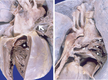
Puerperium was an associated condition in 3 of the 45 female patients (6.7%), with these having a mean age of 27 years, and all presenting clinical and histological signs of pulmonary hypertension and Eisenmenger’s syndrome. An additional patient with chronic cyanotic heart disease died at the 30th week of gestation because of massive thrombosis of systemic to pulmonary collateral arteries (Fig. 4). The fetus showed a large ventricular septal defect.
The heart from a female patient who died during pregnancy due to thrombosis of systemic-to-pulmonary collateral vessels. The left panel shows atresia of the pulmonary trunk (arrow) and branches and a large ventricular septal defect (*). In the right panel, a large branching collateral vessel from the descending aorta, is completely blocked with thrombus (arrow heads).

Morphological features compatible with sequels of rheumatic heart disease were found in 4 patients having a mean age of 39 years, 2 with septal defects, one with sub-pulmonary stenosis, and the other having aortic coarctation. In 2, there had been previous valvar replacement.
Genetic disorders were clinically diagnosed in 3 patients, one each with Noonan’s, Marfan’s, and Turner’s syndromes.
Discussion
Our study is a retrospective analysis of the necropsy reports from the files of the Pathology Laboratory from a tertiary centre devoted to the treatment of cardiovascular disease, and which is located in a developing country. Several aspects must be taken into consideration when reviewing this data. First of all, some words about the rate of necropsies relative to the period analysed. The continuous decrease in the rate of necropsies in University Hospitals all over the World is a well recognized phenomenon.Reference Aiello, Debich-Spicer and Anderson7 As a consequence, the sample may not be representative all over the period or, in other words, in periods when the necropsy rate is low, only selected cases go to the post-mortem evaluation. This aspect may have contributed to the difference detected regarding age of death according to the period analysed, with younger patients in the last quarter of the study. For the same reason, the highest number of peri-operative deaths in the present study can be interpreted as a possible bias, since there is a strong interest in determining the cause of death and complications in the recent post-operative period. There were different clinical, surgical and post-operative findings in this group, but all of these conditions could be not taken apart of the time-relation to surgery.
The occurrence of cases of congenital defects in adults not operated or specifically treated reflects the existence of underserved populations with little access to health care in a developing country. Data from the Ministry of Health of Brazil points to a mean of 5746 hospital admissions a year to treat patients with congenital heart defects at the age of 15 years or more, during the period of 1998 through 2007.8 Considering the estimate of 0.7% for congenital heart defects in live births, we can expect more than new 20,000 cases occurring in Brazil every year. There is no local data, however, about the proportion of children with congenital heart defects who reach adulthood.
Another point to discuss is the difference in the age of death among patients operated or non-operated, with lower mean and median values in the operated group. It is tempting to conclude that non-operated cases have clinically milder defects, while the corrected or palliated ones would have died much before, even in infancy or childhood, if surgery was not available. Yet, the proportion of complex diagnoses according to the British Cardiac Society Working Party did not differ significantly between groups.
The most prevalent cause of death among the not recently operated individuals was cardiac failure, found in one-third, including chronic failure and cardiogenic shock. Half of the patients had been previously operated. Progressive heart failure was reported as the second cause of death, accounting for one-fifth, in a large clinical series of adult patients with congenital heart disease.Reference Oechslin, Harrison, Connelly, Webb and Siu2 Intrinsic or acquired gross and histological myocardial alterations have progressively been recognized as an important cause of cardiac failure in patients with congenital heart defects.Reference Bolger, Coats and Gatzoulis9 Several papers have demonstrated that the distorted cardiac anatomy leads to myocardial adaptation appearing in the form of fibrosis, hypertrophy and/or reduced capillary numbers.Reference Chowdhury, Sathia, Ray, Singh, Pradeep and Venugopal10, Reference Babu-Narayan, Kilner and Li11 If conditions such as pressure or volume overload persist for long periods of time before corrective surgery, or in cases where only palliation is possible, or else in the presence of residual lesions after surgery, myocardial disease or remodelling supervenes, impairing cardiac function over time.Reference Aiello and Binotto12 Although some specific congenital defects such as those presenting the right ventricle as the systemic pumping chamber are the most vulnerable group for developing cardiac failure, some other supposedly minor defects, if left without correction, can lead to poor myocardial function.
Even though pulmonary thromboembolism was significantly associated to histological signs of pulmonary hypertension, it was also detected in cyanotic patients with chronically reduced pulmonary flow. “In situ” thrombosis is a common finding in peripheral pulmonary arteries from patients with pulmonary hypertension of any aetiology. Moreover, episodes of pulmonary thromboembolism have been reported in one-eighth of a series of patients with Eisenmenger’s syndrome.Reference Daliento, Somerville and Presbitero13 In a clinical study of 20 such patients from our institution, age was found to be the main variable related to the development of pulmonary arterial thrombosis, with an odds ratio of 1.204 per year.Reference Caramuru, Maeda, Bydlowski and Lopes14 The present study shows that this complication may also be fatal, representing the second cause of death among non-operated and not recently operated patients.
The problem of infective endocarditis in the population of adolescents and adults with congenital cardiovascular defects has been extensively discussed in the literature.Reference Li and Somerville15 Concerns have emerged regarding the increasing use of mechanical and bioprosthetic materials in corrective or palliative surgeries. These materials would potentially increase the risk for development of endocarditis. In only half of our patients with acute endocarditis were the vegetations found on prosthetic material. The finding of a significantly greater prevalence of endocarditis among chronic hypoxaemic patients is in accordance with other clinical series studying adults with congenitally malformed hearts.Reference Knirsch, Haas, Uhlemann, Dietz and Lange16
The occurrence of acute arterial dissection has been described in patients with aortic coarctation, and also in Eisenmenger’s syndrome.Reference Westaby, Evans and Ormerod17 The pathogenetic mechanism of arterial dissections includes a hypertensive state and structural abnormalities of the arterial wall, with accumulation of mucopolysaccharides and elastic fragmentation in medial arterial layer. Acute dissection of a common arterial trunk, present in one patient from our series, was the first case described in the literature.Reference Gutierrez, Binotto, Aiello and Mansur6
Among the associated conditions, we call attention to the presence of a puerperal state in just over one-twentieth of our female patients. Despite the specific clinical counselling regarding contraception in the group of patients with Eisenmenger’s syndrome. some of them seek for medical care already during advanced pregnancy. As pointed out previously by Somerville,Reference Somerville18 “being female carries special risks when there is serious pulmonary vascular disease in comparison to males with comparable disease and defects”. In fact, pregnancy and delivery increases the risk of death in the population of adolescents and adults with congenital heart disease. Another occurrence in our series was maternal and fetal death in a patient with cyanotic congenital heart disease. In this regard, Presbitero et al,Reference Presbitero, Somerville, Stone, Aruta, Spiegelhalter and Rabajoli19 studying the outcome of pregnancy in a large group of cyanotic patients, finding that less than half produced live births.
Another finding that deserves discussion is the association of rheumatic heart disease and congenital cardiovascular defects. Although this complication is not common in developed countries, it has been described and is a concern in children and adolescents from underserved areas, causing impairment in the evolution of the original defects because of myocardial and/or valvar involvement.Reference Mansur, Grinberg and Lopes20
Supporting previous statement of cardiologists and surgeons, the management of adolescents and adults with congenital heart defects requires a multidisciplinary team. If not specifically treating these patients, the pathologist is part of the team, and should be trained to recognize the possible lesions and complications secondary or associated to operated and non-operated defects. Additionally, the contribution from the Pathology laboratory has the potential to be even broader, if it includes the histological study of the hearts. The ultimate goal of the partnership between cardiologists, surgeons and pathologists in this scenario will be to improve the medical care of adult patients with congenitally malformed hearts.