I. INTRODUCTION
Oxides that crystallize in rutile structures exhibit a broad array of functional properties; they include important transparent semiconductors, catalysts, half metals, and correlated electron systems that exhibit metal–insulator transitions. Epitaxy is the oft-chosen avenue to achieve high-quality thin-film materials. For isostructural epitaxial growth, the only commercially available oxide substrate is rutile TiO2, limiting the number of possible heterostructure combinations. Nonisostructural combinations offer vastly more options, but the surface symmetry and lattice matching at the heterointerface are more complicated. It has been known that (0001) Al2O3 substrates can be exploited for epitaxial growth of (100) TiO2 films.Reference Hotsenpiller, Wilson, Roshko, Rothman and Rohrer1–Reference Gao, Thevuthasan, McCready and Engelhard3 Because the (0001) Al2O3 surface has 3-fold rotational axes and the (100) TiO2 plane contains only 21 screw axes, though the films are “epitaxial” in that the substrates and films have preferred in-plane orientational alignment, they are certainly not single crystalline. In fact, they should contain large-angle (120°) tilt boundaries that connect adjacent rotational domains; the epitaxial variants are related to one another by the surface symmetry elements of the substrates that are nonexistent in the thin-film materials themselves.Reference Flynn and Eades4,Reference Grundmann5 More recently, (0001) Al2O3 substrates have been used for the epitaxial growth of (010) VO2,Reference Wu, Yamamoto, Miyashita, Zhang, Narumi and Naramoto6,Reference Zhou, Chisholm, Yang, Pennycook and Narayan7 which is a monoclinically distorted rutile-type compound and is of interest because of its metal–insulator transition near room temperature. Furthermore, it has been shown that (010) VO2 films can also be grown on (111) perovskite, spinel, and rocksalt substrates; the key is identifying substrates with eutactic planes of (mainly) anions parallel to the surface to match the (010) VO2 plane.Reference Wong, Zhou and Ramanathan8 On eutactic planes, ions occupy the pseudo-close-packed positions but are not necessarily touching one another. The oxygen anions of spinel and rocksalt occupy face-centered cubic positions, which are occupied by the oxygen anions plus the A-site cations in the ABO3 perovskite; the oxygen sublattice of Al2O3 approximately resembles a hexagonal close-packed structure. The perovskite structure is the only one presented here in which the eutactic planes are composed of both anions and cations. We will refer to the corresponding pseudo-close-packed axes (directions) of these substrates as “eutactic axes (directions).”
There are many binary dioxides—including VO2, WO2, and NbO2—that crystallize into distorted versions of the higher-symmetry parent rutile structure. In this invited feature paper, we report the first epitaxial growth of WO2 and NbO2 thin films. We show that the heteroepitaxial relationships of rutile-type films with (0001) corundum Al2O3, (111) perovskite SrTiO3, (111) spinel MgAl2O4, and (111) rocksalt MgO substrates are robust by demonstrating the growth of textured (010) WO2 and (110) NbO2 films with epitaxial rotational domains. The Miller indices of the planes used for WO2 and NbO2 correspond to their room-temperature distorted phases, which have larger unit cells than higher-symmetry rutile structures because of their distortions; in fact, (010) VO2, (010) WO2, and (110) NbO2 all correspond to the (100) pseudorutile orientation. The lattice transformation will be presented in Sec. III. We selected this set of rutile-type compounds for three reasons. First, all have cation dimerization along the rutile c-axis, which is the main reason for their structural distortions to reduced symmetry phases at room temperature. We will show that the epitaxy and orientational alignment are not appreciably affected by these distortions. We find that the axes along which the dimers form in WO2 and NbO2 are aligned parallel with respect to the eutactic axes of the aforementioned substrate surfaces, as in the case for VO2 films.Reference Wong, Zhou and Ramanathan8 Second, all of these compounds contain cations with lower oxidation states than are most favorable under standard conditions, and stabilizing the correct stoichiometric phases is therefore extremely sensitive to deposition conditions, including the choice of substrate. It has been shown that for VO2 films, improved composition control can be achieved via heteroepitaxy.Reference Yang and Ramanathan9 Finally, to the best of the authors' knowledge, this is the first report of epitaxial growth of WO2 and NbO2.
Rutile TiO2 films of different orientations can be grown on various cuts of Al2O3 substrates: (101)TiO2||(10 $\bar 1$2)Al2O3 (r-plane) and (001)TiO2||(10
 $\bar 1$0)Al2O3 (m-plane).Reference Hotsenpiller, Wilson, Roshko, Rothman and Rohrer1–Reference Gao, Thevuthasan, McCready and Engelhard3,Reference Engel-Herbert, Jalan, Cagnon and Stemmer10 Similar results have been observed for rutile SnO2.Reference Dominguez, Fu and Pan11,Reference Pan, Fu and Dominguez12 The control of growth orientations of VO2 films on various cuts of Al2O3 appears to also hold.Reference Zhao, Lee, Zhu, Nazari, Chen, Wang, Bernussi, Holtz and Fan13,Reference Yang, Aggarwal, Gupta, Zhou, Narayan and Narayan14 We show that these relationships are transferable to NbO2 thin films. Because of the lower surface symmetry of (10
 $\bar 1$2) and (10
 $\bar 1$0) Al2O3, symmetry does not require the formation of rotational variants; therefore in principle, nearly single-crystalline films can be achieved. Our (111) NbO2 film on (10
 $\bar 1$2) Al2O3 has considerably better in-plane despite worse out-of-plane orientational alignment than the (110) film on (0001) Al2O3. We propose that the in-plane alignment and overall crystallinity of the (110) NbO2 film are constrained by the formation of nanograin rotational variants that are induced by symmetry mismatch between the film and substrate surfaces. On the other hand, in the case of (111) NbO2 on (10
 $\bar 1$2) Al2O3, the overall quality of crystallinity is primarily limited by film/substrate lattice mismatch. The general results presented are of relevance to thin-film synthesis of other rutile-type compounds, with and without distortions, for emerging solid-state devices.
II. METHODS
WO2 films were grown by DC magnetron reactive sputtering from a ¼ inch thick W metal target at 80 W, 10 mTorr, 42.5–45.0 sccm Ar, 5–7.5 sccm O2, and 650 °C. NbO2 films were grown by DC reactive sputtering from a ¼ inch thick Nb metal target at 200 W, 10 mTorr, 42.25–42.5 sccm Ar, 7.5–7.75 sccm O2, and 650 °C. The substrates used in this study include (111) SrTiO3, (111) MgAl2O4, (111) MgO, (0001) Al2O3, (10 $\bar 1$2) Al2O3, and (10
 $\bar 1$0) Al2O3. X-ray diffraction (XRD)
 $\phi$ scans were performed in a four-circle diffractometer with a Cu source (Kα1 = 1.541 Å); in scans of asymmetric substrate and film peaks, the asymmetry off the plane normal was introduced in χ, and we kept ω = 2θ/2. 2θ–θ scans were taken in a diffractometer with a Ge(220)×2 monochromator. X-ray photoelectron spectroscopy (XPS) measurements were taken with an Al Kα x-ray source, a 400-μm spot size, and a neutralizing flood gun to prevent sample charging. In a reference Au sample, the 4f 7/2 peak energy was measured at 84.1 eV. The energy values of the raw XPS data were shifted by 0.1 eV to have a Au4f 7/2 reference energy at 84.0 eV.
III. RESULTS AND DISCUSSION
A. WO2 films
In our previous work, we showed that monoclinically distorted (010)-oriented VO2 thin films can be grown on a variety of oxide substrates with (mainly) anionic eutactic planes parallel to the surface.Reference Wong, Zhou and Ramanathan8 However, at typical deposition temperatures, the VO2 films should possess an undistorted rutile structure, and they subsequently undergo a monoclinic distortion upon cooling to below 67 °C.Reference Kawakubo and Nakagawa15–Reference Andersson17 On the other hand, WO2 possesses a monoclinically distorted rutile structure at all reported temperature ranges of the stoichiometric phase; therefore, we can directly investigate the viability of growing distorted rutile-structure thin films on oxide substrates of cubic (perovskite, spinel, and rocksalt) and rhombohedral (corundum) symmetry.
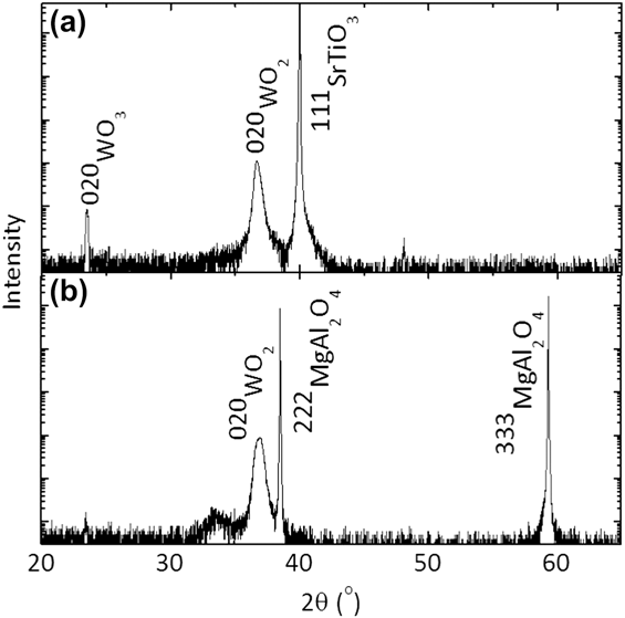
WO2 has a monoclinic structure and W–W dimer pattern similar to those of VO2.Reference Palmer and Dickens18 Its structure is shown in Fig. 1. One can envisage the lattice vector transformation from a monoclinic (M) to a pseudorutile (R) unit cell: aM ≈ 2cR, bM ≈ aR, and cM ≈ bR − cR, where aM = 5.56 Å, bM = 4.90 Å, cM = 5.56 Å, and β = 120.47°.Reference Palmer and Dickens18 The lattice transformation is analogous to that of VO2. We do indeed see that (010)-oriented WO2 films can be grown on (111) SrTiO3 and (111) MgAl2O4 [Fig. 2], and similarly for films on (0001) Al2O3 and (111) MgO (data not shown). In many of our films, a second phase of WO3 is seen, as in the film on the SrTiO3 substrate at 23.5° [Fig. 2(a)]; because both WO2 and WO3 are line compounds that do not allow for much nonstoichiometric defects, tungsten oxides are prone to coexistence of stoichiometric phases.Reference Wriedt19 With XRD, we checked that the WO3 phase in our WOx films is randomly oriented. We will be concerned with only the structure and crystal orientation of the WO2 phase, and the existence of a WO3 second phase does not affect our analysis.
Structure of monoclinic (M) WO2 (space group #14 P21/c). The pseudorutile (R) unit cell is drawn in with dashed lines. The Wyckoff positions and lattice parameters used were reported by Palmer and Dickens.Reference Palmer and Dickens18 A cation dimer pair is circled.

XRD 2θ–θ scans of (010) WO2 films on (a) (111) SrTiO3 and (b) (111) MgAl2O4. The films on SrTiO3 and MgAl2O4 contain peaks at 23.5° and 33.6°, respectively, which match reflections of WO3, a second phase.
For the case of VO2 films on (0001) Al2O3, there has been some uncertainty of whether the films are (010)- or (001)-oriented, or whether they contain a mixture of both orientations.Reference Zhou, Chisholm, Yang, Pennycook and Narayan7 In the case of WO2, the monoclinic distortion from the pseudorutile structure is larger; therefore, the difference in Bragg angles of the 020 and 002 reflections in WO2 are greater than in VO2. The WO2 reflections from the films on the four aforementioned substrates have 2θ Bragg angles ranging from 36.64 to 36.90°, closer to the 020 (36.68°) than to the 002 (37.48°) peak of WO2.Reference Palmer and Dickens18 Our discussion continues assuming the presence of (010)-oriented WO2 grains in the WOx films. Even the existence of (001)-oriented grains would not change the fundamental symmetry arguments presented here but would alter the Miller indices of some of the planes and directions.
Because the (010) plane of WO2 contains only 21 screw axes, whereas (111) SrTiO3, (111) MgAl2O4, (111) MgO, and (0001) Al2O3 contain 3-fold rotational axes, three thin-film rotational structural variants are expected. In addition, the SrTiO3, MgAl2O4, and MgO surfaces also contain mirror planes, which can lead to twin variants as well; in principle, in these cases, a total of six epitaxial variants of WO2 grains related by the surface symmetry elements of the underlying substrates are anticipated.Reference Wong, Zhou and Ramanathan8 To show the in-plane crystalline alignment,  $\phi$ scans of asymmetric peaks were taken. Figure 3(a) shows the 004-type reflections of the MgAl2O4 substrate and the 011-type reflections of the WO2 film. For a cubic crystal in reciprocal space, g
 ${}_{004}$ = 4(g
 ${}_{111}$ + g
 ${}_{\bar 1\bar 12}$)/3, where 4g
 ${}_{111}$/3 is the out-of-plane component and 4g
 ${}_{\bar 1\bar 12}$/3 is the in-plane component. In subsequent references to other asymmetric reflections, they will likewise be decomposed into their out-of-plane and in-plane reciprocal lattice components. From the close alignment of the 004-type MgAl2O4 and 011-type WO2 reflections in Fig. 3(a), we can deduce the film/substrate crystallographic relationship to within a degree of in-plane alignment:
 $$(010)[100]{\rm{W}}{{\rm{O}}_{\rm{2}}}||(111)\left\langle {1\bar 10} \right\rangle {\rm{MgA}}{{\rm{l}}_{\rm{2}}}{{\rm{O}}_{\rm{4}}}\quad .$$
XRD  $\phi$ scans of asymmetric substrate and film peaks. (a)
 $\phi$ scan of the 011-type reflections of WO2 (2θ = 25.85° and χ = 45.6°) with the MgAl2O4 substrate 004-type reflections (2θ = 44.82° and χ = 54.7°) overlaid. (b)
 $\phi$ scan of the 011-type reflections of WO2 (2θ = 25.90° and χ = 45.6°) with the MgO substrate 002-type reflections (2θ = 42.93° and χ = 54.7°) overlaid.
Since there are three <1 $\bar 1$0> axes on the (111) MgAl2O4 plane, we expect three rotational variants of WO2 grains, consistent with the
 $\phi$ scans. However, because a M ≈ c M and β is close to 120° in WO2, we cannot unambiguously claim that rotational variants are absolutely necessary to explain the
 $\phi$ scan of Fig. 3(a), as it should also contain intensity from 110-type and
 $\bar 1$11-type peaks with nearly identical Bragg angles and orientations. Note that this is not a problem for (010) VO2 films,Reference Wong, Zhou and Ramanathan8 nor is it for NbO2 films presented in Sec. III. B. Regardless, our measurements definitively demonstrate well-defined in-plane film/substrate orientational alignment; symmetry considerations lead us to believe that rotational variants do exist. Moreover, we cannot distinguish whether there are three or six variants; three more twin variants can be induced by the mirror planes of (111) MgAl2O4. Figure 3(b) shows
 $\phi$ scans of the 011-type reflections of the WO2 film overlaid with 002-type reflections of the (111) MgO. Similar scans for the films on (111) SrTiO3 and (0001) Al2O3 (data not shown) allow us to determine the following film/substrate crystal orientations to within ±1° in in-plane alignment:
 $$(010)[100]{\rm{W}}{{\rm{O}}_2}||(111)\left\langle {1\bar 10} \right\rangle {\rm{SrTi}}{{\rm{O}}_3}\quad ,$$
 $$(010)[100]{\rm{W}}{{\rm{O}}_2}||(111)\left\langle {1\bar 10} \right\rangle {\rm{MgO}}\quad {\rm{,}}$$
 $$(010)[100]{\rm{W}}{{\rm{O}}_2}||(0001)\left\langle {1\bar 100} \right\rangle {\rm{A}}{{\rm{l}}_{\rm{2}}}{{\rm{O}}_{\rm{3}}}\quad .$$
The axis of the W–W dimers, corresponding to the pseudorutile c-axis, is aligned parallel to the eutactic axes of the substrates.
WO2 is a metallic 5d 2 system, whereas the more stable tungsten oxide phase WO3 is an insulating 5d 0 system. This difference in electronic filling and oxidation state can be observed by XPS. Figure 4(a) shows the W4f core level spectrum of WOx films that contain epitaxial WO2 grains on (0001) Al2O3 compared with a WO3 film as a reference sample. The two WOx films were grown at 5 (45) sccm and 7.5 (42.5) sccm of flowing O2 (Ar). We also include spectra of a W metal film for comparison; we show the spectrum of a W film without surface treatment, which includes signatures of surface oxidation, and also that of the film after in situ sputter cleaning of the sample surface. From the spectra of the W film before and after surface cleaning, we determine that the W4f 7/2 and 4f 5/2 binding energies of the elemental metal are 31.5 and 33.6 eV, respectively, and the peaks at 36.1 and 38.2 eV are from W cations in the surface tungsten oxide, consistent with previously reported values.Reference Colton and Rabalais20,Reference Katoh and Takeda21 In the WO3 sample, the W6+ 4f 7/2 and 4f 5/2 peaks are at 35.55 and 37.7 eV, respectively. The spectral lineshapes of the two WOx films on (0001) Al2O3 are noticeably unique from those of W metal and WO3 and are similar to other XPS studies of WO2.Reference Khyzhun22 However, a combination of the small spin–orbit coupling of the W4f states, possible presence of satellite peaks, and the contributions of both W4+ and W6+ cations make direct identification of W4+ peak positions challenging. Likewise, previous work on WO2 also showed difficulties in discriminating the exact positions of the W4f core levels.Reference Colton and Rabalais20–Reference Sarma and Rao23
X-ray photoelectron spectra of (a) W4f core and (b) valence band states of a WO3 reference, two WOx films on (0001) Al2O3 that contain (010) WO2 grains grown at different O2 flow rates, and a W metal film before and after surface cleaning. In the core spectrum of the W metal film before surface cleaning (a), solid (open) symbols correspond to the W4f 7/2 and 4f 5/2 positions of the elemental metal (surface oxide) peaks. The core spectra were normalized by their respective pre- and post-edge intensities. The valence band spectra were normalized by their respective peak emission intensities in the given energy range.
Examination of the valence band spectrum is another means of illustrating the effects of electron filling. Because WO3 is a 5d 0 insulating oxide, its valence band is composed of O2p-like states. In Fig. 4(b), we see the electron emission increases sharply above a binding energy of ∼2.5 eV, representing the valence band edge of WO3, and emission is negligible at lower energies. Both WOx films also have the spectral features with a large rise in emission above 2.5 eV and peak at about 7 eV (correspondingly 6.5 eV for WO3). This broad peak corresponding to O2p-like bonding states also exist in the W metal before surface treatment; the disappearance of emission in this region after surface cleaning verifies that these states are of the O2p-like bonding orbitals. For the postcleaned W metal film, emission comes from broad bands formed by metallic bonding of W (6s and 5d levels). One the other hand, for the WOx film grown at 7.5 sccm O2, there is emission that extends to zero binding energy, arising from the occupied states of bands formed by W5d-like antibonding levels of WO2, a 5d 2 system. In the film grown at 5 sccm O2, we can see distinctly the two-peak spectral lineshape below 2.5 eV (peaks split by ∼1.3 eV); this signature is attributed to the splitting of W5d-like bands as reported by Jones et al.Reference Jones, Egdell, Brown and Wondre24 and Gulino et al.Reference Gulino, Parker, Jones and Egdell25 Although both WOx films contain epitaxial WO2 grains, the phase is more dominant in the sample grown at the lower O2 flow rate.
B. NbO2 films
NbO2 undergoes a structural transition from rutile to distorted tetragonal structure with Nb–Nb dimers at 850 °C.Reference Sakata, Sakata and Nishida26,Reference Bolzan, Fong, Kennedy and Howard27 The pattern of cation dimerization is different from those of VO2 and WO2, and the unit cell has different symmetry. The lattice vector transformations between its low-temperature tetragonal (T) and its high-temperature rutile (R) unit cells are given by aT ≈ 2(aR ‒ bR), bT ≈ 2(aR + bR), and cT ≈ 2cR, where aT = 13.70 Å and cT = 5.98 Å [Fig. 5].Reference Bolzan, Fong, Kennedy and Howard27,Reference Gannon and Tilley28
Structure of tetragonal (T) NbO2 (space group #88 I41/a). The rutile (R) unit cell is drawn in with dashed lines. The Wyckoff positions and lattice parameters used were reported by Bolzan et al.Reference Bolzan, Fong, Kennedy and Howard27 A cation dimer pair is circled.
In a 2θ–θ scan of a NbO2 film on (0001) Al2O3, we see that the predominant film peak is 440 at 37.09° [Fig. 6(a)], which translates to the 200 (or 020) reflection in the rutile unit cell. There is a small secondary peak at 35.05° that is close to the 222 reflection of NbO2 (35.22°),Reference Bolzan, Fong, Kennedy and Howard27 and it has no preferred orientation. We will focus on the (110)-oriented NbO2 epitaxial variants, which constitute most of the film. Figure 6(b) shows  $\phi$ scans of the asymmetric 400-type NbO2 film reflections (g
 ${}_{400}$ = g
 ${}_{220}$ + g
 ${}_{2\bar 20}$) overlaid with 11
 $\bar 2$3-type Al2O3 substrate reflections, and their match tells us that the in-plane [001] axis of NbO2 is aligned with the three symmetry-equivalent <1
 $\bar 1$00> axes on the (0001) Al2O3 plane to within ±0.2° accuracy. Furthermore, we have similarly verified the following orientational relationships with the (111) MgAl2O4 and (111) MgO substrates (data not shown):
 $$(110)[001]{\rm{Nb}}{{\rm{O}}_2}||(0001)\left\langle {1\bar 100} \right\rangle {\rm{A}}{{\rm{l}}_{\rm{2}}}{{\rm{O}}_{\rm{3}}}\quad ,$$
 $$(110)[001]{\rm{Nb}}{{\rm{O}}_2}||(111)\left\langle {1\bar 10} \right\rangle {\rm{MgA}}{{\rm{l}}_{\rm{2}}}{{\rm{O}}_{\rm{4}}}\quad ,$$
 $$(110)[001]{\rm{Nb}}{{\rm{O}}_2}||(111)\left\langle {1\bar 10} \right\rangle {\rm{MgO}}\quad .$$
XRD (a) 2θ–θ scan of the NbO2 film on c-plane (0001) Al2O3. (b)  $\phi$ scan of the 400-type reflections of NbO2 (2θ = 26.00° and χ = 45.0°) with the Al2O3 substrate 11
 $\bar 2$3-type reflections (2θ = 43.35° and χ = 61.2°) overlaid.
By investigating the growth of WO2 and NbO2 thin films, we have determined that the structural relationships of epitaxial variants of different distorted rutile-structure compounds on various substrates with 3-fold rotational surface planes are analogous to those of TiO2Reference Hotsenpiller, Wilson, Roshko, Rothman and Rohrer1 and VO2Reference Wu, Yamamoto, Miyashita, Zhang, Narumi and Naramoto6 films grown on (0001) Al2O3 when considering the pseudorutile unit cells. In particular, we have found that the axes along which the cations dimerize in WO2 and NbO2, i.e., pseudorutile c-axis, always lie along the eutactic axes of the substrate planes, as is for (010) VO2 films.Reference Wong, Zhou and Ramanathan8
Thus far, we have shown that WO2 and NbO2, two distorted rutile compounds with different cation dimerization patterns, can be grown on different oxide substrates with eutactic planes parallel to the surface plane; because of the surface symmetry of (111) perovskite, spinel, and rocksalt and (0001) corundum, the WO2 and NbO2 form structural variants related by the 3-fold rotational and mirror symmetry elements of the underlying substrates. The growth of epitaxial rutile TiO2 on other cuts of Al2O3 substrates has also been observed.Reference Hotsenpiller, Wilson, Roshko, Rothman and Rohrer1–Reference Gao, Thevuthasan, McCready and Engelhard3,Reference Engel-Herbert, Jalan, Cagnon and Stemmer10 Further epitaxial relationships exist between rutile and corundum because both their anions occupy approximately hexagonal close-packed positions.Reference Vanlanduyt, Gevers and Amelinckx29,Reference Burdett30 We now show that epitaxial relationships exist in NbO2 thin films on (10 $\bar 1$2) and (10
 $\bar 1$0) Al2O3.
Figure 7(a) shows a 2θ–θ scan of a (111)-orientated NbO2 film (222 reflection at 35.05°), which corresponds to the (101) orientation in the rutile unit cell, grown on r-plane (10 $\bar 1$2) Al2O3. Figure 7(b) shows
 $\phi$ scans of the 402-type reflections of NbO2 and 11
 $\bar 2$3-type reflections of Al2O3, and they overlap. For NbO2 in reciprocal space, g
 ${}_{402}$ = g
 ${}_{222}$ + g
 ${}_{2\bar 20}$, and for Al2O3, g
 ${}_{11\bar 23}$ = 1/2(3g
 ${}_{10\bar 12}$ + g
 ${}_{\bar 12\bar 10}$), which means the [1
 $\bar 1$0] axis of NbO2, corresponding to [100] in the rutile unit cell, is aligned along the one in-plane [
 $\bar 1$2
 $\bar 1$0] axis of the (10
 $\bar 1$2) Al2O3 plane: (111)[1
 $\bar 1$0]NbO2||(10
 $\bar 1$2)[
 $\bar 1$2
 $\bar 1$0]Al2O3. With this in-plane orientational relationship, let us examine more closely the lattice matching that occurs at the heterointerface. Figure 8 shows the (111) plane of NbO2 and the (10
 $\bar 1$2) plane of Al2O3, with oxygen drawn as the top surface, and a cation plane is also shown below. The surface unit cells are shown in Fig. 8 by the dashed lines. Because of the lattice distortions in NbO2, the surface unit cell is rather large. An ideal heterointerface would have lattice-matched in-plane axes that allow for the continuation of the oxygen sublattice across the heterointerface, and we define lattice mismatch by focusing on the oxygen sublattices of both surfaces. It can be seen that though along one axis the lattice mismatch is a small +1.75% [(19.38 − 4 × 4.76)/19.38], it is extremely large at +9.9% [(3 × 22.77 − 4 × 15.39)/(3 × 22.77)] along the orthogonal axes [Fig. 8]. In principle, the (10
 $\bar 1$2) A12O3 surface contains glide planes orthogonal to the [
 $\bar 1$2
 $\bar 1$0] axis (If one considers only the oxygen anions of the (10
 $\bar 1$2) Al2O3 plane shown in Fig. 8, it would have perfect glide symmetry, but the precise positions of the cations technically break this symmetry), which can result in variants related by glide symmetry.Reference Flynn and Eades4 We cannot discriminate the (non)existence of such variants from the
 $\phi$ scan of the 402-type NbO2 reflections. However, we note the twinning observed in TiO2 and VO2 films on (10
 $\bar 1$2) Al2O3 reported by Engel-Herbert et al.Reference Engel-Herbert, Jalan, Cagnon and Stemmer10 and Zhao et al.,Reference Zhao, Lee, Zhu, Nazari, Chen, Wang, Bernussi, Holtz and Fan13 respectively, and (101) TiO2 on (1
 $\bar 1$20) Al2O3 by Chang et al.Reference Chang, You, Gao, Guo, Foster, Chiarello, Zhang and Lam31 are not all strictly induced by the film/substrate interface symmetry mismatch as in the cases of VO2, WO2, and NbO2 films on substrates with 3-fold rotational surface symmetry.
XRD (a) 2θ–θ scan of the NbO2 film on r-plane (10 $\bar 1$2) Al2O3. (b)
 $\phi$ scan of the 402-type reflections of NbO2 (2θ = 39.96° and χ = 27.7°) with the Al2O3 substrate 11
 $\bar 2$3-type reflections (2θ = 43.35° and χ = 26.0°) overlaid. The inset shows the substrate and film peaks near
 $\phi$ = 180° more clearly.
Schematic of the (111) NbO2 and (10 $\bar 1$2) Al2O3 surfaces with the oxygen anions drawn on top and showing the next layer of cations below. The surface unit cells are shown by the dashed lines.
Figure 9(a) shows a 2θ–θ scan of a NbO2 film deposited on m-plane (10 $\bar 1$0) Al2O3; the majority orientation is (001) (004 reflection at 61.88°), corresponding to the (001) orientation in the rutile unit cell, but there is also a secondary peak at 65.67° that is close to the reported 662 reflection angle of NbO2 (65.64°)—as deduced from the peak positions and relative diffracted intensities given in Ref. Reference Bolzan, Fong, Kennedy and Howard27. We will analyze the epitaxial relationships between only the (001) NbO2 grains and (10
 $\bar 1$0) Al2O3. The existence of a second orientation does not affect the following discussion. In this case, the (001) NbO2 plane contains 2-fold rotational axes and 41 screw axes, whereas the (10
 $\bar 1$0) Al2O3 plane contains no rotational symmetry. Therefore, we again do not expect the formation of thin-film rotational variants. Figure 9(b) shows
 $\phi$ scans of the 222-type reflections of NbO2 and 3
 $\bar 3$00-type reflections of Al2O3. For NbO2 in reciprocal space, g
 ${}_{222}$ = g
 ${}_{002}$ + g
 ${}_{220}$, and for Al2O3, g
 ${}_{3\bar 300}$ = 3/2(g
 ${}_{10\bar 10}$ + g
 ${}_{1\bar 210}$), indicating that the [110] axis of NbO2 is aligned along the [1
 $\bar 2$10] axis of Al2O3—akin to (222) NbO2 on (10
 $\bar 1$2) Al2O3. From similar analysis, we determine that the in-plane lattice mismatch of two orthogonal directions is +1.75% and +10.6%. Like (10
 $\bar 1$2) Al2O3, the (10
 $\bar 1$0) Al2O3 surface also contains glide planes orthogonal to the [
 $\bar 1$2
 $\bar 1$0] axis, and variants related by glide symmetry are expected, but their (non)existence cannot be directly deduced by the
 $\phi$ scan of the 222-type reflections.
XRD (a) 2θ–θ scan of the NbO2 film on m-plane (10 $\bar 1$0) Al2O3. (b)
 $\phi$ scan of the 222-type reflections of NbO2 (2θ = 35.19° and χ = 31.7°) with the Al2O3 substrate 3
 $\bar 3$00-type reflections (2θ = 68.20° and χ = 60.0°) overlaid.
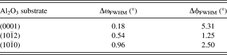
Let us examine the differences in crystallinity of the NbO2 films on the various cuts of Al2O3; we do this by comparing the peak breadths of the ω scans of the out-of-plane film reflections in Figs. 6(a), 7(a), and 9(a), yielding information about out-of-plane texturing, and those of the  $\phi$ scans of the asymmetric peaks of Figs. 6(b), 7(b), and 9(b), yielding information about in-plane alignment (Table I). We note that for our set of samples, the NbO2 films on various Al2O3 substrates have higher degrees of crystallinity than the films on (111) MgAl2O4 and (111) MgO, as quantified by the breadth of the ω scans. Here, we consider only the films grown on Al2O3. With the optics used in these scans, the corresponding substrates would have full width at half maximum (FWHM) values <0.05°. We see that while the (110) NbO2 film deposited on c-plane (0001) Al2O3 is extremely textured out-of-plane, with a small 440 peak ω FWHM value of 0.18°, its in-plane alignment is considerably worse, with a 400 peak
 $\phi$ FWHM value of 5.31°. On the contrary, despite having a lower degree of out-of-plane crystalline texturing (ω FWHM of 0.54°), the (111) NbO2 on r-plane (10
 $\bar 1$2) Al2O3 has dramatically better in-plane alignment (
 $\phi$ FWHM of 1.25°); furthermore, from their respective FWHM values, its in-plane alignment and out-plane texturing are more comparable than for the (110) film on (0001) Al2O3. A likely reason for the difference between these two cases is that the formation of rotational variants in (110) NbO2 on (0001) Al2O3 requires planar defects (120° tilt boundaries) to connect the different epitaxial grains of symmetry-related structural variants, reducing the level of in-plane alignment. On the contrary, for (111) NbO2 on (10
 $\bar 1$2) Al2O3, symmetry does not induce the formation of these defect planes, and the factor limiting the degree of crystallinity should be mainly the lattice mismatch at the heterointerface and the possibility of variants related by glide symmetry. By symmetry considerations and comparing the film/substrate interfacial lattice mismatches, the (001) epitaxial variants of NbO2 on m-plane (10
 $\bar 1$0) Al2O3 should have comparable crystallinity and alignment to (111) NbO2 on (10
 $\bar 1$2) Al2O3; however, the growth of a competing (331) orientation may have reduced its crystallinity, as suggested by the larger FWHM values (Table I). Despite a noticeable second orientation, the in-plane alignment of the (001) NbO2 grains on (10
 $\bar 1$0) Al2O3 is still better than that of (110) NbO2 grains on (0001) Al2O3. Similar trends have been reported on TiO2 films grown on different cuts of Al2O3.Reference Gao, Thevuthasan, McCready and Engelhard3
The FWHM values of the ω scans (ΔωFWHM) of the out-of-plane 440, 222, and 004 and of the  $\phi$ scans (Δ
 $\phi$FWHM) of the asymmetric 400, 402, and 222 NbO2 film reflections on (0001), (10
 $\bar 1$2), and (10
 $\bar 1$0) Al2O3 substrates, respectively.
IV. CONCLUSIONS
We have shown that epitaxial films of WO2 and NbO2 can be grown on a variety of oxide substrates. Thin-film crystallinity is affected by a combination of surface symmetry of the films and substrates and their lattice mismatch. (010) WO2 and (110) NbO2 films grown on substrates with 3-fold rotational surface symmetry form epitaxial rotational variants. These out-of-plane orientations correspond to pseudorutile {100}; furthermore, the epitaxial orientational relationships observed here are analogous to those found in rutile TiO2 and monoclinic VO2 films grown on (0001) Al2O3. In NbO2 films deposited on (10 $\bar 1$2) and (10
 $\bar 1$0) Al2O3 substrates of lower surface symmetry, rotational variants do not form, but variants related by glide symmetry are possible; they have better in-plane orientational alignment with the substrate surfaces than the film on (0001) Al2O3. Our studies lay the foundation for further work on heteroepitaxial rutile-type films on various substrates of a range of crystal structures and suggest many possibilities for the investigation of novel oxide heterostructures.
ACKNOWLEDGMENTS
This work was funded by AFOSR Grant No. FA9550-12-1-0189. Sputter deposition and x-ray photoelectron spectroscopy measurements were performed at the Center for Nanoscale Systems (CNS), a member of the National Nanotechnology Infrastructure Network (NNIN), which is supported by the National Science Foundation under NSF Grant No. ECS-0335765. CNS is part of Harvard University. The XRD 2θ–θ scans were performed on a facility instrument at the MRSEC Shared Experimental Facilities at MIT, supported by the National Science Foundation under Grant No. DMR-08-19762.