Introduction
Aortic arch hypoplasia is found in approximately 30% of patients with aortic coarctation. Reference Brown, Rodefeld and Ruzmetov1 Although extended end-to-end anastomosis via left posterolateral thoracotomy is the most commonly used technique for coarctation with distal arch and isthmus hypoplasia, Reference Kaushal, Backer and Patel2,Reference Mery, Guzman-Pruneda and Trost3 debates persist regarding the optimal treatment for patients with hypoplasia of the proximal arch. Reference Kotani, Anggriawan and Chetan4–Reference Tsang, Haapanen and Neijenhuis7 Some authors suggest that after repairing coarctation and distal arch hypoplasia via thoracotomy, the proximal arch will expand over time, eliminating the need for additional intervention. Reference Kotani, Anggriawan and Chetan4 Others argue that in patients with a hypoplastic proximal arch, if radical repair is not performed during the initial surgery, there would be a risk of long-term restenosis, and they recommend that the proximal arch should also be widened through sternotomy. Reference Mery, Guzman-Pruneda and Carberry6,Reference Bernabei, Margaryan, Arcieri, Bianchi, Pak and Murzi8–Reference Sakurai, Stickley and Stumper11
Direct anastomosis of the ascending and descending aorta is a commonly preferred method for repairing aortic coarctation and aortic arch hypoplasia via sternotomy. However, in some patients, after resection of the pulmonary ductus arteriosus and the coarctated aortic segment, a significant gap may arise between the ascending and descending aorta. It is believed that directly anastomosing these tissues can lead to excessive tension along the anastomosis line which may cause restenosis. Additionally, the descending aorta may compress on the left main bronchus and left pulmonary artery. To avoid these complications, many clinics use autologous, homologous, xenograft patches, or vascular conduits in the repair. Reference Brown, Rodefeld and Ruzmetov1,Reference Bernabei, Margaryan, Arcieri, Bianchi, Pak and Murzi8,Reference Bechtold, Purbojo and Schwitulla12 –Reference Lee, Yang and Jun15 Although debates on this matter continue, both approaches have been reported to yield successful postoperative outcomes. Reference Gray, Wells, Starnes and Kumar9,Reference Bechtold, Purbojo and Schwitulla12,Reference Lee, Yang and Jun15–Reference Roussin, Belli and Lacour-Gayet19
The aim of this study is to compare the bovine pericardial patch augmentation and direct native tissue anastomosis techniques in terms of postoperative outcomes, including morbidity, in-hospital mortality, and the risk factors for reintervention.
Materials and method
Patients
The sample of the study consists of paediatric patients (< 18 years old) who underwent aortic arch hypoplasia and coarctation repair with the midline sternotomy approach at Ankara Bilkent City Hospital between August 2019 and August 2023.
Patients with hypoplastic left heart syndrome who underwent Norwood stage 1 were not included in the study due to the high postoperative intensive care mortality and morbidity, as well as the prolonged need for intensive care support.
Methods
Three types of procedures were performed during the study period: (1) isolated aortic arch hypoplasia and coarctation repair, (2) arch reconstruction with pulmonary banding, and (3) arch reconstruction with single-stage intracardiac repair. The diagnosis of aortic arch hypoplasia was made by evaluating the entire aortic arch using echocardiography, angiography, and, when available, CT imaging. A segment of the aortic arch was considered hypoplastic if its Z score was < 2 and/or it measured below the patient’s body weight + 1 mm. All patients with a hypoplastic proximal arch were approached via median sternotomy.
In addition to the aortic arch, the details of the intracardiac anatomy were also retrospectively examined. Patients were classified based on their suitability for single-stage or two-stage ventricular repair. The decision for single or two-stage repair in patients with double ventricles was made according to the patient’s overall clinical status and the final discretion of the primary surgeon. The details of the patients' operations were retrieved from digital records or operation notes in the patient files.
Morbidity factors and in-hospital mortality rates during postoperative intensive care follow-up were examined, along with their relationship to the applied surgical technique. Additionally, the early-term effectiveness of the operation was evaluated through postoperative transthoracic echocardiogram measurements and upper-lower extremity blood pressure measurements. A maximum systolic gradient > 20 mm Hg in the reconstructed aortic arch on postoperative transthoracic echocardiogram, or an invasive systolic gradient > 20 mm Hg between the upper and lower extremities, was defined as an ineffective repair. The mid-term effectiveness of the procedure was assessed based on the need for reintervention (endovascular or surgical) after discharge.
Surgical technique
In all patients, invasive pressure monitoring was performed via the right radial artery and the right or left femoral artery. After midline sternotomy, the ascending aorta, all branches of the aortic arch, pulmonary ductus arteriosus, right and left pulmonary arteries, and descending aorta were dissected. Dual arterial cannulation (greater curvature of ascending aorta beneath the innominate artery and main pulmonary artery) and selective venous cannulation were performed in all patients to ensure effective and uniform cooling of the brain and the entire body. After initiation of cardiopulmonary bypass), patients were cooled to 24 degrees. When the desired temperature was reached, cross clamp was placed and del-Nido cardioplegia was given. The arterial cannula in the ascending aorta was directed towards the innominate artery and antegrade selective cerebral perfusion was initiated. A C-clamp was placed on the distal descending aorta. The pulmonary ductus arteriosus and the coarcted segment were completely resected, without leaving any residue behind. Then, the aortic arch was opened from its lower aspect, starting from the base of the left subclavian artery and proximally until 0.5–1 cm proximal to the the origin of the innominate artery. Both vessels were then directly anastomosed with 7/0 polipropylene suture. In patients where tension was thought to occur in the anastomosis, a half circumference native tissue anastomosis was initially performed between the greater curvature of the transverse arch and the posterior wall of the descending aorta, in accordance with the decision of the primary surgeon. Then, an interdigitating incision was made to the anterior aspect of the descending aorta. The entire incision line was then patched using bovine pericardium. After arch reconstruction, antegrade selective cerebral perfusion was withdrawn and pulmonary band or intracardiac repair was performed depending on single or two stage repair. With the discretion of the primary surgeon, in selected patients with well-developed ascending aorta proximal to innominate artery, arch reconstruction was performed on beating heart using antegrade continuous coronary perfusion.
Statistical analysis
Normality assumptions of the variables were examined with the Shapiro–Wilk test. Median and interquartile range in descriptive statistics of continuous variables; in defining categorical variables, frequency (n), and percentage (%) values are given. Mann–Whitney test was used to compare between two groups. Relationships between categorical variables were examined with Chi-square/Fisher exact analysis. IBM SPSS.25 program was used in all analyses, and p < 0.05 was accepted as the significance level.
Results
The median age of the cohort consisting of a total of 38 patients was 40 days (15–157.5 days, interquartile range), and body weight was 3.78 kg (3.2–6.0 kg, interquartile range). 16 patients (42.1%) were newborns. The majority of the patients (97.4%) had biventricular anatomy. The proximal and distal arch Z-Scores of the cohort was −4.73 (−5.42 – −4.23, interquartile range) and −4.29 (−5.10 – −3.43, interquartile range), respectively. While 35 (92.1%) patients were operated for the first time, 3 patients (7.9%) had had coarctation resection and extended end-to-end anastomosis via left thoracotomy.
While completely native tissue anastomosis was applied in 18 of the patients (47.4%), bovine pericardial patch was used in arch reconstruction in 20 patients (52.6%). No statistically significant difference was observed between the age, body weight, transverse arch Z scores, cardiopulmonary bypass, and antegrade selective cerebral perfusion durations of the patients between the two groups. However, cross-clamp duration (min) in patients who underwent arch repair with bovine pericardium was found to be significantly higher than in patients who underwent direct tissue anastomosis (p = .016). The number of patients in both groups who underwent biventricular repair, redo operations, retrograde coronary perfusion, and the number of patients admitted to intensive care with an open chest were found to be similar (p > .05). (Table 1)
Preoperative and operative variables according to the surgical methods

*Mann–Whitney test; #Chi-squared test; †Fisher’s exact test; ASCP = antegrade selective cerebral perfusion; CPB = cardiopulmonary bypass; VSD = ventricular septal defect.
The average ICU stays of patients who underwent repair using direct tissue anastomosis and those who underwent repair using bovine pericardium were similar and were 7.0 and 8.0 days, respectively (p = .450). No statistically significant difference was observed between the two groups regarding the postoperative morbidity factors such as extracorporeal membrane oxygenator need, postoperative bleeding and duration of open chest. During the study period, 9 patients died and the overall in-hospital mortality was 23.6%. There was no significant difference between the two surgical procedures in terms of in-hospital mortality (p = .914).
25 (65.7%) patients remained under follow-up after discharge. 8 interventional procedures were performed in 7 patients (18.4%) during a mean follow-up period of 16.5 months (3.75 – 31.50, interquartile range). Average intervention time was 6 months (4–20.5, interquartile range). All patients underwent catheterisation and balloon dilatation with the preliminary diagnosis of restenosis. 3 (7.9%) patients has succesfull balloon dilatation alone and 1 (2.6%) patient underwent stent implantation in addition to balloon dilatation. 3 patients (7.9%) did not have succesful balloon dilatation and underwent recurrent surgical repair due to multiple stenosis in the anastomosis line. In all operated patients, anastomotic areas were augmented with a bovine pericardial patch.
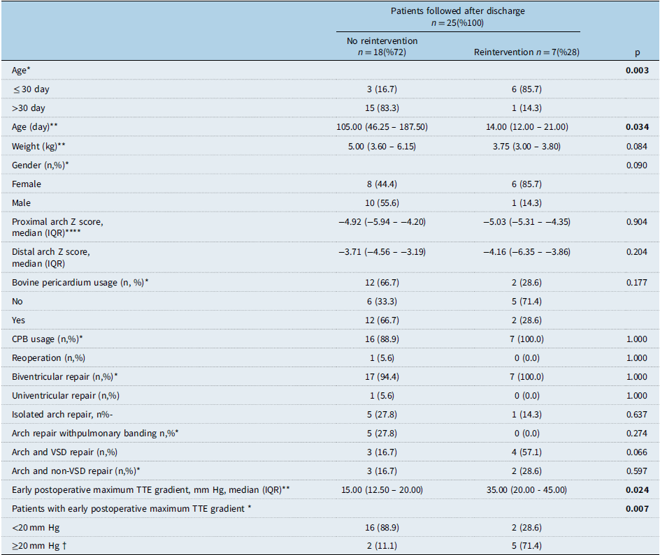
As shown in Table 2, 6 of the 7 patients (85.7%) who underwent reintervention during follow-up were newborns (p = 0.03), and the average age of these patients was significantly lower than those who did not undergo intervention (14.00, 12.00–21.00, interquartile range) (p = 0.034). No significant difference was detected between patients who underwent intervention and those who did not, in terms of aortic arch Z scores, use of bovine pericardial patch, basic cardiac morphologies, and additional cardiac repair during the operation. When 7 patients who underwent reintervention were examined, it was determined that the 5 patients (71.4%) had a maximum transthoracic echocardiogram gradient of ≥ 20 mm Hg with no tail extension to diastole in the early transthoracic echocardiogram follow-ups. This situation was found to be a risk factor for re-intervention (p = 0.07). Similarly, patients who underwent re-intervention had significantly higher average of TEE gradients than in those without intervention (p = 0.24).
Risk factors for reintervention during follow-up

*Fisher’s exact test; **Mann–Whitney test; †Patients who have not diastolic tail in descending aorta; CPB = cardiopulmonary bypass; TTE = transthoracic echocardiography; VSD = ventricular septal defect.
Discussion
Different surgical techniques can be used in aortic arch reconstructions. The successful long-term outcomes of the “aortic advancement” method, which utilises entirely autologous tissues, have contributed to the growing popularity of this technique. Notably, it has been reported that no patient required reintervention during the 5-year follow-up period for interrupted aortic arch patients treated with this method. Reference Morales, Scully and Braud17 Additionally, in a cohort of 275 patients with complex anomalies involving hypoplastic arches repaired using the aortic advancement method, the need for reintervention was observed in only 3% of cases during a 6-year follow-up. Reference Mery, Guzman-Pruneda and Carberry6 Similar to many centres worldwide, our clinic primarily prefers direct native tissue anastomosis as the first choice for anatomically suitable cases. However, the potential for left main bronchus and left pulmonary artery compression, as well as the need for advanced dissection of the descending aorta to achieve a tension-free anastomosis, has led some clinics to adopt the use of patches in arch reconstruction.
In patch augmentation techniques, after radical resection of the coarcted segment and ductal tissues, native tissue anastomosis is performed between the descending aorta and the outer curvature of the transverse arch. A patch is then applied to the lesser curvature of the aortic arch. The native tissue anastomosis at the outer curvature facilitates the development of the aortic arch, thereby preventing restenosis in this region. Reference Ashcraft, Jones and Border20 Furthermore, the patch applied to the lesser curvature helps preserve the retroaortic space and reduces the risk of compression of the left main pulmonary artery and left main bronchus. Reference Patukale, Shikata and Marathe10 Moreover, patch augmentation is considered beneficial in achieving a smooth “Romanesque” form of the aortic arch. It has been noted that steeply angled anastomosis lines, which may result in “Gothic” or “Crenel” forms often seen in end-to-side anastomoses, disrupt aortic flow dynamics in the long-term postoperatively, and are associated with persistent hypertension. Reference Donazzan, Crepaz, Stuefer and Stellin21
Bovine pericardial patches are frequently used in cardiovascular surgery owing to their advantageous features such as accessibility compared to homografts, durability, ease of handling, non-thrombogenic nature, resistance to immune reactions and infections, and minimal bleeding due to needle holes. Reference De Martino, Milano and Bortolotti22–Reference Li, Guo and Ziegler24 Today, numerous bovine pericardial patches are commercially available. Reference De Martino, Milano and Bortolotti22,Reference Li, Guo and Ziegler24 While most of these patches are fixed with glutaraldehyde, alternative fixation methods have also been developed. Reference Baird, Myers and Piekarski16 Experience to date indicates that bovine pericardium treated with glutaraldehyde is prone to structural degradation caused by calcification, leading to reduced clinical efficacy. To address this issue, various decalcification techniques have been introduced in recent years, with promising results reported. Reference De Martino, Milano and Bortolotti22 However, the medium- and long-term outcomes of using bovine pericardium treated with modern anti-calcification methods in neonatal and infant patients—where calcium metabolism is particularly rapid—remain uncertain and under investigation. In a study by Bell et al., only 1 out of 11 neonates who underwent arch reconstruction using a tissue-engineered bovine pericardium (CardioCel®, Admedus Regen Pty Ltd, Perth, WA, Australia) required intervention for anastomotic stenosis during a 24-month follow-up period. Reference Bell, Prabhu and Betts25 Similarly, Morell et al. observed an 18.2% recurrence rate of arch stenosis in neonates undergoing the Norwood stage 1 procedure with bovine pericardium (Shelhigh, Inc., Millburn, NJ). They concluded that this rate was acceptable and that bovine pericardium could be suitable for this application. Reference Morell and Wearden18 Conversely, van Beynum et al. reported that in newborn and infant patients undergoing arch reconstruction with either homografts or CardioCel®, 70% of the bovine pericardium group required reintervention within an average of 12 months. Ashcraft et al. further noted that the use of bovine pericardium in arch reconstruction for patients with HLHS increased the risk of restenosis by 1.8 times. Reference Ashcraft, Jones and Border20 Another study demonstrated that while bovine pericardium showed success in repairs involving the atrial and ventricular septum, valvular structures, and pulmonary artery, its use in aortic arch reconstruction in neonates and infants led to intense neointimal proliferation, causing the graft to lose functionality rapidly. Reference Pavy, Michielon, Robertus, Lacour-Gayet and Ghez26 This limitation is thought to stem from the stiffness of bovine pericardium compared to the native aorta. The disparity in biomechanical properties likely generates high shear stress along the anastomosis lines of grafts placed in the aortic position. Reference Tremblay, Zigras and Cartier27 Unfortunately, prospective controlled studies focusing on the use of bovine pericardium produced with current technologies in the aortic arch position for neonatal and infant patients are still lacking.
In our clinic, we have been using XenoLogiX™ (Edwards Lifesciences LLC, Irvine, CA, USA) bovine pericardium, which is fixed with glutaraldehyde and incorporates a proprietary anticalcification method, for arch reconstructions since 2019. An in vitro comparison of this pericardium with other bioscaffolds demonstrated that it offers higher tensile strength but may lead to reduced pliability and loss of compliance in vivo. Histological examinations revealed giant cell formation, indicating a foreign body reaction to the glutaraldehyde-treated bovine pericardium. Reference Neethling, Puls and Rea28 Although these findings suggest that this product may lack the characteristics of an ideal patch for arch reconstructions in newborn and infant patients, there is currently no conclusive evidence-based study recommending against its use in this patient group. Furthermore, alternative options such as new-generation, tissue-engineered bovine pericardial bioscaffolds, Reference van Beynum, Kurul and Krasemann29 and equine pericardium have also been reported to be associated with high restenosis rates. Reference Vitanova, Cleuziou, von Ohain, Burri, Eicken and Lange30 Additionally, pulmonary or aortic homografts are difficult to obtain, and the cost–benefit comparisons of other bioscaffolds remain unclear. Autologous pulmonary arterial patch or autologous pericardium treated with glutaraldehyde can also be used in arch reconstructions. However, we did not prefer this tissue due to lack of decalcification process and concerns about glutaraldehyde related toxicity. Given these challenges and its ready availability in our clinic, we preferred to use bovine pericardium in arch reconstructions when the patient’s anatomy was not suitable for complete direct native tissue anastomosis.
In our study, 7 out of 25 patients (28%) who were followed after discharge required reintervention, with a median postoperative follow-up of 6 months (interquartile range 4.0–20.25 months). Of these, arch reconstruction was performed using bovine pericardium in 2 patients (28.5%) and direct tissue anastomosis in 5 patients (72.5%) (p = .177). In patients with direct tissue anastomosis, discrete stenosis at the anastomosis was detected through imaging methods during follow-up. On the other hand, among the two patients who underwent bovine pericardial repair, one developed distal anastomosis stenosis, while the other exhibited multiple stenoses on the patch. Balloon angioplasty was initially attempted in all patients. Partial success was observed in 3 patients (who are still under close follow-up), while 4 patients did not achieve satisfactory results. In one of the latter cases (bovine pericardium group), an expandable stent was implanted at the distal anastomosis site. In the remaining 3 cases (direct anastomosis group), patchplasty with bovine pericardium was performed during reoperation. Intraoperative findings revealed significant fibrotic thickening along the anastomosis line in patients with direct tissue anastomosis stenosis. In patients with restenosis following bovine pericardium augmentation, thickened neointimal hyperplasia and peeling along the anastomosis line were noted.
We observed a considerable rate of reintervention in the early postoperative period following discharge in patients who underwent direct tissue anastomosis (5 out of 11 patients, 45.4%). This rate was higher than the outcomes reported in similar series in the literature. Reference Mery, Guzman-Pruneda and Carberry6,Reference Gray, Wells, Starnes and Kumar9,Reference Morales, Scully and Braud17 We believe that surgical errors and inappropriate patient selection may have contributed to this outcome. Intraoperative findings of reoperation of native tissue anastomosis revealed intense fibrotic thickening at the anastomosis sites in the patients. This may be attributed to excessive tension on the anastomosis due to insufficient mobilisation of the descending aorta from the surrounding tissues. Additionally, the insufficient removal of residual ductal tissues from the descending aortic wall might have also contributed to this outcome. Although we paid close attention to the removal of ductal remnants and the dissection of the descending aorta from surrounding tissues, we may not have recognised the excessive tension that could have occurred at the anastomosis during the operation. The absence of needle hole bleeding from the anastomosis and the satisfactory external appearance of the surgical procedure may have led us to this misconception.
Notably, in the 14 patients who underwent repair with bovine pericardium, only 2 patients (14.2%) required early reintervention after discharge. This may be attributed to the effectiveness of the surgical technique, which was recommended by Langley and colleagues. According to this technique, following resection of entire ductal tissue, we extend the incision exclusively along the anterior aspect of the descending aorta. Reference Langley, Sunstrom, Reed, Rekito and Gerrah5 After performing a native tissue anastomosis in the transverse arch, we augment the lesser curvature of the entire aortic arch with bovine pericardium. Thus, we were able to confidently ensure that no tension developed at the anastomosis line, and we also successfully separated the connections of the ductal remnants in the descending aorta, which were not detectable upon external examination.
As a result of our study, both direct native tissue anastomosis and the use of bovine pericardium for augmentation in patients undergoing arch reconstruction were not statistically associated with early reintervention (p = .117). However, given that the longest follow-up period in our study was 31.5 months, it is not possible to draw definitive conclusions about the mid- and long-term outcomes of both techniques. The discouraging results reported in the literature regarding bovine pericardium in the aortic arch position underscore the importance of close monitoring of these patients for restenosis.On the other hand, a significant number of patients in our sample who underwent entirely native tissue anastomosis also developed restenosis. When compared with the large and highly successful series reported in the literature, we believe this outcome may be attributed to technical errors during the surgical procedure. Therefore, we will continue to advocate for direct native tissue anastomosis in arch reconstructions for patients who are deemed clinically and anatomically suitable. However, in cases where the use of an additional patch is required, we intend to continue using bovine pericardium patches without hesitation.
Conclusion
In paediatric patients with hypoplastic aortic arch, no significant differences were observed between direct anastomosis and bovine pericardial patch augmentation techniques regarding postoperative mortality and the need for reintervention. While early results are promising for both techniques, further investigation into the durability and reliability of both techniques in arch reconstruction is warranted through larger, prospective controlled studies.
Acknowledgements
None.
Financial support
This research received no specific grant from any funding agency, commercial, or not-for-profit sectors.
Competing interests
None.
Ethical standarts
The study was evaluated and approved by the institution’s ethics committee under approval number E2-23-5325 on 25.10.2023.