High entropy alloys (HEAs) are a relatively new group of materials[Reference Yeh, Chen, Lin, Gan, Chin, Shun and Tsau1,Reference Cantor, Chang, Knight and Vincent2] with a wide range of potential applications. Specifically, face centered cubic (FCC) HEAs have been shown to maintain their ductility and strength at cryogenic temperatures.[Reference Zou, Wheeler, Ma, Okle and Spolenak3–Reference Chang and Yeh5] In addition to compelling mechanical properties, their corrosion resistance can make certain HEAs ideal candidates for structural applications in more demanding settings such as marine environments.[Reference Fu, Schuh and Olivetti6] HEAs contain five or more unique principal elements, with the content of each alloying component in the range of 5–35 at.% and with a configurational entropy of 1.5R or higher, where R is the gas constant.[Reference Zhang, Zuo, Tang, Gao, Dahmen, Liaw and Lu7]
Cantor alloys[Reference Cantor, Chang, Knight and Vincent2] and Cantor-like alloys have received significant interest within the research community.[Reference Zhang, Zuo, Tang, Gao, Dahmen, Liaw and Lu7–Reference Ye, Wang, Lu, Liu and Yang9] These HEAs typically contain some mix of the elements Mn, Fe, Ni, Co, Cu, and/or Cr. Addition of other alloying elements such as Al, or deviation from an equiatomic composition, has been explored as well.[Reference Chang and Yeh5,Reference He, Liu, Wang, Wu, Liu, Nieh and Lu10–Reference Tang, Gao, Diao, Yang, Liu, Zuo, Zhang, Lu, Cheng, Zhang, Dahmen, Liaw and Egami12] A variety of resultant crystal structures can be achieved by minor changes to alloy compositions. Kao et al. have studied HEA crystal transformations from FCC to body centered cubic (BCC) induced by minor additions of Al to the alloy FeNiCoCr,[Reference Kao, Chen, Chen and Yeh13] which significantly affected the mechanical properties of the alloy. Furthermore, relatively few studies have explored the addition of Cu to this family of alloys.[Reference Li, Xue, Hua, Cao, Ma and Wang14,Reference Zhang, Yang and Liaw15] The current study focuses on the MnFeCoNiCu alloy system, in order to screen for candidate non-equiatomic single-phase HEAs.
Unlike conventional alloys, HEAs do not have a single majority element that serves as a solvent, and this can complicate the development of new HEAs when using conventional techniques. Conventional alloy development approaches typically involve iteration within the context of general guidelines. When applied to HEAs, this approach can include the consideration of atomic size differences (δ) and the ratio between enthalpy and entropy of mixing (Ω).[Reference Zhang, Yang and Liaw15,Reference Youssef, Zaddach, Niu, Irving and Koch16] Another popular approach within the research community is to develop computational models to predict the formation of a single-phase HEA.[Reference Guo and Liu17–Reference Troparevsky, Morris, Kent, Lupini and Stocks21] However, these techniques can require extensive effort, whether experimental or computational, and are relatively time consuming and expensive.
The current project is based on a more efficient experimental approach to develop new HEA systems via combinatorial synthesis and screening of thin film samples. This involves the creation of a 2D compositional gradient over a relatively large area. Discrete regions within this gradient have different compositions that can lead to the formation of a different phase (or phases). Therefore, by screening for crystal structure over a wide combinatorial range in a thin film sample, one should be able to predict which phases will form in the bulk for a particular composition. By screening properties in a similar manner, an enhanced understanding of composition–phase–property relations will be achieved.
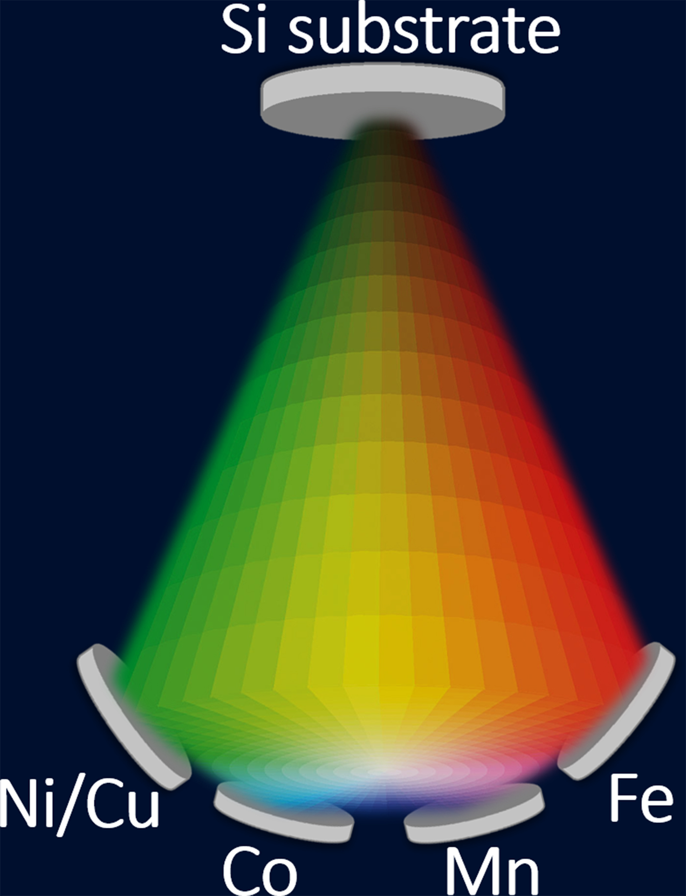
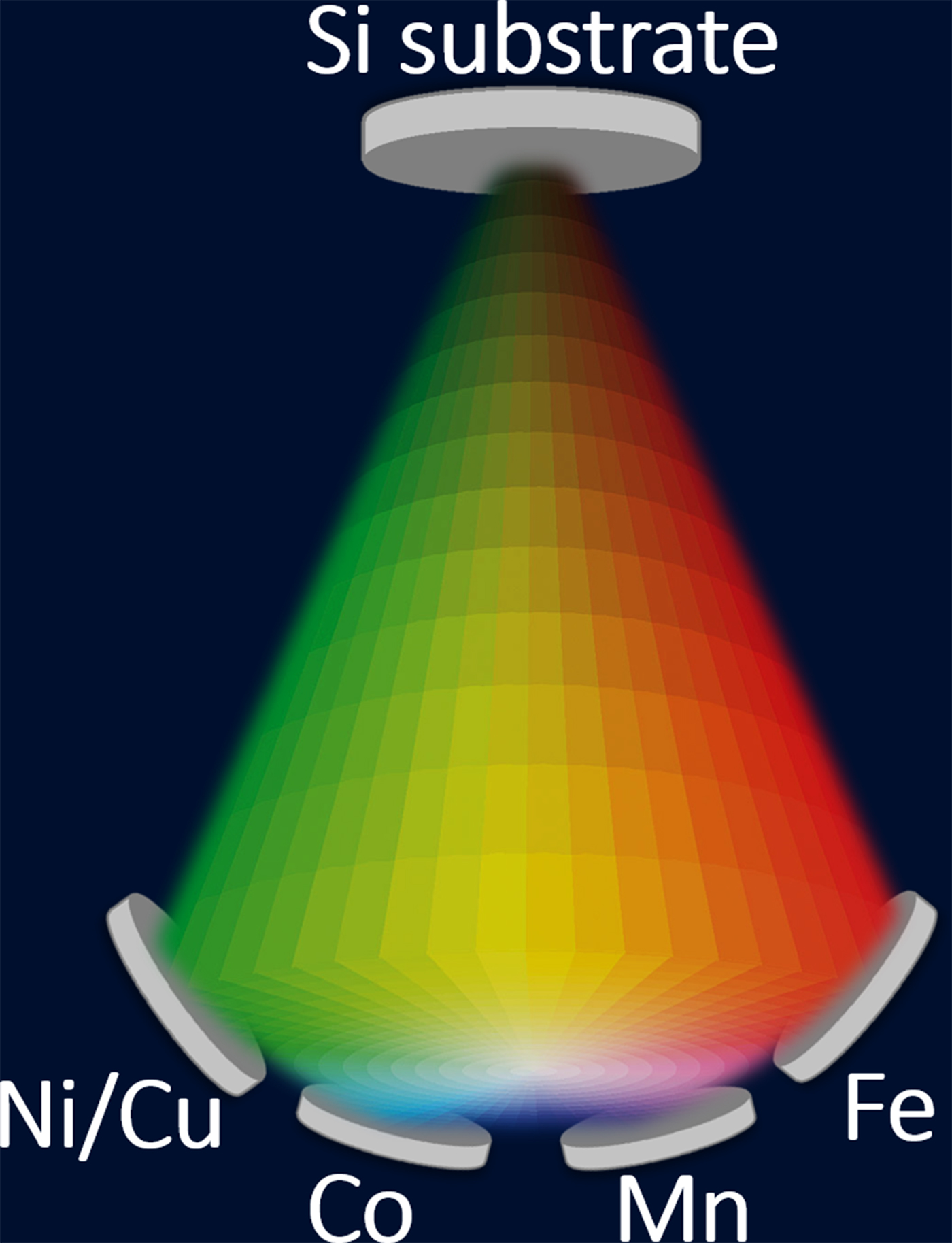
In the present work, magnetron sputtering (ORION system, AJA International, North Scituate, MA) was used to fabricate gradient thin films on single-crystal Si wafers, with MnFeCoNiCu serving as the basis of the alloy system. The gradient in composition was achieved by deposition from multiple sputtering targets arranged concentrically around the substrate, as shown in Fig. 1. For film deposition, elemental targets of Mn, Fe, and Co (99.95+% purity for each metal) were used. Additionally, a mixed Cu/Ni target (50 at.% for each metal) was prepared via powder pressing and sintering for this study. Magnetron sputtering was performed using a separate DC power supply for each target, with power settings of 80, 110, 80, and 160 Watts for the Mn, Fe, Co, and Cu/Ni targets, respectively. These power levels were optimized to achieve an equiatomic composition in the center of the substrate. Total deposition time was 20 min.
Sputtering arrangement for thin film deposition, with the confocal target arrangement shown at the bottom. By holding the substrate (at the top) stationary during deposition, a compositional gradient film is generated, due to decreasing amounts of elemental deposition with increasing distance (measured from the target) along the substrate.
Three-inch diameter Si(001) wafers with compositional gradient films were cut into smaller pieces ~10 mm × 10 mm in size and mounted on aluminum stubs with conductive double-sided carbon tape, to facilitate observation in the scanning electron microscope (SEM; FEI Quanta FEG 250, Hillsboro, OR). The composition of each smaller sample was measured using energy-dispersive x-ray spectroscopy (EDS; Oxford X-MaxN 80 mm2 detector, Concord, MA) in a dual-beam microscope (FEI Helios NanoLab 660, Hillsboro, OR). The typical error associated with EDS measurement of composition depends on which element is measured, but for the elements measured in this study, it is typically ±1 at.%. X-ray diffraction (XRD; Siemens D500, Germany) was used to determine the crystal structure of the corresponding region at the center of each smaller sample. For additional characterization through the film thickness in certain film regions, thin lamellae were prepared with the lift-out technique, using the focused ion beam (FIB) capabilities of the Helios dual-beam.
Candidate single-phase alloys, identified by screening the composition and crystal structure in combinatorial thin films, were melted from elemental pieces and cast in bulk form (Edmund Buhler compact arc-melter MAM-1, Hechingen, Germany). In order to obtain a more uniform sample, the as-cast piece was flipped and remelted several times. Arc-melted bulk samples were rolled to 50% of the original thickness and then annealed in a furnace with reducing atmosphere (Ar95H5). Furnace temperature was increased to 900 °C at a rate of 5 °C/min, and the samples were held at this temperature for 1 h. Subsequently, the temperature was raised to 1000 °C at a rate of 10 °C/min and the samples were held for 1 h, followed by furnace cooling to room temperature.
Bulk HEA samples, both as-cast and thermo-mechanically processed, were sectioned and prepared for microstructural characterization. Metallographic sample preparation steps included: mounting in epoxy, grinding with SiC papers, polishing with 9, 6, and 3 µm diamond solutions, followed by ultra-fine vibratory polishing (Buehler Vibromet, Lake Bluff, IL) with 0.02 µm colloidal silica suspension. Bulk alloy composition was also determined by EDS. Additionally, grain orientation was mapped using electron backscatter diffraction (EBSD; Oxford NordlysMax2 detector, Concord, MA) in order to assess the uniformity of the microstructure. EBSD results were processed using the Channel 5 software package. By applying Tango software, ~2.5% noise reduction was applied to the collected data, and grain boundaries were defined using a threshold value of 10° misorientation between neighboring grains.
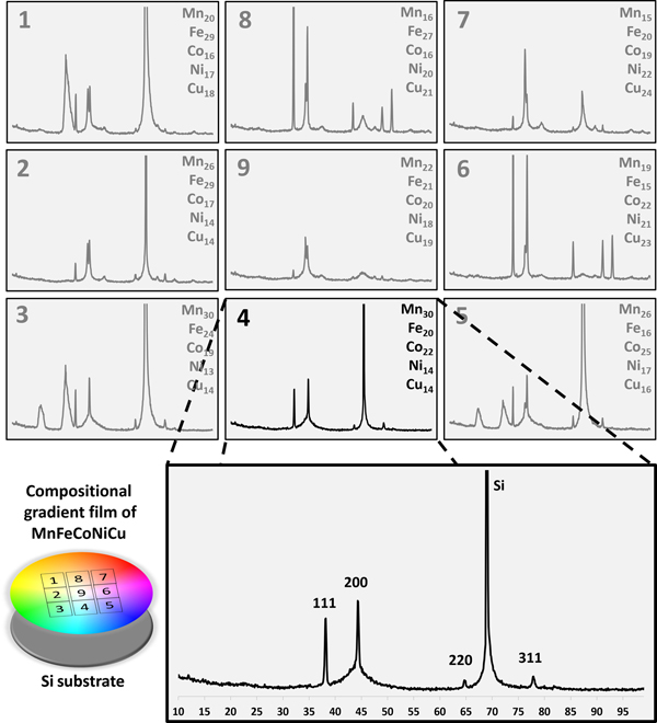
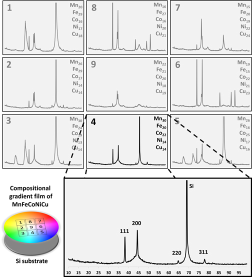
XRD and EDS data were collected from the center of each 10 mm × 10 mm thin film section, in order to construct a map of crystal structure and composition across gradient samples. The content of each element in the HEA samples varied ~15 at.% (e.g., 10–25 at.%) along the 3 in. Si wafer diameter. Figure 2 summarizes the crystal structure data collected from nine different regions of the thin film, spanning an area ~40 mm × 40 mm centered on the Si wafer. Each region had a different composition, which can result in the formation of different phases. The composition for each film region is presented along with the corresponding XRD scan, in a series of panels corresponding to the schematic at lower left. The XRD scan for each region spans the same angular range (10–100° 2θ) as shown in the larger scan at the bottom (region 4). Each region typically exhibited a unique XRD pattern, confirming the formation of multiple phases with different crystal structures for a given chemical composition. Additionally, the particular mixture of phases varied across the 2D compositional gradient. A single-phase FCC structure was observed in region 4, with composition Mn30Fe20Co22Ni14Cu14 (at.%), which made it a promising candidate HEA for further investigation in this study.
XRD scans over the range 10–100° 2θ, collected from nine distinct regions of a gradient film. EDS measurement of composition (in at.%) for each region is listed in the top right corner of the corresponding spectrum. The color schematic indicates approximate relative location of the regions analyzed.

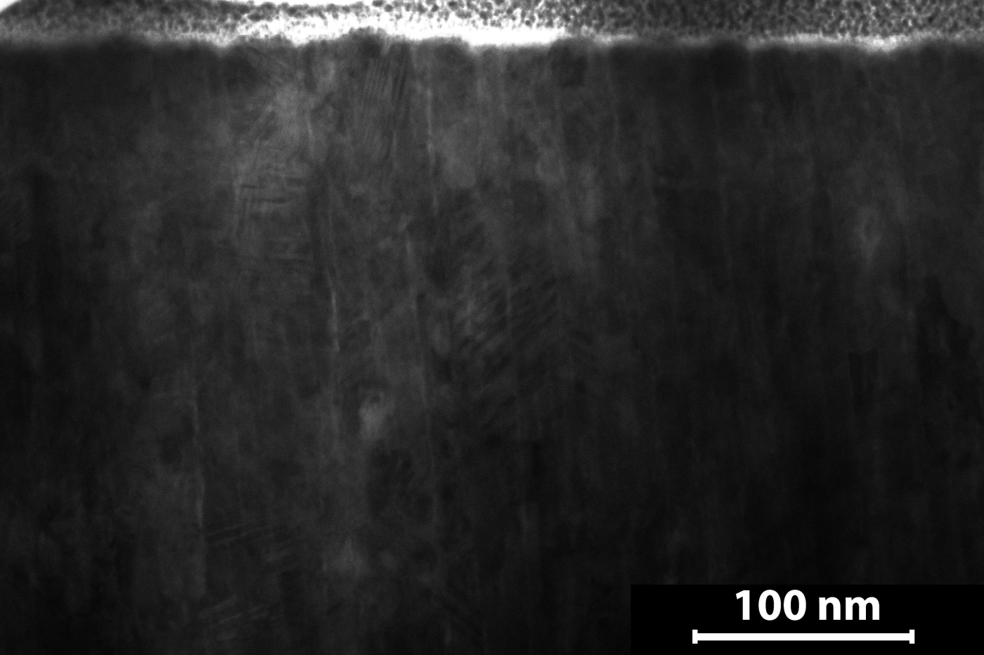
Grain structure in region 4 of the film was imaged via scanning transmission electron microscopy in the Helios NanoLab microscope and is presented in Fig. 3. The top layer visible here is a protective Pt film, with the sputtered HEA film located directly below. This micrograph indicates a columnar grain structure in the film, with grains ~10–30 nm wide. Columnar grains are common in as-deposited thin films fabricated via physical vapor deposition.[Reference Ohring22] The otherwise uniform grain structure and the XRD scan of this region (see. Fig. 2) suggested that this particular alloy was a promising candidate for fabrication and further analysis in bulk form.
STEM micrograph of the single-phase film location (region 4), obtained from a FIB-fabrication lamella. The columnar grain structure of the single-phase area is clearly observed here.

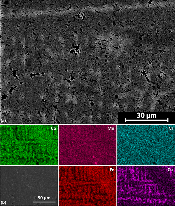
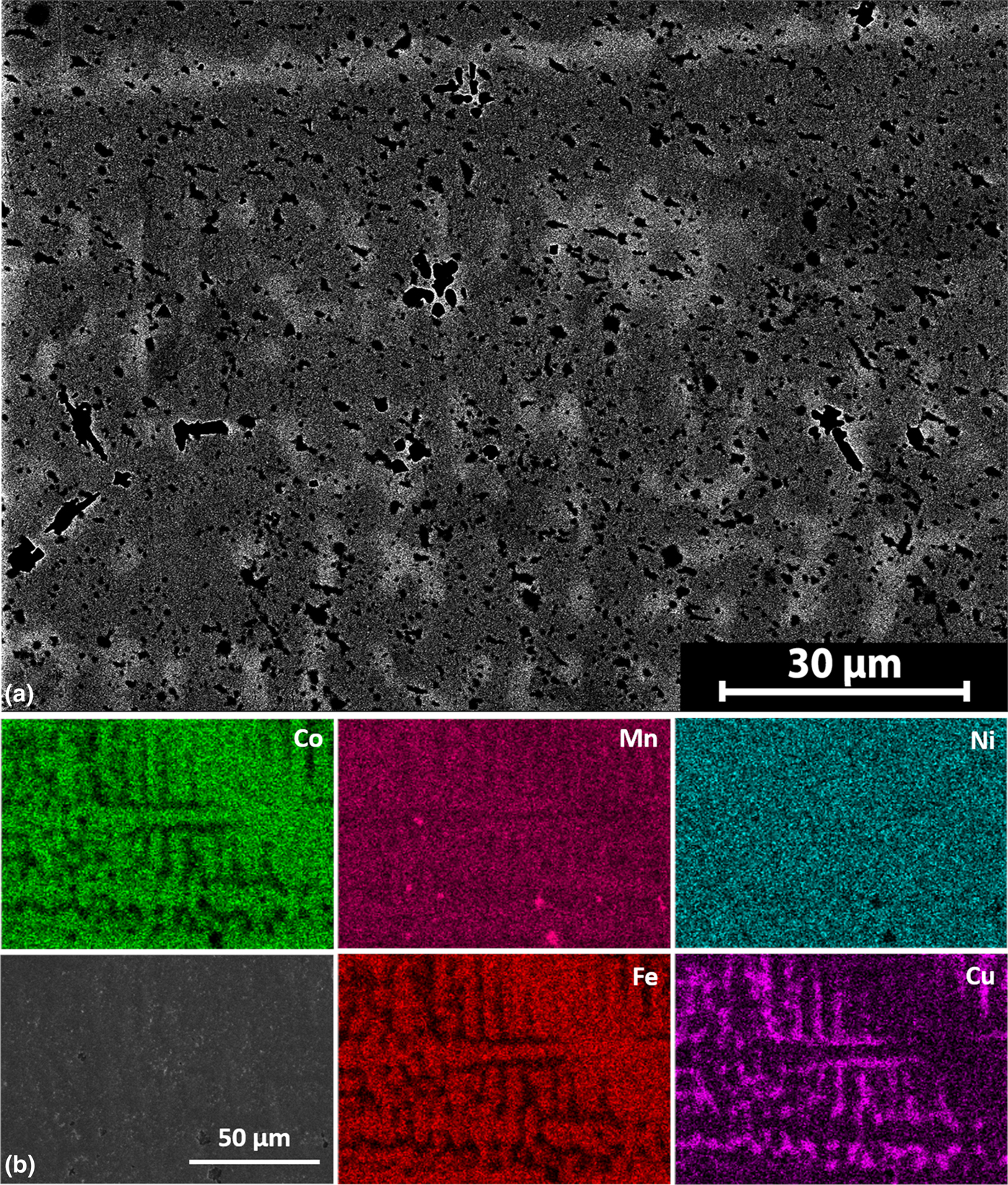
The particular alloy in region 4 satisfied a primary goal of this study, which was to identify and produce a non-equiatomic, single-phase FCC HEA. Therefore, an alloy with composition matching that of region 4 was arc-melted and cast in bulk form. The composition of the as-cast sample was Mn30Fe20Co20Ni15Cu15 (at.%). Considering the standard error inherent to EDS quantification (±1 at.%), the composition of the bulk alloy is effectively equivalent to that of region 4 from the compositional gradient film. Figure 4 presents an SEM micrograph and EDS elemental maps of this as-cast sample after proper metallographic sample preparation, and these observations clearly indicate a dendritic structure. The dendrite arms are richer in Fe and Co, whereas the inter-dendritic phase is richer in Cu. This is as expected, given the comparatively lower melting point of Cu and higher melting points of Fe and Co. Mn and Ni did not exhibit significant elemental segregation within the as-cast material. As discussed in the literature,[Reference Jablonski, Licavoli, Gao and Hawk23] the formation of a dendritic structure results from the different melting points and diffusion rates of elements within the alloy system.
(a) SEM image of as-cast bulk HEA sample, exhibiting a dendritic microstructure. (b) EDS maps of a separate area from the same as-cast bulk sample, showing the segregation of elements that occurred during solidification.
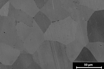
The as-cast samples were subjected to a series of thermo-mechanical processing steps, as described above, in order to achieve a more homogeneous microstructure in the bulk HEA. Figure 5 presents an SEM micrograph of the same bulk alloy (Mn30Fe20Co20Ni15Cu15) after rolling and multiple stages of heat treatment. This processing approach resulted in an equiaxed grain structure that was observed consistently throughout the sample. It is noted that the processed alloy still exhibited pores, which is typical of an arc-melted sample, although the degree of porosity was mitigated by the thermo-mechanical processing (compare Figs. 4 and 5).
SEM image of the bulk HEA sample after thermo-mechanical processing, with equiaxed grain structure.

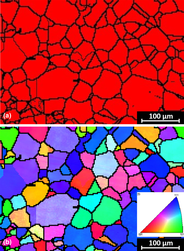
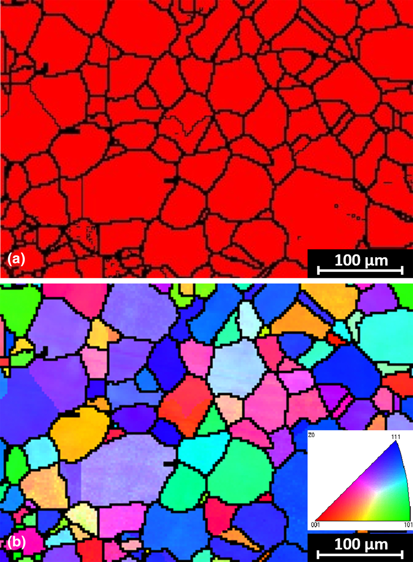
EBSD maps acquired from the processed bulk alloy are presented in Fig. 6, which shows an area that was free of pores. The phase map in Fig. 6a confirms that the HEA is single-phase, with all grains exhibiting the desired FCC structure. Moreover, the grain structure is equiaxed, with an average grain size of ~37 µm. No significant preferential orientation or texture can be seen in the orientation map (Fig. 6b). Although not shown in this manuscript, EDS elemental mapping of the microstructure over a large area (~1 mm2) shows that all elements are distributed uniformly throughout the grain structure. These results indicate that the thermomechanical processing steps involving recrystallization and grain growth were successful in achieving a uniform microstructure.
EBSD results of the processed HEA showing (a) phase map and (b) grain orientation and confirming that a single-phase equiaxed grain structure was achieved by thermo-mechanical processing.
Using the combinatorial thin film approach, a single-phase FCC HEA with non-equiatomic composition was successfully identified and fabricated in bulk form in a single attempt. Fabrication of gradient thin films of MnFeCoNiCu enabled the methodical study of this alloy family over a broad composition range. Screening of film composition and crystal structure using complementary characterization techniques revealed Mn30Fe20Co22Ni15Cu15 as a candidate single-phase FCC HEA. This alloy composition was cast in bulk form and thermo-mechanically processed, with subsequent confirmation that the predicted single-phase alloy with equiaxed grain structure had been achieved.
This technique is more effective and efficient compared to conventional alloy development methods. Due to the versatility of thin film fabrication, this combinatorial approach can be applied to a wide variety of alloy combinations. This should reduce the number of casting attempts and thereby reduce alloy development costs, streamlining the exploration of new alloy systems. Additional studies utilizing this approach are underway to identify new HEAs and multiple principal element alloys with different crystal structures, such as BCC and hexagonal close packed, and including a screening approach to evaluate the properties and performance across the compositional gradient samples.
Acknowledgments
This work was supported by the Kentucky Science and Engineering Foundation under Grant No. KSEF-148-502-15-363. Access to characterization instruments and staff assistance was provided by the Electron Microscopy Center at the University of Kentucky, supported in part by the National Science Foundation/EPSCoR Award No. 1355438 and by the Commonwealth of Kentucky.