Introduction
More than any other species, humans are beneficiaries and victims of their emotional memories. While the adaptive value of these memories for survival and reproductive success is obvious, excessive stress often turns them into a source of anxiety and depression (Roozendaal et al. Reference Roozendaal, McEwen and Chattarji2009). This is best evidenced in post-traumatic stress disorder (PTSD), which is characterized by the clinical triad hypermnesia/re-experiencing symptoms, avoidance behaviour and hyperarousal in response to an inescapable life-threatening stressor. By focusing on the link between stress-related amygdala hyper-responsiveness (Rosenkranz et al. Reference Rosenkranz, Venheim and Padival2010) and overexpression of conditioned fear and avoidance behaviour (Yamamoto et al. Reference Yamamoto, Morinobu, Takei, Fuchikami, Matsuki, Yamawaki and Liberzon2009), the prevailing rodent models of PTSD emphasize fear memory dysregulation in the aetiology of the illness. However, these models fail to account for the hypermnesia/re-experiencing symptoms, which indicate emotional episodic (autobiographical) memory dysfunction (Brewin, Reference Brewin2008) and sometimes dominate the clinical phenotype. To treat or perhaps even prevent these symptoms, a valid neurocircuitry model of how stress influences, and interferes with, emotional episodic memory function is essential.
Current perspectives from functional magnetic resonance imaging (fMRI) studies in healthy human subjects suggest the interaction of two endogenous modulators, noradrenaline (NE) and cortisol (CORT), in mediating the impact of stress on amygdala and hippocampus (van Stegeren et al. Reference van Stegeren, Wolf, Everaerd, Scheltens, Barkhof and Rombouts2007, Reference van Stegeren, Roozendaal, Kindt, Wolf and Joels2010), which together orchestrate the efficient encoding of emotional episodic memories (Dolcos et al. Reference Dolcos, LaBar and Cabeza2004; Richardson et al. Reference Richardson, Strange and Dolan2004). Consequently, a combination of stress-related NE–CORT interactions and exaggerated amygdala-hippocampal responses triggered by these interactions is thought to underlie overactive emotional episodic encoding in PTSD (Hurlemann, Reference Hurlemann2008). Using fMRI in healthy volunteers, we recently showed that combined pharmacological elevation of NE and CORT triggered an amygdala response bias to fearful faces (Kukolja et al. Reference Kukolja, Schläpfer, Keysers, Klingmüller, Maier, Fink and Hurlemann2008a), which experimentally mimics amygdala hyper-responsiveness to fearful faces in PTSD patients (Rauch et al. Reference Rauch, Whalen, Shin, McInerney, Macklin, Lasko, Orr and Pitman2000; Shin et al. Reference Shin, Wright, Cannistraro, Wedig, McMullin, Martis, Macklin, Lasko, Cavanagh, Krangel, Orr, Pitman, Whalen and Rauch2005). A crucial question is whether elevations of NE and CORT also affect hippocampal function. The rationale of the present fMRI experiment was thus to measure – within the established experimental framework of amygdala hyper-responsiveness to fearful faces (Kukolja et al. Reference Kukolja, Schläpfer, Keysers, Klingmüller, Maier, Fink and Hurlemann2008a) – hippocampal activation during encoding of emotional versus neutral material. We hypothesized that NE and CORT – when simultaneously elevated to moderate stress levels – would act together by synergistically enhancing hippocampal function during emotional episodic encoding, a mechanism that may turn maladaptive under circumstances of excessive stress.
Method
Subjects
Altogether, 62 healthy adults (students at the University of Aachen and trainees at the Research Center Jülich, recruited by local advertisement) participated in the present study after providing written, informed consent. The sample characteristics are detailed online in Supplementary Method. The study had full ethical approval. One subject was excluded from further analysis due to excessive head movements during the fMRI session, three subjects were excluded due to a computer failure to record responses at recognition and seven subjects were excluded due to salivary CORT measurement failure. Thus, the data from 51 subjects were included in the analysis.
Encoding session
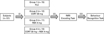
The experiment consisted of an encoding and a recognition session 1 week apart (Fig. 1). FMRI was measured while subjects were engaged in an episodic encoding task (Fig. 1) based on emotional and neutral stimuli selected from the International Affective Picture System (IAPS; Lang et al. Reference Lang, Bradley and Cuthbert2008). Stimulus delivery and response recording during encoding and recognition was carried out using Presentation11 (Neurobehavioral Systems, Inc., USA). Specifically, 72 neutral pictures [defined by low arousal ratings ranging from 2.32 to 4.19 and valence ratings ranging from 4.14 to 7.97, according to the IAPS manual (Lang et al. Reference Lang, Bradley and Cuthbert2008)] and 72 emotional pictures [defined by high arousal ratings ranging from 5.35 to 7.34 and valence ratings ranging from 1.31 to 4.79, according to the IAPS manual (Lang et al. Reference Lang, Bradley and Cuthbert2008)] were randomly displayed in the centre of an LCD screen behind the scanner, visible to the subjects via a mirror system (viewing distance 254 cm). Each stimulus was presented for 4000 ms, followed by an inter-stimulus interval lasting 1710 ms. In total, 40 null-events in which a white fixation cross was displayed on a black background were interspersed. This resulted in variable stimulus-onset asynchronies and allowed a comparison of the blood-oxygen-level-dependent signal of the event types of interest with a no-stimulus baseline. In order to ensure proper stimulus processing, subjects were asked to indicate via button presses whether or not a given stimulus was emotional. The encoding session lasted 22 min.
The randomized double-blind placebo-controlled parallel-group design and the timeline of the study are illustrated. (i) Random allocation to one of four treatment groups; (ii) functional magnetic resonance imaging (fMRI) scan of encoding 2 h 25 min after treatment; (iii) behavioural recognition test 1 week later. PLC, placebo; CORT, cortisol; RBX, reboxetine.

Drug administration
In a between-subjects, double-blind study design, participants were randomly allocated to one of four treatment groups. Importantly, these groups did not differ in demographic and cognitive characteristics (i.e. age, education, IQ, general memory functioning, mood and valence and arousal ratings, see Supplementary Material). The first group (PLC) received four tablets of saccharose placebo, the second (CORT-only) received three tablets of hydrocortisone (10 mg each) and one tablet of saccharose placebo, the third (RBX-only) received one tablet of reboxetine (4 mg) and three tablets of saccharose placebo and the last (RBX-CORT) received one tablet of reboxetine (4 mg) and three tablets of hydrocortisone (10 mg each) (Fig. 1; for randomization methods, see Supplementary Material). Reboxetine is a highly selective inhibitor of presynaptic NE reuptake (Kent, Reference Kent2000; Scates & Doraiswamy, Reference Scates and Doraiswamy2000). A 4-mg single oral dose of reboxetine was administered in analogy to our previous pharmacological fMRI studies of amygdala response changes following noradrenergic stimulation (Kukolja et al. Reference Kukolja, Schläpfer, Keysers, Klingmüller, Maier, Fink and Hurlemann2008a; Onur et al. Reference Onur, Walter, Schlaepfer, Rehme, Schmidt, Keysers, Maier and Hurlemann2009). A 30-mg single oral dose of hydrocortisone was administered to elevate CORT activity to moderate levels ranging between low (20 mg) and high (40 mg) stress levels (Abercrombie et al. Reference Abercrombie, Kalin, Thurow, Rosenkranz and Davidson2003). Drugs were administered 2.5 h prior to scanning. Further details are available online in Supplementary Material. For an assessment of CORT levels, saliva samples were collected immediately before drug administration (CORT1) as well as before (CORT2) and after (CORT3) scanning from each subject using Salivette collection devices (Sarstedt, Germany). After scanning, a venous blood sample was taken for the measurement of reboxetine serum levels.
Recognition session
The recognition session was performed 1 week after encoding to exclude confounding effects of drug action (Fig. 1). Subjects were placed in a quiet test room equipped with a computer set-up and presented with all 144 pictures previously shown during encoding. A total of 72 new pictures were intermixed (36 emotional and 36 neutral), resulting in an old/new ratio of 2:1. Subjects underwent a 45-min remember-know recognition test, requiring button presses to indicate whether: (i) they had seen the presented picture (remember); (ii) the picture looked familiar (know); or (iii) they had never seen the picture before. Prior to further data analysis, χ2 tests were calculated, ensuring that subjects had not been guessing when performing the recognition test. To analyse recognition performance, the detection sensitivity index d-prime (d') was calculated in accordance with the signal detection analysis (Green & Swets, Reference Green and Swets1966; Macmillan, Reference Macmillan, Keren and Lewis1993; Stanislaw & Todorov, Reference Stanislaw and Todorov1999), relating the number of correctly recognized items with the sum of all positive responses, including false positives. This enabled us to estimate a performance score that was unaffected by any individual response bias. d′ scores were analysed using repeated-measures three-way analysis of variance (ANOVA) including the between-subjects factors RBX (RBX, no RBX) and CORT (CORT, no CORT) and the within-subjects factor emotion (emotional, neutral). Gender (male, female) was added as a covariate to ensure that effects were generalizable, irrespective of gender. Adding gender as a fourth factor would not have added valuable information to this analysis because sample sizes of male and female subjects were too small (n=5–8) within each treatment group. Significant main effects and interactions were further investigated using post-hoc t tests, with the significance level p being Bonferroni-adjusted for multiple comparisons. To assess potential dose–response relationships, d′ scores for emotional and neutral pictures were correlated separately with salivary CORT and serum RBX levels. For the closest estimate of CORT levels during scanning, we used the mean of pre- and post-scan CORT levels [mCORT level=(CORT2+CORT3)/2]. CORT levels were determined in all subjects, such that data from all subjects (irrespective of treatment group, reflecting either endogenous or endogenous plus exogenous CORT) were included in the correlation analysis. Due to large inter-individual variability, mCORT levels [which originally were not normally distributed (Kolmogorov–Smirnov test n=51, Z=1.802, p<0.005)] were log-transformed (mCORTlog) to obtain normal distribution (Kolmogorov–Smirnov test n=51, Z=1.194, p=0.116), meeting requirements for a correlation analysis. An analogous analysis was performed when correlating serum RBX-levels with d′ scores. In this case, only subjects receiving reboxetine (n=24) were included in the analysis, because inclusion of non-medicated subjects would have distorted the correlation analysis. After the recognition session, subjects were asked to rate the pictures with regard to arousal and pleasure on a scale ranging from 1–9 (not arousing – very arousing; very unpleasant – very pleasant, respectively). Arousal and valence ratings were analysed using repeated-measures ANOVA, with the between-subjects factor treatment group (PLC, CORT-only, RBX-only, RBX-CORT) and the within-subjects factor emotion (emotional, neutral). Valence and arousal ratings are reported online in Supplementary Material. Significant results of post-hoc tests are reported after Bonferroni-correction for multiple comparisons.
fMRI hardware and image processing
Functional magnetic resonance images were acquired using a TRIO 3-T whole-body scanner (Siemens, Germany) equipped with a standard head coil for radio frequency transmission and signal reception. Sequence parameters are documented online in Supplementary Material. The image pre-processing was performed using Matlab7 (The MathWorks Inc., USA) and SPM5 (http://www.fil.ion.ucl.ac.uk/spm) (for details, see online Supplementary Material). The data were analysed using a general linear model as implemented in SPM5 (Kiebel & Holmes, Reference Kiebel, Holmes, Frackowiak, Friston, Frith, Dolan, Price, Ashburner and Penny2003) in an event-related fashion. In accord with previous studies (van Stegeren et al. Reference van Stegeren, Goekoop, Everaerd, Scheltens, Barkhof, Kuijer and Rombouts2005), all trials containing subsequently recognized items (i.e. pictures categorized as ‘remembered’ or ‘known’) were collapsed in the fMRI analysis in order to capture all neural activity related to successful encoding of these items. For each subject, simple main effects for each condition were computed using baseline contrasts. These first-level individual contrasts were then entered into a second-level group analysis using an ANOVA with the between-subjects factors RBX (RBX, no RBX) and CORT (CORT, no CORT) and the within-subjects factors emotion (emotional, neutral) and memory (recognized, forgotten) employing a random effects model (Penny & Holmes, Reference Penny, Holmes, Frackowiak, Friston, Frith, Dolan, Price, Ashburner and Penny2003). In analogy to the behavioural data analysis, gender was added as a covariate to the ANOVA. Violations of sphericity were accounted for by modelling non-independence across parameter estimates from the same subject and by allowing unequal variances both between conditions and subjects. Due to our a priori anatomical focus on amygdala and hippocampus, we restricted the analyses to these structures by defining regions of interest (ROI) based on cytoarchitectonic maximum probability maps (Amunts et al. Reference Amunts, Kedo, Kindler, Pieperhoff, Mohlberg, Shah, Habel, Schneider and Zilles2005) as available in the SPM Anatomy toolbox (Eickhoff et al. Reference Eickhoff, Stephan, Mohlberg, Grefkes, Fink, Amunts and Zilles2005; see also Goossens et al. Reference Goossens, Kukolja, Onur, Fink, Maier, Griez, Schruers and Hurlemann2009; Hurlemann et al. Reference Hurlemann, Walter, Rehme, Kukolja, Santoro, Schmidt, Schnell, Musshoff, Keysers, Maier, Kendrick and Onur2010; Onur et al. Reference Onur, Schlaepfer, Kukolja, Bauer, Jeung, Patin, Otte, Shah, Maier, Kendrick, Fink and Hurlemann2010). To identify regions differentially active as a function of emotion, subsequent memory and treatment, we used F-contrasts to test for main effects and interactions. Activations yielded by the resulting SPM(F) maps are reported at p<0.05, corrected for family-wise error (FWE) by reference to the Gaussian random field theory (Worsley et al. Reference Worsley, Marrett, Neelin, Vandal, Friston and Evans1996). To disentangle these effects, we performed subsequent tests for simpler interactions or simple main effects, whenever applicable, within the clusters of voxels found active in the primary analysis. For these post-hoc tests, FWE correction was applied only for the respective cluster of voxels at which the analysis was performed because, on the one hand, post-hoc tests are only meaningful in voxels where higher-order effects are found and, on the other, post-hoc tests to voxels are not justified where no higher-order effect is present.
Results
Behavioural data
Recognition memory
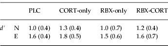
Repeated measures ANOVA on d′ detection sensitivity (Greenhouse–Geisser corrected for non-sphericity) revealed a main effect of emotion (mean±s.d.) [d′: F(1, 46)=45.22, p<0.0001]. This influence of emotion was extremely robust and not further modulated by treatment, i.e. there were neither main effects of RBX or CORT nor interaction effects on d′ (Table 1). Post-hoc paired t tests including the four treatment groups revealed that the main effect of emotion was due to enhanced detection sensitivity [d′: t(50)=−8.13, p<0.0001] for emotional relative to neutral items (Table 1).
Behavioural results

E, emotional; N, neutral; CORT, cortisol; RBX, reboxetine; PLC, placebo.
d-prime (d′) scores were gathered 1 week after pharmacological intervention.
Values are shown as mean (s.d.).
Correlation analyses
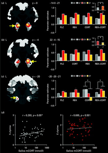
There was a positive correlation between d′ detection sensitivity and mCORTlog for neutral items, i.e. the detection sensitivity improved as a function of increasing CORT levels (n=51, r=0.293, p<0.05). For emotional items, no such relationship was found (n=51, r=0.085, p=0.55) (Fig. 2 d). There were no significant correlations between RBX and d′ for either neutral or emotional items (neutral: n=24, r=0.012, p>0.05; emotional: n=24, r=−0.061, p>0.05).
Brain activation clusters projected upon maximum probability maps provided by the Anatomy toolbox (see text). (a) Main effect of emotion; (b) main effect of memory; (c) three-way interaction between reboxetine (RBX), cortisol (CORT) and emotion. For illustration purposes only, activations are shown at a threshold of p<0.001, uncorrected. Signal plots show parameter estimates for each condition. Small plots in (a) and (b) show mean parameter estimates comprising the main effects of emotion and memory, respectively. (d) Correlation between log-transformed mean CORT levels (mCORT) and d-prime (d′) scores for neutral (N) and for emotional (E) pictures. PLC, placebo; ER, emotional recognized; EF, emotional forgotten; NR, neutral recognized; NF, neutral forgotten; R, recognized; F, forgotten; L, left; R, right.

Imaging data
Main effects
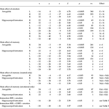
The ROI analysis showed a main effect of emotion, irrespective of the factors subsequent memory or treatment, which encompassed both amygdala and hippocampus bilaterally (Table 2, Fig. 2 a). Subsequent paired t tests revealed that activations of amygdala and adjacent anterior hippocampus were greater for emotional than for neutral items. In contrast, more distant posterior parts of the hippocampus (especially the cornu ammonis subregion) showed the reverse pattern, with greater responses to neutral than to emotional items (p<0.05, FWE-corrected) (Table 2). Moreover, amygdala and hippocampus bilaterally showed a main effect of subsequent memory. Post-hoc paired t tests confirmed that, in both regions, activation was greater for subsequently remembered relative to subsequently forgotten items, irrespective of the factors emotion or treatment (p<0.05, FWE-corrected) (Table 2, Fig. 2 b). As collinearity between the observed main effects is likely, we additionally performed separate analyses of subsequent memory effects for emotional and neutral items. These analyses document a main effect of subsequent memory for emotional items in the amygdala bilaterally and a main effect of subsequent memory for neutral items in both the right amygdala and hippocampus (Table 2).
Functional magnetic resonance imaging data

E, Emotional; N, neutral; r, recognized; f, forgotten; L, left; R, right; RBX, reboxetine; CORT, cortisol.
Brain activations at p<0.05 family-wise error-corrected.
Listed are the effects that yielded significant results and post-hoc tests in the respective clusters.
Interaction effects
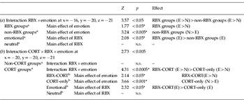
Crucially, the ANOVA showed a two-way interaction between RBX and emotion in the left hippocampus. Post-hoc t tests revealed that this interaction effect was based upon opposite main effects of emotion in the RBX groups (RBX-only, RBX-CORT) and in the non-RBX groups (PLC, CORT-only). In the RBX groups, greater hippocampal activation was elicited by emotional items (Table 3 a); whereas in the non-RBX groups, neutral items elicited greater hippocampal activation (Table 3 a). In a neighbouring cluster, a three-way interaction between RBX, CORT and emotion was found. This interaction effect appeared to be driven by a stronger two-way interaction between RBX and emotion in the CORT groups (CORT-only, RBX-CORT) than in the non-CORT groups (PLC, RBX-only) (Fig. 2 c). Indeed, the presence of a two-way interaction between RBX and emotion in the CORT groups (CORT-only, RBX-CORT) contrasted with the absence of such interaction in the non-CORT groups (PLC, RBX-only) (p>0.05) (Table 3 b). Post-hoc t tests revealed that, in the RBX-CORT group, greater hippocampal responses were elicited by emotional items, whereas neutral items elicited greater hippocampal responses in the CORT-only group (Table 3 b, Fig. 2 c). There were no further two-, three- or four-way interactions between the factors RBX, CORT, emotion and subsequent memory (all p values >0.05). Taken together, these results suggest that treatment with hydrocortisone selectively decreased hippocampal responses to emotional relative to neutral items, whereas treatment with reboxetine potentiated hippocampal responses to these stimuli. Importantly, the inhibitory influence of hydrocortisone was reversed by co-administration of reboxetine.
Functional magnetic resonance imaging data: interaction RBX×emotion (left hippocampus) – post-hoc tests

E, Emotional; N, neutral; n.s., not significant.
(a) Sequential post-hoc tests decomposing the reboxetine (RBX)×emotion interaction in the left hippocampal formation (x=−16, y=−20, z=−21).
(b) Sequential post-hoc tests decomposing the RBX×cortisol (CORT) emotion interaction in the left hippocampal formation (x=−20, y=−20, z=−21).
a and b Indicate second-order and third-order post-hoc tests, respectively.
* Indicates the significance levels of the post-hoc tests adjusted for the size of the respective cluster where the main interaction effect was found.
RBX groups comprised the RBX-only group and the RBX-CORT group. Non-RBX groups comprised the placebo group (PLC) and the CORT-only group. Conversely, CORT groups comprised the CORT-only group and the RBX-CORT group and non-CORT groups comprised the PLC group and the RBX-only group.
Discussion
Imaging results
The present pharmacological fMRI study in 51 healthy subjects showed that experimental elevation of NE and CORT differentially modulated hippocampal responses in an emotional episodic encoding task, depending on whether NE and CORT signalling were challenged separately versus in combination. While elevated NE levels enhanced hippocampal responses to emotional items, elevated CORT levels required the presence of elevated NE levels to promote processing of emotional items, suggesting that CORT alone decreased hippocampal function, whereas it increased the effects of noradrenergic stimulation. In a related experiment, we have shown that elevation of NE and CORT to moderate stress levels induced an amygdala response bias to fear (Kukolja et al. Reference Kukolja, Schläpfer, Keysers, Klingmüller, Maier, Fink and Hurlemann2008a), which is equivalent to the amygdala hyper-responsiveness to fear in patients with PTSD (Rauch et al. Reference Rauch, Whalen, Shin, McInerney, Macklin, Lasko, Orr and Pitman2000; Shin et al. Reference Shin, Wright, Cannistraro, Wedig, McMullin, Martis, Macklin, Lasko, Cavanagh, Krangel, Orr, Pitman, Whalen and Rauch2005). When switching the task to emotional episodic encoding, we observed a similar response bias in the hippocampus, which is known to orchestrate, together with the amygdala, the efficient episodic encoding of emotional memories (Burgess et al. Reference Burgess, Maguire and O'Keefe2002; Phelps, Reference Phelps2004; LaBar & Cabeza, Reference LaBar and Cabeza2006).
In the present study, both amygdala and hippocampus showed robust subsequent memory effects, i.e. their activation during encoding of items predicted later memory for these items, irrespective of whether these items were emotional or neutral. While this result is suggestive of a collinearity between emotion and subsequent memory effects, being the most likely reason for the lack of an interaction between these factors, it is important to note that subsequent memory effects were preserved even when emotional and neutral items were analysed separately, confirming the central role of amygdala and hippocampus, and their interplay, in episodic encoding (Burgess et al. Reference Burgess, Maguire and O'Keefe2002; Phelps, Reference Phelps2004; LaBar & Cabeza, Reference LaBar and Cabeza2006). To some degree, our results imply a functional architecture related to the emotionality of items, with a preferential response bias of the amygdala to the encoding of emotional items and a preferential response bias of the hippocampus to the encoding of neutral items. However, the absence of an interaction between emotion and subsequent memory effects is reflective of largely overlapping amygdala and hippocampal activations, suggesting that episodic encoding engages, to a large extent, the same subset of amygdala-hippocampal neurons.
The observed two-way interaction between the factors emotion and reboxetine treatment suggests that noradrenergic stimulation facilitates hippocampal processing of emotional items at the cost of neutral items. Presuming that enhanced hippocampal responses are reflective of increased encoding efficiency, the present results – on a neural level – support the hypothesis that privileged encoding of emotional information is mediated by elevations of NE and CORT signalling. Originally, Cahill et al. (Reference Cahill, Prins, Weber and McGaugh1994) have shown, within the experimental context of an emotional story paradigm, that β-noradrenergic blockade with propranolol specifically eliminated the consolidation advantage of emotional episodic memories. Consistent with this landmark finding, accumulating evidence from pharmacological fMRI studies suggests that propranolol, when administered prior to episodic encoding, blocks the amygdala response to subsequently remembered emotional information (Strange & Dolan, Reference Strange and Dolan2004) and to emotional information, irrespective of subsequent memory (van Stegeren et al. Reference van Stegeren, Goekoop, Everaerd, Scheltens, Barkhof, Kuijer and Rombouts2005). Here, we demonstrate a reciprocal neural effect by using a strategy of noradrenergic stimulation instead of inhibition.
In the present study, elevation of CORT shifted encoding-related hippocampal activity towards increased responses to neutral information and decreased responses to emotional information. This result replicates our previous findings (Kukolja et al. Reference Kukolja, Schläpfer, Keysers, Klingmüller, Maier, Fink and Hurlemann2008a) and is consistent with observations that elevations of CORT levels reduce fear responses in humans (Soravia et al. Reference Soravia, Heinrichs, Aerni, Maroni, Schelling, Ehlert, Roozendaal and de Quervain2006; Putman et al. Reference Putman, Hermans, Koppeschaar, van Schijndel and van Honk2007), an inhibitory effect implying a protective negative feedback regulation (Soravia et al. Reference Soravia, Heinrichs, Aerni, Maroni, Schelling, Ehlert, Roozendaal and de Quervain2006). However, when NE levels were simultaneously elevated, the inhibitory effect of CORT was reversed, such that CORT synergistically interacted with noradrenergic stimulation in jointly amplifying hippocampal activity. Thus, from a functional perspective, the presence or absence of elevated NE signalling quintessentially determines whether CORT activates or deactivates the hippocampus, suggesting a molecular ‘switch’ by which hippocampal function is physiologically controlled. This hypothesis is in line with rodent studies documenting a noradrenergic dependency of CORT effects on emotional memory (Quirarte et al. Reference Quirarte, Roozendaal and McGaugh1997; McGaugh & Roozendaal, Reference McGaugh and Roozendaal2002; Roozendaal et al. Reference Roozendaal, Okuda, van der Zee and McGaugh2006, Reference Roozendaal, McEwen and Chattarji2009).
Behavioural results
As expected, emotional items were much better recognized than neutral items. Interestingly, however, stress levels of CORT predicted better encoding of neutral items. This result does not only corroborate evidence of the memory-enhancing potential of CORT (Buchanan & Lovallo, Reference Buchanan and Lovallo2001; Abercrombie et al. Reference Abercrombie, Kalin, Thurow, Rosenkranz and Davidson2003; Het et al. Reference Het, Ramlow and Wolf2005; Joels et al. Reference Joels, Pu, Wiegert, Oitzl and Krugers2006; Diamond et al. Reference Diamond, Campbell, Park, Halonen and Zoladz2007; Kukolja et al. Reference Kukolja, Thiel, Wolf and Fink2008b), but also illustrates that neutral items, when paired with (pharmacologically) elevated CORT levels, which usually tag emotional items, can achieve the privileged memory status of these items. This finding underlines that physiological mechanisms underlying the memory advantage of emotional events can, in principle, be mimicked by pharmacological means. In contrast, no additional effect of CORT on the encoding of emotional items was observed, suggesting that their emotional salience was sufficient for driving amygdala and hippocampus to optimal performance, without additional benefit from elevated CORT signalling.
The neural effects of reboxetine are not reflected in the behavioural data, which is consistent with the absence of memory-modulating effects following noradrenergic stimulation with RBX (Papps et al. Reference Papps, Shajahan, Ebmeier and O'Carroll2002; Chamberlain et al. Reference Chamberlain, Muller, Blackwell, Clark, Robbins and Sahakian2006) or the α2-receptor antagonist yohimbine in other fMRI studies (van Stegeren et al. Reference van Stegeren, Roozendaal, Kindt, Wolf and Joels2010). While reboxetine has been shown to modulate emotional episodic encoding in the short term (Hurlemann et al. Reference Hurlemann, Hawellek, Matusch, Kolsch, Wollersen, Madea, Vogeley, Maier and Dolan2005, Reference Hurlemann, Matusch, Hawellek, Klingmuller, Kolsch, Maier and Dolan2007), long-term effects may require much more intense and/or prolonged noradrenergic stimulation to evolve. Thus, the present study was successful in modelling the adaptive neural mechanisms by which moderate stress may shape amygdala and hippocampal function but could, of course, not model the effects of excessive NE release associated with traumatic stress.
Conclusions
The present pharmacological fMRI study translates evidence of stress-related NE–CORT interactions in the modulation of amygdala and hippocampal function from experimental animals to the human. Together with our previous study, the present findings illustrate the adaptive influence of NE and CORT on amygdala-hippocampal responses. These responses may turn maladaptive under circumstances of traumatic stress, leading to pathological hyperencoding and hyperconsolidation of episodic memories of trauma.
Note
Supplementary material accompanies this paper on the Journal's website (http://journals.cambridge.org/psm).
Acknowledgements
This work was supported by a German Research Foundation (DFG) grant (HU1302/2–2) and by a Starting Independent Researcher Grant jointly provided by the Ministry of Innovation, Science, Research and Technology of the State of North Rhine-Westphalia (MIWFT) and the University of Bonn (to R.H.). The authors wish to thank C. Frahnert, H. Kölsch, and P. Wegener for analysing blood samples, K. Linkert for assisting in neuropsychological testing and A. Patin for proofreading the manuscript. In addition, the authors acknowledge the invaluable help of the MRI and Cognitive Neurology groups located at the Institute of Neuroscience and Medicine, Research Center Jülich.
Declaration of Interest
None.