I. INTRODUCTION
It is well known that the textile, painting, leather, printing, and photography industries would release some organic pollutants, which could seriously contaminate the water environment. Reference Domínguez, Beltrán and Rodríguez1,Reference Zhang and Fang2 The removal of the pollutants based on photocatalysis, before they are released to water system, has attracted extensive concerns over the last few decades. Reference Inoue, Fujishima, Konishi and Honda3–Reference Chen, Bae and Oh5 TiO2, a popular semiconductor-based photocatalyst, has been extensively investigated for the degradation of dyes in aqueous solution. Reference Panniello, Curri, Diso, Licciulli, Locaputo, Agostiano, Comparelli and Mascolo6 Due to the broad band gap of 3.0–3.3 eV, however, TiO2 can only work under the ultraviolet (UV) light irradiation. The UV light only occupies about 5% in the normal sunlight. Therefore, utilization and development of photocatalysts' working in the visible light range are of great interest.
Goethite (α-FeOOH), an important member of limonite with a narrow band gap of 2.1 eV, is one of the most common and reactive crystalline iron oxide phases found in the soils and sediments. It is environment friendly and acts as natural regulators of nutrients or pollutants. Reference Manning, Kiser, Kwon and Kanel7–Reference Wang, Ma and Chen9 Recently, α-FeOOH has been reported as a visible light photocatalyst owing to its nontoxicity, low cost, and relatively good stability. Reference Amstaetter, Borch, Larese-Casanova and Kappler10–Reference Zhou, Yang, Wang, Mao, Wang and Liu13 Under UV–visible (UV–vis) irradiation, α-FeOOH easily produces hydroxyl radical (⋅OH) and surface iron complex (e.g., surface Fe(III) species). The ⋅OH is a nonspecific oxidant which can react with most of organic contaminants. Reference Xu and Wang14–Reference Kwan and Voelker16 The highly oxidized and unstable surface iron complex could also react with water to form ⋅OH. Reference He, Ma, He, Zhao and Yu17 So, these characteristics of α-FeOOH make it a promising photocatalyst in the heterogeneous system. Reference Panniello, Curri, Diso, Licciulli, Locaputo, Agostiano, Comparelli and Mascolo6,Reference Murakami, Matsuo, Tsubota and Ohno18,Reference Tong, Guan and Zhang19 To date, various nanostructured α-FeOOH have been fabricated including nanowires, Reference Wang, Ma and Chen9,Reference Amstaetter, Borch, Larese-Casanova and Kappler10 nanobelts, Reference Krýsa, Jirkovský, Bajt and Mailhot12 etc. For instance, Krýsa et al. synthesized α-FeOOH nanorods by a solution method and showed that the photocatalytic degradation of salicylate and oxalate was about 20 h under Philips HPW 125 W lamps. Reference Krýsa, Jirkovský, Bajt and Mailhot12 Zhou et al. fabricated α-FeOOH nanobelts by an assembly method within ferritin and demonstrated that photocatalytic degradation to Rhodamine B is about 70 min with H2O2 additive under a 300 W xenon lamp. Reference Zhou, Yang, Wang, Mao, Wang and Liu13 However, thermodynamically, the powders consisting of such nanoobjects are easily tending to aggregate if they are not chemically surface-modified, which would decrease reusability and weaken the photocatalytic performances. The micro/nanostructured photocatalysts, which consist of microsized objects with nanostructures, could provide an effective way to solve this problem, due to their high activity of nanostructure and stability of the bulk. Reference Krýsa, Jirkovský, Bajt and Mailhot12,Reference Zhou, Yang, Wang, Mao, Wang and Liu13,Reference Kwan and Voelker16 There are some reports about fabrication of micro/nanostructured α-FeOOH by a hydrothermal route. Reference Wang, Wu, Yu, Xu, Lim and Lou8,Reference Wang, Ma and Chen9,Reference Hojamberdiev, Zhu, Eminov and Okada15,Reference Tong, Guan and Zhang19 For instance, Wang et al. synthesized α-FeOOH hollow spheres with hierarchical structures by glycerol-assisted one-pot reaction at 120 °C Reference Wang, Wu, Yu, Xu, Lim and Lou8 and one-pot hydrothermal treatment of Fe2(SO4)3 aqueous solution at 160 °C. Reference Wang, Ma and Chen9
Herein, we present a facile route to fabricate the micro/nanostructured α-FeOOH via a low-temperature water bath treatment of the precursor containing urea and ethanediol, in addition to FeSO4•7H2O, at 95 °C. The obtained α-FeOOH consists of the microsized particles with nanostructure, which are urchin-like in morphology and of high specific surface area (∼118 m2/g). Importantly, such micro/nanostructured α-FeOOH has shown much higher photocatalytic activity to the organic pollutants, such as Rhodamine 6G (R6G), and better re-usable performances than the nanostructured goethite, exhibiting the good material for the environmental treatment.
II. EXPERIMENTAL
All the used chemical reagents, such as ferrous sulfate (FeSO4•7H2O), ethanol (C2H5OH), urea (CO(NH2)2), glycol (C2H6O2), and hydrogen peroxide solution (H2O2), were of analytical grade and obtained from National Medicines Co. Ltd. of China. These reagents were used as-received without further purification.
A. Preparation
The micro/nanostructured α-FeOOH (or MNF for short) was prepared based on a low-temperature water bath treatment at 95 °C. In a typical procedure, the ferrous sulfate solution was first prepared in a three-neck flask by dissolution of FeSO4•7H2O (7 mmol) in 45 mL deionized water and mixing with 45 mL C2H6O2. Subsequently, the urea solution was prepared in a 50 mL conical flask in nitrogen atmosphere by mixing CO(NH2)2 (25 mmol), 15 mL deionized water, and 15 mL C2H6O2. The three-neck flask with the ferrous sulfate solution was put into a thermostatic water tank with the constant temperature at 95 °C and mechanically stirred for 15 min in nitrogen atmosphere. The urea solution in the conical flask was then added into the three-neck flask with a flow rate of 1 mL min−1 by a peristaltic pump in nitrogen atmosphere. After that, the atmosphere was imported into the mixed solution with a flow rate of 3 mL min−1 by the peristaltic pump. After 3 h, the solution was taken out from the water bath and naturally cooled in air. Finally, the powder products were obtained by filtering, washing with distilled water and ethanol several times, and then drying in the vacuum chamber at 55 °C for 6 h.
For reference, the α-FeOOH nanorods were also prepared via a chemical route, as previously described. Reference Tang, Wang, Zhuo, Ge and Cui20 Briefly, a mixture of FeSO4•7H2O (0.5 mmol) and anhydrous CH3COONa (1 mmol) was dissolved in 20 mL of distilled water in a beaker, followed by stirring vigorously for about 20 min. A yellow suspension was thus obtained, and then transferred into a Teflon-lined autoclave up to 80% of the total volume, sealed, and heated at 100 °C for 8 h, before cooling to room temperature naturally. The final yellow solid products were collected by centrifuging, washing with distilled water and absolute ethanol, and then drying at 40 °C under a vacuum for 4 h.
B. Characterization
Phase structure of the products was characterized by x-ray diffraction (XRD, Philips X'Pert Pro MPD) with Cu Kα radiation (1.5406 Å). The morphological observation and microstructural examination were conducted on a field emission scanning electron microscope (FESEM; FEI Sirion-200) and a transmission electron microscope (TEM; JEOL JEM-2010, 200 kV). Thermal gravimetric (TG) analysis and differential thermal analysis (DTA) were carried out on a Pyris 1 thermal analyzer at a heating rate of 10 °C/min from room temperature to 500 °C under the nitrogen environment. The spectral measurements were performed by Fourier transform infrared spectroscopy (FTIR, Nexus). The diffuse reflectance spectra were measured on an UV–visible–near infrared spectrophotometer (Shimadzu, UV3600). The N2 isothermal sorption was measured on a sorption apparatus (Omnisorp 100 CX, Coulter).
C. Evaluation of the photocatalytic activity
The photocatalytic activity of the as-prepared products was evaluated by measuring the optical absorbance of the UV–vis irradiated pollutant-contained solution. Rhodamine 6G (R6G, Alfa Aesar) aqueous solution was selected as a model contamination. Prior to irradiation, 30 mg photocatalyst was mixed with R6G solution (30 mL, 1 × 10⁻⁵ mol L−1) in a 100 mL sample bottle and stirred for 5 min in the dark. Subsequently, 100 μL of hydrogen peroxide solution (H2O2, 30 wt%) was added to the mixed solution (the H2O2 concentration in the mixed solution was 33 mmol L−1). The pH value of the mixed solution was adjusted to 7 by hydrochloric acid or sodium hydroxide. Then, the solution was exposed to a 300 W xenon lamp at the distance of 15 cm with stirring. The irradiated solution was sampled in each interval of 8 min for optical absorbance measurement. The photocatalytic degradation conversion was calculated by the formula:
 $$\eta = {{{A_0} - {A_{\rm{t}}}} \over {{A_0}}}\quad ,$$
where η is the degradation ratio of R6G, A 0 and A t are the absorbances at 525 nm for the R6G solution before and after irradiation for time t, respectively.
III. RESULTS AND DISCUSSION
A. The morphology and structure
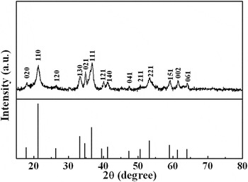
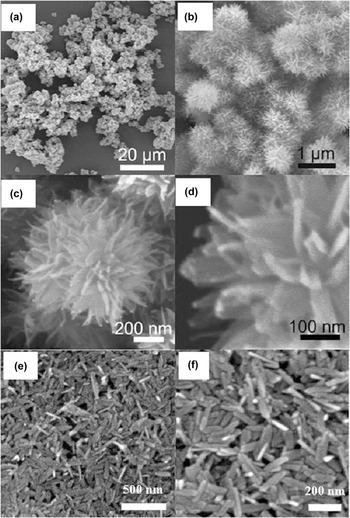
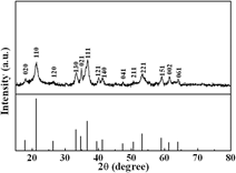
Figure 1 shows the XRD pattern of the as-prepared products. All the diffraction peaks can be indexed to the pure orthorhombic structure of α-FeOOH with lattice constants of a = 4.6188 Å, b = 9.9528 Å, and c = 3.0236 Å (JCPDS No. 81-0462). The reference sample shows the same structure, as similarly seen in Fig. 1. The corresponding FESEM observation has revealed that the as-prepared products or the α-FeOOH powders consist of nearly spherical particles with about 0.5–1 μm in diameter [Fig. 2(a)]. The high magnification image has shown that the microsized α-FeOOH particles are urchin-like in morphology and composed of nanosized leaf-like building blocks in radial arrangement, as clearly seen in Figs. 2(b)–2(d). The reference sample consists of rod-like particles which are 30–50 nm in diameter and 130–220 nm in length, as shown in Figs. 2(e)–2(f).
XRD pattern of the as-prepared products. The line spectrum is the standard diffraction pattern of α-FeOOH powders.

(a–d) FESEM images of the as-prepared products and (e–f) reference sample.

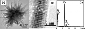
Further, TEM examination was performed to reveal the microstructure of the urchin-like α-FeOOH objects. Figure 3(a) gives the TEM image of a single urchin-like particle. We can see that the particle is built of nanoscaled leaves with about 150–200 nm in length and about 30–50 nm in width. The high resolution TEM has revealed that there is a distinct lattice fringe observed in the nanoleaves, corresponding to the (101) plane of α-FeOOH, as illustrated in Fig. 3(b). The growth of the nanoleaves was preferentially along the direction [101]. The energy-dispersive x-ray (EDX) spectroscopic analysis has confirmed that the products are only comprised of Fe and O elements (the hydrogen could not be observed due to the light element), as demonstrated in Fig. 3(c). The atomic ratio of Fe to O elements is approximately 1:2, which is in good agreement with the elemental ratio in FeOOH. The signals of Cu and C should come from the Cu grid. No impurity such as sulfur was observed in the sample.
Microstructure and composition of urchin-like α-FeOOH particles. (a) TEM image of a single α-FeOOH particle. (b) High resolution TEM image of the segment of a single leaf. The arrow is along the axial direction of the leaf or [101]. (c) EDX spectrum.

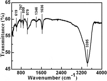
The FTIR spectral measurements were carried out for the urchin-like α-FeOOH, as shown in Fig. 4. The absorption bands, centered at 1346, 979, and 619 cm−1, correspond to the Fe–O vibrational modes. Reference Cwiertny, Hunter, Pettibone, Scherer and Grassian21 The bands at 899 and 797 cm−1 are assigned to the characteristic Fe–O–OH bending vibration modes in α-FeOOH, Reference Cwiertny, Hunter, Pettibone, Scherer and Grassian21,Reference Musi, Krehula and Popovi22 indicating that the surfaces of the urchin-like α-FeOOH particles were hydroxylated. The hydroxylated surface could adsorb water molecules, which has been confirmed due to the existence of the bands at 3395 and 1636 cm−1, in the spectrum, corresponding to the H2O stretching and bending modes, respectively. Reference Morterra, Chiorlno and Borello23
FTIR spectrum of the as-prepared α-FeOOH.

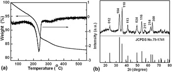
Furthermore, the nitrogen sorption isotherms of the as-prepared urchin-like micro/nanostructured α-FeOOH were measured, as illustrated in Figure S1. The specific surface area was thus estimated to be about 118 m2/g, much larger than that of the reference sample or the α-FeOOH nanorods (only 52 m2/g). The TG and DTA results show that there are two main weight loss processes in the temperature ranges from 30 to 200 °C and from 200 to 380 °C, respectively, as illustrated in Fig. 5(a). The weight loss (∼6.0%) from 30 to 200 °C should be attributed to the desorption of physically adsorbed water, whereas that in the range of 200–380 °C (∼10%) could be ascribed to the decomposition of α-FeOOH due to the reaction:
 $$2\alpha {\hbox-} {\rm{FeOOH}} \to {\rm{F}}{{\rm{e}}_2}{{\rm{O}}_3} + {{\rm{H}}_2}{\rm{O}}\quad .$$
(a) TG and DTA curves of the as-prepared α-FeOOH. (b) The XRD pattern of the α-FeOOH after calcination at 500 °C. The line spectrum is the standard diffraction of α-Fe2O3.

No significant weight loss occurred when the temperature is higher than 380 °C. According to Eq. (2), the weight loss induced by decomposition of α-FeOOH can be estimated to be 10.1%, which is consistent with the experimental measurement. As a consequence, the stable residue could reasonably be ascribed to α-Fe2O3. For further confirmation, α-FeOOH was calcined at 500 °C for 5 h in air before XRD measurement. Figure 5(b) shows the corresponding result and indicates the formation of α-Fe2O3.
B. Morphological evolution with reaction time
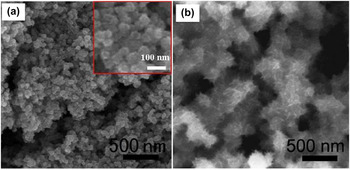
To understand the formation of such urchin-like micro/nanostructured α-FeOOH, evolution of the products' morphology with reaction time was examined, as shown in Fig. 6. When the reaction was for a short time (say, about 6 min), only spherical α-FeOOH nanoparticles with about 30–50 nm in size were observed [Fig. 6(a)]. If the reaction time was prolonged to 30 min, the larger α-FeOOH particles of about 200–300 nm in size were formed with large number of nanoleaves on the particle surface, as illustrated in Fig. 6(b). Finally, when the reaction was long enough (about 3 h or longer), the urchin-like micro/nanostructured α-FeOOH objects were obtained, as shown in Fig. 2.
(a) FESEM images of the products after reaction for 6 min and (b) 30 min, respectively.

C. Formation of the urchin-like α-FeOOH
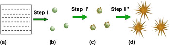
On basis of the morphological evolution with reaction time shown in Fig. 6, the formation process of the urchin-like α-FeOOH could be proposed, involving two steps during the water bath synthesis, or Step I: formation of spherical α-FeOOH nanoparticles, and Step II: growth of nanoleaves on the spherical nanoparticles, as schematically illustrated in Fig. 7.
Schematic illustration of formation process for the urchin-like α-FeOOH particles. (a) The reaction solution. (b) The spherical particles. (c) The particles with initially growing nanoleaves. (d) The urchin-like α-FeOOH particles. Step I: formation of spherical α-FeOOH particles; Step II: growth of nanoleaves on the spherical particles.

During preparation of the precursor solution and subsequent reaction at 95 °C, the following reactions would take place.
 $${\rm{CO}}{\left( {{\rm{N}}{{\rm{H}}_2}} \right)_2} + {{\rm{H}}_2}{\rm{O}} \to {\rm{C}}{{\rm{O}}_2} + 2{\rm{N}}{{\rm{H}}_3}\quad ,$$
 $${\rm{N}}{{\rm{H}}_3} + {{\rm{H}}_2}{\rm{O}} \to \mathop {{\rm{N}}{{\rm{H}}_4}}\nolimits^ + + {\rm{O}}{{\rm{H}}^ - }\quad ,$$
 $${\rm{F}}{{\rm{e}}^{2 + }} + {\rm{O}}{{\rm{H}}^ - } \to {\rm{Fe}}{\left( {{\rm{OH}}} \right)_2}\quad ,$$
 $$4{\rm{Fe}}{\left( {{\rm{OH}}} \right)_2} + {{\rm{O}}_2} \to 4{\rm{FeOOH}} + 2{{\rm{H}}_2}{\rm{O}}\quad {\rm{.}}$$
At the initial stage, the urea was slowly decomposed during heating and ammonia would be released over 70–80 °C according to reaction (3). Reference Wang, Cai, Lin, Wang and Liang24 The ammonia was then hydrolyzed, forming hydroxyl ions, through reaction (4). The hydroxyl ions reacted with Fe2+ to form Fe(OH)2 in nitrogen atmosphere [reaction (5)]. Finally, the Fe(OH)2 reacted with O2, under the ambient conditions, to form FeOOH according to reaction (6). Here, the Fe2+ ions are easily oxidized to Fe3+, as previously reported. Reference Tong, Guan and Zhang19 When the concentration of FeOOH in solution was high enough, the nucleation and fast growth of α-FeOOH can lead to the spherical morphology, which is a kinetic crystallization process as clarified by the chemical bonding theory of single crystal growth Reference Sun and Xue25,Reference Sun and Xue26 [see Fig. 6(a) or Step I in Fig. 7].
As mentioned above, there exist hydroxy groups on α-FeOOH nanoparticles' surface (Fig. 4). After the formation of α-FeOOH nanoparticles, their surface hydroxy groups could adsorb ethylene glycol in the solution. Such adsorption would hinder the further spherical or isotropic growth of the nanoparticles. In this case, preferential growth could take place on some special sites on the nanoparticles' surface, where no ethylene glycol was adsorbed, forming the spokewise nanoleaves on the α-FeOOH nanoparticles, as shown in Fig. 6(b) or Step II of Fig. 7. Finally, the urchin-like micro/nanostructured particles were thus obtained after enough long reaction.
D. Photocatalytic performances
1. Measurements
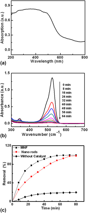
The optical absorption spectrum of the urchin-like α-FeOOH, from the diffuse reflectance spectral measurement, is shown in Fig. 8(a). The absorption edge is at about 550 nm, indicating that α-FeOOH could be used as the potential visible light high efficient photocatalyst. The catalytic activity of the as-prepared α-FeOOH was evaluated by photodegradation of R6G under xenon lamp illumination. Figure 8(b) shows the optical absorption spectral evolution of the R6G solution with irradiation time, in the presence of the as-prepared urchin-like α-FeOOH and H2O2. The strong absorbance band in the visible region around 525 nm is attributed to the monomer and dimmer of R6G in the solution. Reference Igarashi, Maeda, Takao, Oki and Kusama27,Reference Arbeloa, Tapia Estévez, Arbeloa and Arbeloa28 The absorbance band decreases with increasing irradiation time. It was suggested that both chromophores and aromatic rings of R6G have been destroyed. Reference Li and Kikugawa29 After irradiation for 56 min, the R6G was fully degraded and the solution turned into transparency. The absorption peak around 525 nm completely disappeared. For the reference sample or the α-FeOOH nanorods, the R6G was completely degraded in 80 min under the same conditions (see Fig. S2). The catalytic activity of the urchin-like α-FeOOH is higher than that of the rod-like α-FeOOH nanoparticles, as clearly shown in Fig. 8(c). The photocatalytic activity of the urchin-like α-FeOOH is more than three times as high as that of the flower-like nanostructured TiO2 for degradation of R6G, Reference Zhang, Ding and Zhou30 and very close to that of P-25 under the same conditions (see Fig. S4). For the R6G solution with hydrogen peroxide but without catalysts, the R6G degradation ratio was only 19% in 80 min in our experiment [Fig. 8(c)]. Further, under the dark conditions, the R6G concentration was only decreased by 23% in 60 min after addition of the α-FeOOH catalyst in the R6G solution with hydrogen peroxide, as shown in Fig. S3. This should be attributed to the adsorption of R6G on α-FeOOH.
(a) Optical absorption spectrum of the urchin-like α-FeOOH. (b) Time-dependent optical absorbance spectra of the R6G solution (the starting concentration: 1 × 10⁻⁵ M, 30 mL) containing H2O2 (33 mmol L−1) and the urchin-like α-FeOOH (30 mg) after its exposure to the xenon lamp for different durations. (c) Degradation ratio of R6G as a function of time for different catalysts.

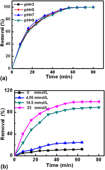
We also studied the effect of initial pH values. The corresponding results are presented in Fig. 9(a). The initial pH values have only insignificant effect on the photodegradation in the range of 3.0–9.0. The removal rates of the R6G in the solution within the pH range of 3.0–9.0 were above 99% after irradiation for about 60 min.
(a) Influence of pH values and (b) H2O2 amount in the R6G aqueous solution on photocatalytic performances.

In addition, it should be mentioned that the presence of H2O2 in the solution is important for the catalysts to degrade R6G in the solution under photoirradiation, as shown in Fig. 9(b). Without addition of H2O2, the degradation ratio of R6G was only about 13%. The photocatalytic degradation ratio of R6G increased with rising in H2O2 concentration, and when H2O2 is up to 33 mmol L−1, the degradation ratio could reach 100% within 60 min.
2. Structurally enhanced mechanism of photocatalytic activity
When the catalyst α-FeOOH was added into the R6G aqueous solution with H2O2 under photoirradiation, the reaction
 $$\alpha {\hbox-} {\rm{FeOOH}} \to \alpha {\hbox-} {\rm{FeOOH}}\left( {{{\rm{e}}^ - },\;{{\rm{h}}^ + }} \right)\quad ,$$
would take place. At this time, there are two electron transfer pathways. On one hand, the photogenerated electrons are trapped by H2O2 on the surface of α-FeOOH photocatalysts to form ⋅OH radicals, or
 $${{\rm{H}}_2}{{\rm{O}}_2} + {{\rm{e}}^ - } \to {\rm{O}}{{\rm{H}}^ - } + \cdot {\rm{OH}}\quad .$$
On the other hand, the electrons can be trapped by the Fe3+ on the surface of α-FeOOH particles to form Fe2+, or
 $${\rm{F}}{{\rm{e}}^{3 + }} + {{\rm{e}}^ - } \to {\rm{F}}{{\rm{e}}^{2 + }}\quad .$$
The reduced Fe2+ ions would react with H2O2 to form ⋅OH radicals in addition to (Fe3+ + OH−), or
 $${\rm{F}}{{\rm{e}}^{2 + }} + {{\rm{H}}_2}{{\rm{O}}_2} \to {\rm{F}}{{\rm{e}}^{3 + }} + {\rm{O}}{{\rm{H}}^ - } + \cdot {\rm{OH}}\quad .$$
The formed radicals would lead to degradation of the R6G, according to the reaction Reference Yu, Yu, Huang, Zhang and Dai31–Reference Li, Chen, Yang, Jin and Liu33
 $${{\rm{C}}_{28}}{{\rm{H}}_{32}}{{\rm{N}}_2}{{\rm{O}}_3} + \cdot {\rm{OH}} \to {\rm{C}}{{\rm{O}}_2} + {{\rm{H}}_2}{\rm{O}} + {\rm{HN}}{{\rm{O}}_3}\;<$$><$$>\left( {{\rm{or}}\;{\rm{small}}\;{\rm{molecules}}} \right)\quad .$$
The radicals ⋅OH are the nonselective strong oxidant, which can oxidize some organic compounds into CO2 and H2O. Reference Jiang, Zhang, Liu and Xu34
The high photocatalytic activity of the urchin-like α-FeOOH particles could be easily understood. It can be attributed to their special structure. The urchin-like α-FeOOH possesses a much higher specific surface area, due to the micro/nanostructure, than the reference sample. Furthermore, the urchin-like micro/nanostructure would effectively prevent aggregation and keep a high active surface area, which is favorable for highly efficient photocatalytic degradation of organic pollutants in wastewater. Reference Lu, Cai and Zhang35 Comparatively, for the nanoparticles' or nanorods' powders, the undesired aggregation during the photocatalytic reaction usually induces a significant decrease in the active surface area and hence the photocatalytic performance. Additionally, the nanoscaled thickness of the leaves on the urchin-like α-FeOOH particles is close to the region where the quantum size effect is prominent. The nanoleaves' band gap broadening, induced by the quantum size effect, would not only bring higher redox potentials, but also promote electrons' transferring from the conductive bands of nanoleaves with high electric-potential to those of the core-part microparticles with low electric-potential. It is well known that band positions would move and higher redox potentials of the free electrons and holes will be achieved accordingly in this region, which reduces the probability of the photogenerated electron/hole pair recombination and in turn enhances the charge-transfer rates in the materials. Reference Ye, Bando, Shen and Golberg36,Reference Chen, Shen, Guo and Mao37 On basis of all these above, the urchin-like α-FeOOH particles would promote formation of the more radicals ⋅OH [see reactions (7–10)] and thus induce an enhanced photocatalytic performance.
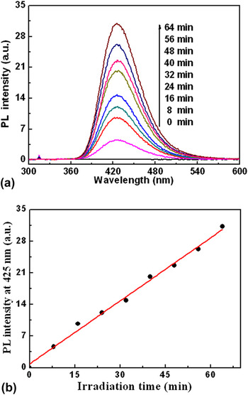
From reaction (11), the existence of radicals in the solution is crucial to R6G's degradation. For further confirmation, irradiation-induced ⋅OH radicals were measured based on the photoluminescence (PL) technique using terephthalic acid (TA) as the probe molecules, which would readily react with ⋅OH to produce highly fluorescent product 2-hydroxyterephthalic acid. Reference Musi, Krehula and Popovi22 The PL intensity of 2-hydroxyterephthalic acid at 425 nm is proportional to the amount of ⋅OH produced on the surface of the photocatalyst. The measurement details are described as previously reported. Reference Xiao, Si, Zhang, Xiao and Tan38 Briefly, 30 mg of α-FeOOH powder sample was dispersed in a 30 mL 5 × 10⁻⁴ mol L−1 TA aqueous solution with 1 mL 2 × 10⁻³ mol L−1 NaOH in a 50 ml quartz tube. Subsequently, 100 μL of hydrogen peroxide solution (H2O2, 30 wt%) was added to the mixed solution and stirred for about 5 min in the dark. The experiment was carried out under irradiation using a 300 W xenon lamp. PL spectra of the generated 2-hydroxyterephthalic acid were thus recorded with excitation by 320 nm light, as illustrated in Fig. 10(a). The PL peak at 425 nm corresponds to 2-hydroxyterephthalic acid. Reference Musi, Krehula and Popovi22 The PL intensity at about 425 nm increases linearly with the irradiation time, as shown in Fig. 10(b). However, such PL peak was not observed without irradiation or in the absence of α-FeOOH powders under irradiation within 60 min. This indicates that the PL peak is from the chemical reaction between TA and ⋅OH occurred at the interface of H2O2/α-FeOOH. The increasing trend of ⋅OH radicals is consistent with the decreasing trend of R6G concentration in Fig. 8. The ⋅OH radicals produced on the urchin-like α-FeOOH particles should be much more than those on the reference sample.
(a) PL spectral evolution with the irradiation time for the TA solution (5 × 10⁻⁴ M) in the presence of the urchin-like α-FeOOH (the details are seen in the text). (b) Plot of PL intensity at 425 nm versus irradiation time [data from (a)].

As for insignificant effect of the initial pH value in the solution on the photodegradation processes, shown in Fig. 9(a), it should be ascribed to the acid-alkaline buffering capacities of the catalyst caused by Fe-polycations [Fe n (OH) m (H2O) x ](3n−m)+, Reference Chen and Zhu39 which were intermediate derivatives between the primary hydrolyzed products and the insoluble FeOOH due to the intrinsic features of Fe-cations in the solution. Reference Li, Li and Xue40,Reference Chen, Chen, Wang, Song and Xue41 Under the alkaline condition, most bases would be consumed by the Fe-polycations, and as a result, the degradation of R6G could occur effectively just as in an acidic condition under irradiation.
3. Reusability
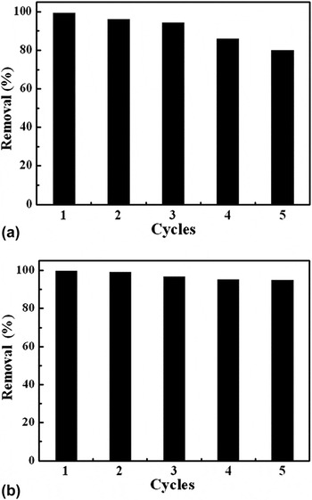
The recycling photocatalytic efficiency of the urchin-like α-FeOOH particles was also tested. Experiments were carried out (the reaction time is for 56 min at pH of 7.0). The solid powders were carefully separated from the treated solution and rinsed with water and nonaqueous ether 3 times respectively to remove any possible remnants on surface of the catalyst before next cycle. The corresponding results are shown in Fig. 11(a) or Fig. S5.
The recycling photocatalytic efficiency of the urchin-like α-FeOOH particles (the details are seen in the text). (a) After reaction time for 56 min for each cycle. (b) After reaction time for 80 min for each cycle.

The R6G degradation was higher than 95% in the first three runs, showing high structural stability of the urchin-like α-FeOOH particles. In the fourth and fifth runs, the efficiency of the catalyst decreased to 86 and 80%, respectively, which could be attributed to unavoidable leaching of iron and possible effect of contaminant adsorption on reactive sites on the catalyst surface. Nevertheless, when the reaction time was prolonged to 80 min, the R6G degradation was above 95% for five cycles, as shown in Fig. 11(b) or Fig. S5.
IV. CONCLUSIONS
In summary, the urchin-like α-FeOOH with micro/nanostructure has been fabricated by a simple low-temperature water bath method based on the reaction of urea and ferric sulfate aqueous solution at 95 °C. The obtained α-FeOOH particles are microsized and composed of nanosized leaf-like objects in radial arrangement, showing high specific surface area (∼118 m2/g). The formation of urchin-like morphology could be attributed to the ethylene glycol-adsorption induced preferential growth of nanoleaves on the preformed spherical particles. Importantly, due to its unique structure, such urchin-like α-FeOOH has shown much higher photocatalytic activity to the R6G, under the existence of H2O2, than the goethite nanorod powders, exhibiting significant structural enhancement of the photocatalytic performance. Also, because of its high structural stability, this material has good recycling photocatalytic performances. Since the α-FeOOH is a mineral substance in natural word and its disturbance to the environment is comparatively small, this work could thus provide a useful candidate of environmental materials for pollution treatment.
Supplementary Material
To view supplementary material for this article, please visit http://dx.doi.org/jmr.2015.103.
ACKNOWLEDGMENTS
This work was financially supported by the National Key Basic Research Program of China (Grant No. 2013CB934303), the China Postdoctoral Science Foundation (2013T60632), and the CAS/SAFEA International Partnership Program for Creative Research Teams.