Introduction
Transorbital endoscopic approaches are becoming increasingly popular in the management of skull base pathologies. They offer better access and exposure of the lateral skull base compared to the well-described transnasal approaches, and have limited morbidity, improved aesthetic outcomes and faster post-operative recovery compared to traditional open approaches.Reference Di Somma, Andaluz, Cavallo, Keller, Solari and Zimmer1
A novel, minimally invasive transorbital endoscopic approach has been described using the superior lateral orbital portal.Reference Moe, Bergeron and Ellenbogen2–Reference Luzzi, Zoia, Rampini, Elia, Del Maestro and Carnevale4 This has been utilised successfully in the treatment of various pathologies, including sphenoid wing meningiomas, and in the repair of cerebrospinal fluid leaks and orbital trauma, and in orbital decompressions.Reference Vural, Carobbio, Ferrari, Rampinelli, Schreiber and Mattavelli5–Reference Lim, Sung, Kim, Yoo, Jung and Choi10
As with any novel surgical approach, further study and innovation are necessary to maximise surgical efficiency and exposure, while minimising morbidity. The transorbital approach includes a narrow corridor and can have limited surgical maneuverability.Reference Kong, Kim and Hong11 Here, we provide a description of the relevant anatomy and tips for better access to the superior lateral orbital portal, to increase surgical efficiency and safety while preserving outcomes. This paper describes the approach taken for a resection of a sphenoid wing meningioma. Written consent was obtained for use of the patient clinical photographs.
Technical description
The transorbital approach is indicated for various orbital and intracranial pathologies. Depending on the treating institute and the extent of surgery required, a multidisciplinary approach to treatment is encouraged.
The approach described below should be performed in conjunction with the ophthalmology and neurosurgery departments. Pre-operative imaging is encouraged, using high-resolution computed tomography of the skull base. Magnetic resonance imaging of the skull base may also be indicated. It is important to obtain pre-operative ophthalmological examination findings in order to document visual function.
The patient should be counselled on all possible complications associated with the approach, including keratoconjunctivitis sicca resulting from injury to the lacrimal gland, diplopia caused by injury to extraocular muscles, and blindness due to injury to the optic nerve.
Surgical preparation
The patient is positioned in a similar manner as in routine functional endoscopic sinus surgery (FESS), lying supine with their head flat or flexed 15 degrees. Their head may be placed in Mayfield pins, as directed by the neurosurgery department.
The eye is cleansed prior to draping, with either normal saline or diluted Betadine®. The surgical site is prepped with 0.5 ml local anaesthetic (1:80 000 adrenaline with 2 per cent lidocaine), injected along the proposed brow incision line. The cornea is kept moist or protected with a corneal protector throughout the duration of the procedure, to prevent corneal ulceration or keratitis.
The following instruments should be available: camera system; 0-degree and 30-degree, 4 mm diameter, standard 18 cm length endoscopes; standard FESS tray; sharp curved pointed iris scissors; fine forceps; ribbon retractors and dural retractors – ensure a variety of sizes are available (8 mm, 10 mm, 15 mm); small cat's paw retractor; Freer suction elevator; Freer elevator; handheld drill that allows only a short length of burr shaft exposed between the drill handle and burr tip; various sizes of cutting and diamond burrs (1 mm, 2 mm, 4 mm); bipolar forceps; and (with or without) a Ligaclip® (ligating clip) applicator.
Procedure
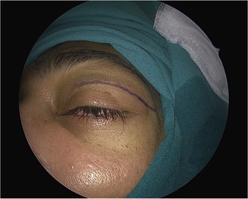
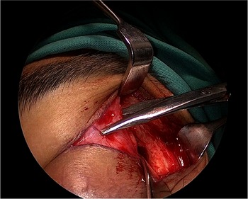
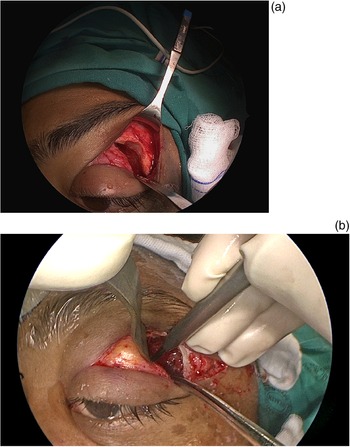
An incision is made within the superior lid crease, as for blepharoplasty surgery. This is extended laterally, sparing the lateral canthus (Fig. 1). Once the skin and orbicularis muscle are transected, scissors are used to dissect onto the bone, just lateral to the lateral orbital rim (Fig. 2). Approximately 20 mm of lateral periosteum and overlying temporalis is stripped to expose the outer aspect of the lateral orbital rim. This allows for identification of the lateral orbital rim, and retraction of the soft tissues of the upper lid and eyebrow to allow palpation of the superior orbital rim. The soft tissue above the superior orbital rim can then be transected, avoiding any potential injury to the levator palpebrae superioris muscle, as this muscle inserts onto the periosteum of the inferior surface of the superior orbital rim (Fig. 3a). This is an important step in gaining quick access to the superior and lateral portal.
Skin incision in natural crease line of the upper eyelid, as for blepharoplasty surgery.

Sharp dissection of soft tissue onto the bone lateral to the lateral orbital rim.

(a) Transecting soft tissue above the superior orbital rim to avoid injury to the levator muscle. (b) Whitnall's tubercle.

The next step entails a subperiosteal dissection of the lateral and then superior orbital rim. Laterally, the first structure to identify is Whitnall's tubercle, a bony prominence onto which the upper and lower canthal tendons attach. (Fig. 3b) Elevating the periosteum off Whitnall's tubercle preserves these ligaments and avoids injury to the lateral canthal angle of the eyelid.Reference Whitnall12 Here, tissue is very adherent to the orbital wall, and the sharp tip of curved iris scissors or a Cottle's elevator is useful. Once the periosteum is elevated, a Freer elevator is used to quickly lift the rest laterally and superiorly. A 10 mm malleable ribbon retractor can then be inserted between the orbit and the lateral wall to retract the orbital contents medially. It is important to keep the ribbon retractor parallel with the lateral orbital wall, to maximise the portal width.
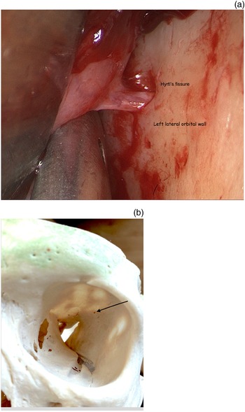
The next constant landmark is a recurrent branch of the middle meningeal artery (Fig. 4). This artery, also called the anastomotic branch to the middle meningeal artery, is an arterial branch arising from the lacrimal or ophthalmic artery that anastomoses with the anterior division of the middle meningeal artery through Hyrtl's canal.Reference Akdemir Aktaş, Ergun, Tatar, Arat and Hayran13–Reference Diamond15 Hyrtl's canal is a foramen within the greater wing of the sphenoid. Once this artery has been ligated using bipolar forceps, subperiosteal dissection continues posteriorly towards the superior orbital fissure, which lies approximately 1 cm posterior to Hyrtl's fissure.
(a) Recurrent branch of the middle meningeal artery. (b) Hyrtl's fissure (arrow).

Attention is then turned to widening the surgical portal. Surgical loupes can be used up to this point. Subsequently, an 18 cm length, 4 mm diameter, 0 degree endoscope allows four hands to comfortably work through the lateral orbital corridor, with better visualisation to identify the temporalis muscle and dura early.
A Freer dissector is used to develop a subperiosteal plane, to medialise the orbital contents. It is important to create space during the initial part of the procedure by thinning the lateral orbital wall using a drill, until the temporalis muscle is visualised beneath the thinned bone (Fig. 5). Special care should be taken within the first 1 cm in the surgical corridor, to ensure that the exposed shaft of the drill bit does not get caught up in the orbital fat or temporalis muscle. Drilling starts just posterior to lateral orbital rim (about 5 mm), leaving the rim intact and obviating the need for any plating, as would be required in the standard orbitotomy approach.
Exposing the temporalis muscle early is important.

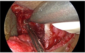
The lacrimal fossa is an area within the superior lateral bony orbit where the lacrimal gland rests. Once the periosteum over the lacrimal gland is elevated, this area of bone can be safely drilled away, enlarging the surgical corridor to create the ‘lacrimal keyhole’ (Fig. 6).Reference Goldberg, Kim and Kerivan16 The endoscope can also rest in this area and it serves as a good stabilisation point during the surgery, similar to the soft tissue triangle during FESS (Fig. 7).Reference Goldberg, Kim and Kerivan16
(a) Lacrimal keyhole. (b) Superior lateral portal in the left eye, showing the proper orientation of orbital retractors.

Superior lateral portal in the left eye, showing distal access and a sagittal crest just medial to the drill.

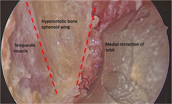
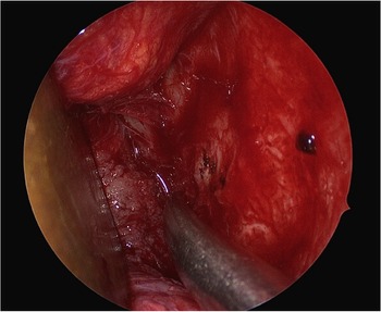
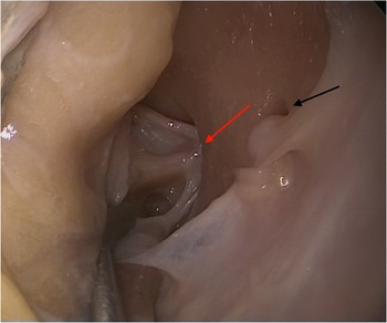
In the case of a sphenoid wing meningioma, the hyperostotic bone and intraosseous meningioma are easily recognised as distinctly different to normal bone (Fig. 8). As soon as the cancellous portion of the greater sphenoid wing is encountered, a bony tunnel is created allowing for a thin wall of bone on either side, protecting the temporalis muscle laterally and the periorbita medially. This allows for a quick debulking of the hyperostotic bone. Navigation is used in the majority of cases but is not essential. It is useful to show the extent of bone that requires resection and the proximity to the superior orbital fissure. The hyperostotic bone and intraosseous meningioma are removed to expose: the temporalis muscle anterior-laterally, the dura of the temporal lobe posterior-laterally, the anterior cranial fossa superiorly and the periorbita medially. The superior orbital fissure can be identified (Fig. 9), and the neurovascular structures can be protected by drilling from a lateral to medial direction. As the lateral wedge of bone of the greater sphenoid wing is drilled away, the final ‘crest’ of bone is removed using a 2 mm drill burr or Kerrison punch. This then exposes the meningo-orbital band.Reference Dallan, Di Somma, Prats-Galino, Solari, Alobid and Turri-Zanoni17,Reference Bly, Ramakrishna, Ferreira and Moe18 Finally, once exposure is complete, addressing the intracranial or orbital pathologies can be performed in conjunction with the neurosurgery or ophthalmology departments.
Hyperostotic bone and meningioma involving the lateral orbital wall.

Superior orbital fissure (red arrow), found 1 cm posterior to the recurrent branch of the middle meningeal artery (black arrow).

Discussion
Maximal exposure within the superior lateral corridor is important to enable manipulation of the endoscope and other instruments comfortably during the surgery. The lateral or superior lateral portal can be compared with the transsphenoidal corridor to the pituitary fossa. In order to allow four-handed surgery, it is essential to have a wide portal, with the endoscope resting in one area during the neurosurgical part of the dissection to obtain optimal access to the middle cranial fossa.
Important tips to ensure maximal and quick access to the middle cranial fossa are as follows. First, the superior eyelid incision should extend in a natural crease line lateral to the lateral canthus, sparing the lateral canthus. Second, dissection lateral to the lateral orbital rim should be performed first, in order to free the soft tissues superior to the superior orbital rim. Once scissors can be passed between the superior orbital rim and the soft tissue, the tissue can be cut in one swift move, without risking injury to the levator muscle. Third, the lateral orbital rim should be preserved to prevent plating post-operatively. Fourth, Whitnall's tubercle must be identified, and the ligaments elevated in a subperiosteal plane. Fifth, the lacrimal keyhole should be drilled away. This widens the corridor significantly and is an ideal place to rest the endoscope. Sixth, the recurrent branch of the middle meningeal artery and Hyrtl's fissure should be identified and cauterised. Seventh, the superior orbital fissure is located 1 cm posterior to Hyrtl's fissure, and care should be taken to avoid directly retracting the tissue exiting the superior orbital fissure, to prevent superior orbital fissure syndrome. Eighth, it is important to use the correctly sized orbital retractor (usually starting with 10 mm), and to bend the retractor so that the blade medialising the orbital contents is parallel to the orbit. Angulating the distal tip posteriorly and medially will narrow the entrance to the corridor and increase pressure on the orbital structures. Finally, the pupil of the eye should be checked periodically to ensure retraction is not causing raised intraocular pressure or vascular congestion. If the pupil changes shape or size, the retractors should be relaxed for 30–60 seconds.
Conclusion
Transorbital endoscopic approaches are becoming increasingly popular in the management of skull base pathologies. The superior and lateral orbital portal is advantageous in allowing access to the middle cranial fossa; however, it can have the disadvantage of a narrow surgical corridor. This technical description of a resection of a sphenoid wing meningioma highlights tips for maximising the surgical portal and minimising morbidity. These include: dissecting onto bone lateral to the orbital rim in order to avoid injury to the levator muscle, elevating periosteum off Whitnall's tubercle to preserve the lateral canthus, identifying the recurrent branch of the middle meningeal artery 1 cm anterior to the superior orbital fissure, and drilling away the lacrimal keyhole to widen the surgical portal.
Competing interests
None declared