Introduction
Facial nerve palsy is a relatively common presentation to an ENT department. The majority are idiopathic.Reference Adour1 Lipomas in the head and neck are common, benign neoplasms. They are mesenchymal in origin and are typically found in the subcutaneous tissues. They are less commonly encountered in internal organs and are usually asymptomatic. Only five previous cases of middle-ear lipomas have been described in the English literature. To our knowledge, none presented with facial palsy.
Case report
A 33-year-old man presented to the ENT emergency clinic with a one-day history of right-sided, lower motor neurone facial palsy. The preceding week, he had suffered from an upper respiratory tract infection, with some right otalgia and clear otorrhoea. As a child, he had undergone insertion of ventilation tubes for bilateral otitis media with effusion, and had also undergone a right modified radical mastoidectomy for cholesteatoma. He had been asymptomatic for 20 years.
On presentation to our unit, the patient had a grade two House–Brackmann palsy and a mildly congested right tympanic membrane. The rest of the ENT examination was normal.
A diagnosis of acute otitis media and right facial palsy was made. The patient was treated with a seven-day course of oral prednisolone 60 mg once daily, ciprofloxacin 500 mg twice daily, acyclovir 800 mg five times daily and Gentisone HC ear drops (Roche, Basel, Switzerland) thrice daily.
At one-week review, the patient's facial palsy had deteriorated to House–Brackmann grade three. The tympanic membrane remained congested and was noted to be retracted with an underlying effusion. The pure tone audiogram revealed a moderate right conductive hearing loss.
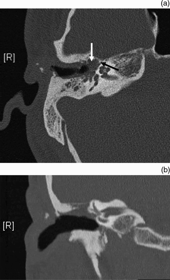
At four-week review, the patient's facial palsy had improved (to House–Brackmann grade two). However, the apparent middle-ear effusion persisted. A computed tomography (CT) scan revealed abnormal soft tissue in the epitympanic recess, extending to the region of the geniculate ganglion (Figure 1).
(a) Axial and (b) coronal computed tomography images demonstrating a soft tissue mass (white arrow) adjacent to the horizontal portion of the facial nerve in the fallopian canal (black arrow).

In view of the patient's history of mastoidectomy for cholesteatoma, recurrence was suspected, and it was decided to undertake a middle-ear exploration. This revealed an encapsulated lump of fatty tissue filling the anterior middle-ear cleft, juxtaposed to the horizontal portion of the facial nerve.
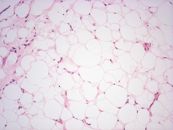
Histological analysis confirmed the presence of a lipoma, with evidence of chronic inflammation within the benign tumour (Figure 2).
Histological section of the mass found in the right middle ear, demonstrating adipose tissue and scanty lymphocytes in a fibrous stroma (H&E; × 100).

Post-operatively, the patient made a good recovery. His facial palsy resolved within a few weeks of surgery.
Discussion
Lipomas in the middle ear are rare, and only five cases have been previously reported. We performed a literature review by searching PubMed and Ovid, using the key words ‘lipoma’, ‘middle ear’ and ‘mastoid’.
Luetje et al. Reference Luetje, Milbrath and Linthicum2 found that 17 per cent of 240 temporal bones examined at the House Ear Institute demonstrated varying amounts of fat juxtaposed to the mastoid vertical portion of the facial nerve. Essentially, two theories exist as to the origin of fat in the middle ear. Luetje et al. postulated that, embryologically, fat marrow which has persisted in the mastoid could lead to the growth of ectopic fat in the middle ear. Their other hypothesis was that mastoid and VIIth nerve development could have dragged fat cell precursors into the middle ear, which could then lead to lipoma development.Reference Luetje, Milbrath and Linthicum2
Stegehuis et al. Reference Stegehuis, Guy and Anderson3 described a most unusual case of a middle-ear lipoma that extended down the eustachian tube into the oropharynx, presenting with airway distress in a 64-year-old woman. Selesnick et al.,Reference Selesnick, Edelstein and Parisier4 Abdullah et al. Reference Abdullah, Williamson, Gallimore and Shah5 and Edmonds et al. Reference Edmonds, Woodroof and Ator6 all reported middle-ear lipomas in children presenting with middle-ear effusions and conductive hearing loss. Abdullah et al. described bilateral middle-ear lipomas which were found to be obstructing the eustachian tube and causing a middle-ear effusion in a five-year-old child.Reference Abdullah, Williamson, Gallimore and Shah5 Luetje et al. reported the case of a 16-year-old adolescent with bilateral lipomas who presented with an incidental, unilateral, high frequency sensorineural hearing loss.Reference Luetje, Milbrath and Linthicum2 None of the previously reported cases of middle-ear lipoma has been associated with facial palsy. Agarwal et al., Reference Agarwal, Mishra and Pratap7 however, described a case of unilateral facial nerve palsy caused by a primary liposarcoma in a four-year-old boy.
Our patient's lipoma had presumably developed since his childhood mastoidectomy, as there was a considerable quiescent period after this procedure. It is possible that the mastoidectomy disturbed fat precursor cells from persisting bone marrow fat, or triggered a growth response in the adipose cells already present in the middle ear, by the mechanisms proposed above. Possible aetiologies for the VIIth nerve palsy include direct nerve compression from the lipoma, or, more likely, the effect of the acute otitis media due to eustachian tube obstruction secondary to the lipoma. This latter theory is supported by the fact that the patient's facial nerve palsy showed signs of improvement prior to surgery.
This case report reminds us to be thorough in our evaluation of patients with facial nerve palsies, especially when there is a history of previous mastoid surgery. Recurrent cholesteatoma is the most likely aetiology, and surgical exploration and decompression would be advised. Computed tomography is the first-line investigation; in this case, magnetic resonance imaging may have shown a mass with similar intensity to fat.
Conclusion
Lipomas are a rare but real differential diagnosis of a mass in the middle ear. When removed, the surgeon should expect any pre-existing facial nerve palsy to recover. To our knowledge, the present case represents the first published report of a facial nerve palsy arising secondary to a middle-ear lipoma.