Implication
Histological observation of testicular structure of cattleyak aged 10, 12 and 14 months in this study identified that germ cell reduction was more obvious and spermatogenic arrest may become more serious as early as the cattleyak were 12-month old. Then, we characterized the testis protein expression pattern of cattleyak by comparing testis proteome of cattleyak aged 12 and 14 months to that of cattleyak aged 10 months, respectively. The differentially expressed testis proteins of cattleyak aged 12 and 14 months did not indicate significant differences from those aged 10 months, respectively, which confirmed the result of comparative histological study. On the other hand, some of the differentially expressed proteins that were coexpressed in the testis of cattleyak aged 12 and 14 months were possibly associated with testis development and spermatogenesis, which could be selected as candidate proteins in future study of the mechanisms of cattleyak infertility.
Introduction
Bos grunniens (yak) are the most important domesticated species with outstanding adaptability to the alpine climates of the Qinghai-Tibetan Plateau (Cai et al., Reference Cai, Mipam, Zhao and Sun2015). Cattleyak are resulted from crossbreeding domestic cattle and yak, and exhibit equivalent adaptability to yak and equivalent performance to cattle. Therefore, cattleyak breeding is one of the most important directions in hybrid improvement of yaks. In the history of animal husbandry on Qinghai-Tibetan Plateau, cattleyak contributed much more to social and economic development than yaks (Li et al., Reference Li, Li, Zhao, Xu, Gu, Zhu, Xie and Liu2010). However, the F1 males of cattleyak are sterile, which greatly restricts the effective utilization of this hybrid. As a result of the infertility of F1 males, many valuable genes cannot be fixed and passed down to next generations. Up to now, the mechanisms of cattleyak sterility have not been revealed.
Over the past decades, much work has been done focusing on the mechanisms of the cattleyak infertility, while little progress has been made. Many researchers have carried out relevant research based on morphological anatomy, histological observation and karyotype analysis. The seminiferous tubules in the testis of cattleyak were found to be much thinner than those in yaks and the numbers of spermatogonia, primary spermatocytes and secondary spermatocytes decreased sharply at each developmental stage (Lu and Zi, Reference Lu and Zi2014). Karyotype analysis showed that cattleyak have the same number of chromosomes (2n=60) with that of cattle and yaks (Guo, Reference Geissler, Davis, Wu, Bradshaw, Patel, Mendonca, Elliston, Wilson, Russell and Andersson1983). A study on relationship between synaptonemal complex (SC) and male infertility of cattleyak indicated that the SC structure of autosomes could be observed only in a few primary spermatocytes of cattleyak and many of them were abnormal in morphology. Meanwhile, no XY bivalents were observed in primary spermatocytes of cattleyak (Hu et al., Reference Hu, Zhong, Chen and Cai2000). In recent years, some researchers attempted to explore the mechanisms of male sterility of cattleyak by means of molecular genetics and epigenetics. The expression level of SYCP3 in cattleyak testis was found to be significantly lower than that in cattle and yak testis, and this gene was considered to be correlated with male sterility of cattleyak. The testicular expression level of SNRPN gene in cattleyak were also lower than that in their parental species, and the transcriptional inhibition of this gene was due to its higher methylation level, which contributed to the meiotic arrest of spermatogenesis in cattleyak (Pan et al., Reference Pan, Liu, Li, Yu, Li, Xie and Li2010). Lower expression level of DDX4 was also suggested to be involved in cattleyak male sterility, and the promoter hypermethylation was correlated to the lower expression of DDX4 in the testes of cattleyak (Zhou et al., Reference Zhou, Luo, Li, Jia, Xie, Zhao, Zhong and Li2013). In recent years, comparative analysis of transcriptomes of testicular tissues between cattleyak and yak identified 9089 differentially expressed genes, in which 5000 were upregulated and 4089 were downregulated in cattleyak (Zeng et al., Reference Zeng, Chai, Wang, Ma, Yang, Song and Zhong2014). Two-dimensional gel electrophoresis combined with MS distinguished 19 differentially expressed testicular proteins between cattleyak and yak, in which three chaperons may be related to the male infertility of cattleyak (Fu et al., Reference Fujihara, Tokuhiro, Muro, Kondoh, Araki, Ikawa and Okabe2014).
However, it is a fundamental work to identify specific time point when spermatogenic arrest occurs in cattleyak. Both cattleyak and yak manifest sexual behavior (mount behavior) without penile erection around the age of 12 months and spermatids initially present in the seminiferous tubules of yak (Lei et al., Reference Lei, Sun, Xu and Liu1964), while no spermatids could be observed in cattleyak due to spermatogenic arrest. In order to examine the abnormal development of testis and spermatogenic arrest of cattleyak, it is essential to study the subtle and sequential changes of histological structure and proteome of testis around this age stage (12 months). The isobaric tag for relative and absolute quantification (iTRAQ) is a quantitative proteomic approach and has the potential to provide novel information with the greater proteome coverage (Pottiez et al., Reference Peskin, Low, Paton, Maghzal, Hampton and Winterbourn2012). All these prompted us to investigate the subtle and sequential differences between the testis structure and proteomes of cattleyak aged 10, 12 and 14 months.
Material and methods
Sample collection
F1 males of cattleyak sampled are the crossbreed between Tibetan taurine (♂) and Maiwa yak (♀). Nine cattleyak individuals from three developmental stages (10, 12 and 14-month old) were sampled on a pasture in Hongyuan county of Sichuan province. Each developmental stage comprised three biological repetitions of different individuals. Testis tissue of each animal was obtained by veterinary surgical operation. Caudal epididymis, fat and fascia tissues surrounding the testis, and part of rete testis in the testis were eliminated. Then, the obtained testicular samples were immediately frozen in liquid nitrogen and transported to laboratory.
Histology and histochemistry of testicular samples
We applied hematoxylin–eosin method to examine the histological features of testicular samples of cattleyak from different developmental stages. Each sample was processed routinely for paraffin embedding, and sections were cut and stained with hematoxylin–eosin for morphogenetic analysis. Micrographs of each section was obtained with the BA400Digital camera system and analyzed by the software Motic Images Advanced (Motic Electric Group Co. Ltd, Xiamen, China). The image was magnified 100 times.
Protein preparation
Testis tissue sample of each cattleyak individual was ground into powder by pestle in the mortar with liquid nitrogen, extracted with lysis buffer (7 M urea, 2 M thiourea, 4% 3-[(3-cholamidopropyl)dimethylammonio]-1-propanesulfonate, 40 mM Tris-HCl, pH 8.5) containing 1 mM phenylmethylsulfonyl fluoride (Sigma-Aldrich, Buchs, SG, Switzerland) and 2 mM ethylenediaminetetraacetic acid (Sigma-Aldrich) (final concentration). After 5 min, 10 mM dithiothreitol (DTT; Sigma-Aldrich) (final concentration) was added to the samples. After being sonicated at 200 W for 15 min, the suspension was centrifuged at 30 000×g for 15 min at 4°C. The 5× volume of chilled acetone containing 10% (v/v) trichloroacetic acid was homogenized with the supernatant and incubated at −20°C overnight. The mixture was centrifuged at 30 000×g for 5 min at 4°C and the supernatant was removed. The precipitate was washed with chilled acetone for three times, then the pellet was air-dried for 5 to 10 min and dissolved in lysis buffer (7 M urea, 2 M thiourea, 4% NP40, 20 mM Tris-HCl, pH 8.0 to 8.5). After being sonicated at 200 W for 15 min, and the suspension was centrifuged at 30 000×g for 15 min at 4°C. The supernatant was moved to a new tube, was mixed with 10 mM DTT (final concentration) and was incubated at 56°C for 1 h to reduce disulfide bonds in proteins of the supernatant. To block the thiol groups associated with cysteine residues, 56 mM iodoacetamide (final concentration) was added and the sample was incubated for 1 h in the darkroom. Chilled acetone of 55× volume was well mixed with the supernatant for 2 h at −20°C to precipitate proteins. After centrifuged at 4°C, 30 000×g, the supernatant was removed. The pellet was air-dried for 5 min and then dissolved in 500 μl 0.5 M tetraethylammonium bromide (TEAB; Applied Biosystems, Milan, Italy). The sample was sonicated at 200 W for 15 min and was centrifuged at 30 000×g for 15 min at 4°C. The supernatant was transferred to a new tube and quantified, protein concentration was determined using the modified Bradford assay protocol (Bio-Rad, Berkeley, CA, USA). The supernatant proteins were kept at −80°C until analysis. Then the proteins from 10, 12 and 14-month-old cattleyak were pooled, respectively. The protein pool of each developmental stage comprised equivalent amount of protein from each cattleyak individual.
Isobaric tag for relative and absolute quantification labeling
From each sample pool solution, 100 μg of total protein was taken out and then was digested with Trypsin Gold (Promega, Madison, WI, USA) with the ratio of protein : trypsin=30 : 1 at 37°C for 16 h. Subsequently, peptides were dried by vacuum centrifugation and reconstituted in 0.5 M TEAB and mixed with 24 μl isopropanol by vortexing for 1 min. iTRAQ experiment was performed in double duplex manner using the iTRAQ Reagent-8 Plex Multiplex Kit (Applied Biosystems, Foster City, CA, USA). Samples were labeled with the iTRAQ tags as follow: P1 (114 tag), P2 (116 tag) and P3 (119 tag), and incubated at room temperature for 2 h. Following labeling and quenching, the eight iTRAQ-labeled peptides were pooled together and vacuum concentration to dryness (Pereira et al., Reference Pottiez, Wiederin, Fox and Ciborowski2011).
Strong cation exchange fractionation
iTRAQ-labeled peptides were fractionated by strong cation exchange (SCX) chromatography using the LC-20AB HPLC Pump system (Shimadzu, Kyoto, Japan). The dried peptide mixture was reconstituted and acidified with 4 ml buffer A (25 mM NaH2PO4 in 25% acetonitrile (ACN), pH 2.7) and loaded onto a 4.6×250 mm Ultremex SCX column containing 5-μm particles (Phenomenex, Shanghai, China). The peptides were eluted at a flow rate of 1 ml/min with a gradient of 5% to 56% buffer B (25 mM NaH2PO4, 1 M KCl in 25% ACN, pH 2.7) for 27 min, 60% to 100% buffer B for 1 min. After maintained at 100% buffer B for 1 min, the system was equilibrated with buffer A for 10 min before the next injection. The elution was monitored by absorbance at 214 nm, and fractions were collected every 1 min. The eluted peptides were pooled into 20 fractions and then desalted with a Strata XC18 column (Phenomenex) and vacuum-dried.
Liquid chromatography electrospray ionization tandem mass spectrometry analysis based on TripleTOF 5600
Each of the above-mentioned peptide fraction was resolved in buffer A (0.1% formic acid in 5% ACN) and centrifuged at 20 000×g for 10 min, with a final concentration of 0.5 μg/μl on average for the peptide; 10 μl supernatant was injected in an LC-20AD nanoHPLC (Shimadzu). Then, 10 μl supernatant was loaded onto a 2 cm C18 trap column installed on a LC-20AD nanoHPLC by the autosampler. Then, the peptides were eluted onto a 10 cm analytical C18 column (inner diameter 75 μm) packed in-house. The samples were loaded at 8 μl/min for 4 min onto the trap column, then the 35 min gradient was run at 300 nl/min starting from 2% to 35% buffer B (0.1% formic acid in 95% ACN), followed by 5 min linear gradient to 60%, then, followed by 2 min linear gradient to 80%, and then maintained in 80% buffer B for 4 min before finally returning to 5% in 1 min.
The TripleTOF 5600 settings were as follows: spray voltage, 2.5 kV; curtain gas, 30 psi; nebulizer gas, 15 psi; and an interface heater temperature, 150°C. The mass spectrometry was operated with an reversed phase of ⩾30 000 full-width at half-maximum for time-of-flight (TOF) MSscans. For information dependant acquistion, survey scans were acquired in 250 ms and as many as 30 product ion scans were collected if exceeding a threshold of 120 counts/s and with a 2+ to 5+ charge-state. Total cycle time was fixed to 3.3 s. Q2 transmission window was 100 Da for 100%. Four time bins were summed for each scan at a pulser frequency value of 11 kHz through monitoring of the 40 GHz multichannel time-to-digital converter detector with four-anode channel detect ion. A sweeping collision energy setting of 35±5 eV coupled with iTRAQ adjust rolling collision energy was applied to all precursor ions for collision-induced dissociation. Dynamic exclusion was set for 1/2 of peak width (15 s), and then the precursor was refreshed off the exclusion list.
Data analysis
For peptide data analysis, raw data files acquired from the Orbitrap were converted into MGF files using Proteome Discoverer 1.2 (PD 1.2; Thermo). Proteins were identified using Mascot server engine (version 2.3.02; Matrix Science, London, UK) against Bos_taurus_ncbi database containing 63 379 non-redundant sequences (http://www.ncbi.nlm.nih.gov/protein?term=txid9913[Organism]).
Searches of protein identification were performed using the following criteria: allowing up to one missed cleavages, Gln→pyro-Glu (N-term Q), oxidation (M) and deamidated (NQ) were set as variable modifications; carbamidomethyl (C), iTRAQ8plex (N-term), iTRAQ8plex (K) were set as fixed modifications, and peptide and fragment mass tolerance values were 15 ppm and 0.02 Da, peptide charges of 2+ and 3+. Specifically, an automatic decoy database search was performed in Mascot by choosing the decoy checkbox in which a random sequence of database is generated and tested for raw spectra as well as the real database. Protein identification was accepted at a false discovery rate of 1% at protein and peptide level, with at least two unique peptides. All reported data were based on 95% confidence intervals for protein identification as determined by a Mascot probability analysis greater than ‘identity’ were counted as identified. Protein identification was supported by at least one unique.
For protein quantitation, it was required that a protein contains at least two unique spectra. Quantitative protein ratios were weighted and normalized using the median ratio in MASCOT. We only used ratios with P-values<0.05, and only fold changes of >1.2 were considered as significant.
Gene ontology analysis
The list of proteins obtained from the iTRAQ data was exported to Excel and contained protein-specific information such as accession numbers assigned to each identified protein, protein quant number matching individual proteins. Gene ontology (GO) analysis of the differentially expressed proteins was performed by searching Geneontology (http://www.pantherdb.org/index.jsp) for protein annotation (Wu et al., Reference Wang, Saxe, Tanaka, Chuma and Lin2006).
The PANTHER classification and function analysis
Differential expressed proteins were submitted to PANTHER classification system (http://www.pantherdb.org/index.jsp) to identify biological processes. The function of differentially expressed proteins were retrieved in Genecards system (http://www.genecards.org) (Faulkner et al., Reference Faulkner, Elia, Mullen, O’Boyle, Dunn and Morris2012).
Results
Histological features of testicular samples of cattleyak
Histological section of testicular samples of cattleyak from three developmental stages (10, 12 and 14-month old) indicated similar morphology. The base membrane of seminiferous tubule was shrunk and there were no germ cells in the highly vacuolated seminiferous tubule. Only a small number of germ cells (spermatogonia) could be observed around the regions of the base membrane of seminiferous tubule. The number of germ cells significantly decreased in seminiferous tubule of cattleyak aged 12 and 14 months in comparison with those aged 10 months (Figure 1). Therefore, germ cell reduction was more obvious and spermatogenic arrest may become more serious in 12-month-old cattleyak.
Hematoxylin–eosin-stained testicular samples of cattleyak aged 10 (a), 12 (b) and 14 months (c). The basement membrane and lumen of seminiferous tubule were indicated by the arrow and asterisk, respectively. The spermatogenic cell was indicated by arrowhead.

Differentially expressed proteins between different developmental stages
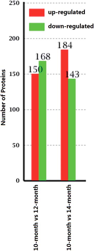
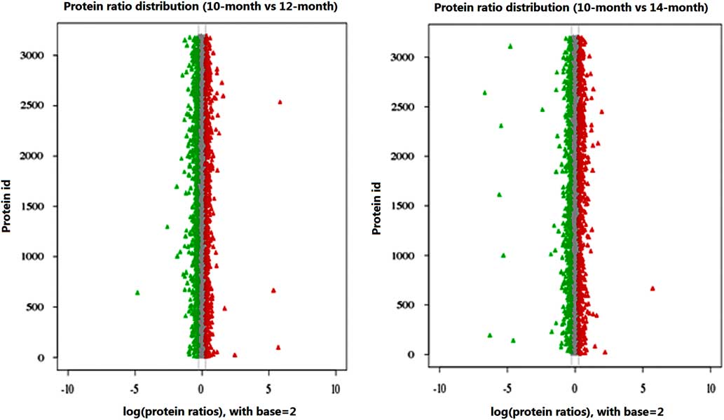
A total of 645 differentially expressed proteins were identified between different developmental stages using the iTRAQ-labeled proteomic approach. Compared with testis proteome at 10-month stage, 150 proteins were significantly upregulated and 168 downregulated at 12-month stage, 184 were significantly upregulated and 143 downregulated at 14-month stage (Figures 2 and 3), which was consistent with comparative histological features of testicular samples of cattleyak from different developmental stages (Figure 1).
Differentially expressed proteins between different developmental stages. In 10-month stage v. 12-month stage, 150 protein expressions were significantly upregulated and 168 were downregulated. In 10-month stage v. 14-month stage, 184 protein expressions were significantly upregulated and 143 were downregulated.

Protein ratio distribution. X-axis presents log2 value of multiple fold differences. Y-axis presents identified proteins. Red (up) and green (down) dot mean the protein has significant difference (false discovery rate⩽0.001, twofold difference).

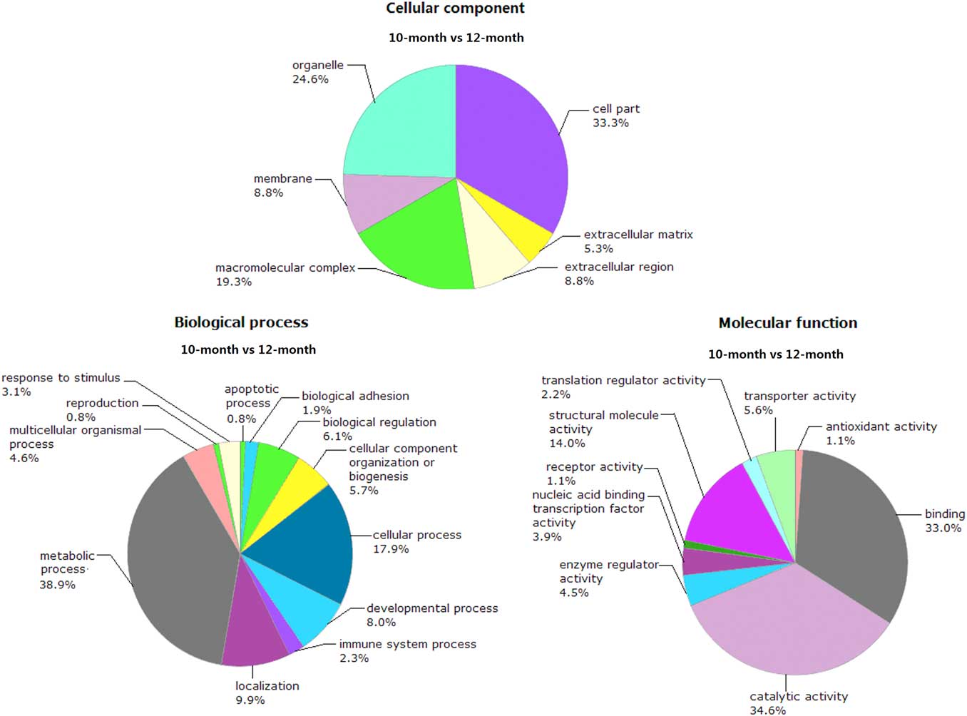
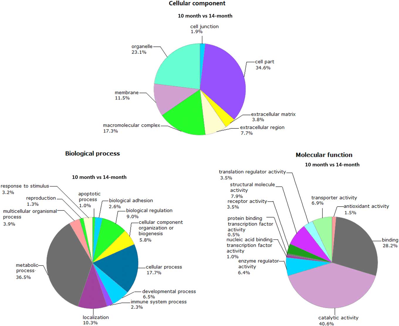
The result on the GO annotations of the 318 differentially expressed proteins in 10-month v. 12-month stage was shown in Figure 4. Regarding cellular component, most of the proteins were located cell part (33.3%), organelle (24.6%), macromolecular complex (19.3%), extracellular matrix (5.3%) and extracellular region (8.8%). Regarding biological process, the majority of the proteins were involved in metabolic process (38.9%), cellular process (17.9%), localization (9.9%), developmental process (8.0%), biological regulation (6.1%) and cellular component organization or biogenesis (5.7%). Regarding molecular function, 34.6% of the proteins participated in catalytic activity, 33.0% in binding, 14.0% in structural molecule activity, 5.6% in transporter activity and 4.5% in enzyme regulator activity. The GO annotations of the 327 differentially expressed proteins in 10-month v. 14-month stage were shown in Figure 5. In regard of cellular component, most of the proteins were located cell part (34.6%), organelle (23.1%), macromolecular complex (17.3%), extracellular region (7.7%) and extracellular matrix (3.8%). Regarding biological process, the majority of the proteins were involved in metabolic process (36.5%), cellular process (17.7%), localization (10.3%), biological regulation (9.0%), developmental process (6.5%) and cellular component organization or biogenesis (5.8%). Regarding molecular function, 40.6% of the proteins participated in catalytic activity, 28.2% in binding, 7.9% in structural molecule activity, 6.9% in transporter activity and 6.4% in enzyme regulator activity.
The annotation of cellular component, biological process and molecular function of differentially expressed proteins in testis at 12-month stage compared with 10-month stage. The percentages are the total hits divided by the number of annotated proteins for each gene ontology category.

The annotation of cellular component, biological process and molecular function of differentially expressed proteins in testis at 14-month stage compared with 10-month stage. The percentages are the total hits divided by the number of annotated proteins for each gene ontology category.

The co-differentially expressed proteins at 12 and 14-month stage compared with 10-month stage
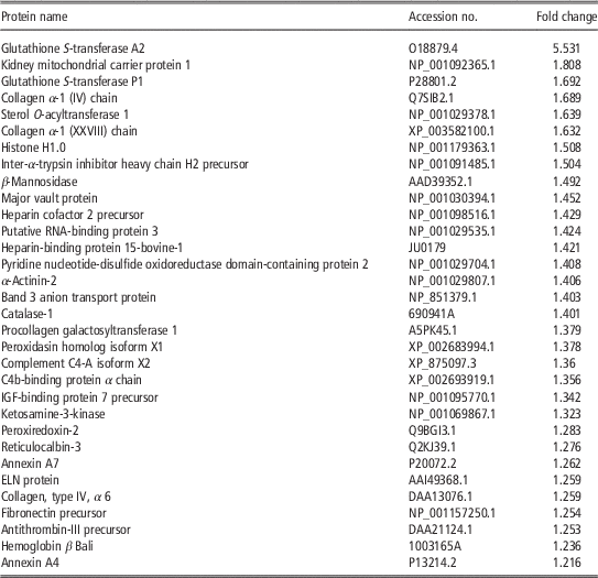
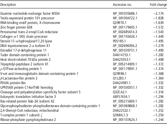
Some differentially expressed proteins were found to be coexisted at 12 and 14-month stage compared with 10-month stage, which may play important roles in testicular development and reproduction with the individual growth and development. Compared with the proteome 10-month stage, 32 proteins were identified to be commonly upregulated and 24 downregulated at 12 and 14-month stage (Tables 1 and 2). The fold change of the upregulated proteins ranged from 1.216 to 5.531, in which glutathione S-transferase A2 (GSTA2), kidney mitochondrial carrier protein 1 and glutathione S-transferase P1 (GSTP1) were the three top listed ones (Table 1). Among the upregulated proteins, some were necessary for detoxification and neutralization of reactive oxygen species (ROS) (GSTA2, GSTP1), reproduction and development (RNA-binding protein, heparin-binding proteins (HBPs), elastin protein, etc.) or involved in membrane fusion and exocytosis (Annexin A4 and Annexin A7). The fold change of the downregulated proteins ranged from −1.244 to −2.174, in which guanine nucleotide exchange factor MSS4 (MSS4), testis-expressed protein 101 (TEX101) precursor and RNA-binding motif protein, X chromosome (RBMX) were the three top listed (Table 2). Among the downregulated proteins, some were involved in basic metabolic processes (ribose-phosphate pyrophosphokinase 2,2,4-dienoyl-CoA reductase, MSS4) or individual reproduction and development (TEX101, RBMX, tudor domain containing). The downregulation of these proteins in the testis of cattleyak may affect the development of seminiferous epithelium and lead to infertility.
The common proteins upregulated at 12 and 14-month stage compared with 10-month stage

The common proteins downregulated at 12 and 14-month stage compared with 10-month stage

Function analysis of the co-differentially expressed proteins at 12 and 14-month stage compared with 10-month stage
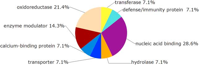
The co-differentially expressed proteins (56) at 12 and 14-month stage compared with 10-month stage were categorized by the PANTHER classification system (http://www.pantherdb.org/index.jsp) into eight biological processes, which is shown in Figure 6.
PANTHER classification of testis protein according to biological function. Proteins were categorized into eight biological processes, including transferase (7.1%), defense/immunity protein (7.1%), nucleic acid binding (28.6%), hydrolase (7.1%), transporter (7.1%), calcium-binding protein (7.1%), enzyme modulator (14.3%) and oxidoreductase (21.4%).

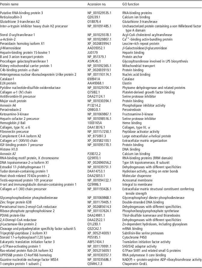
The above-mentioned 56 proteins were found to be upregulated or downregulated simultaneously at 12 and 14-month stage compared with 10-month stage. These proteins were most likely to be related to important cellular pathways in cattleyak spermatogenesis, which could be shown by their functions listed in Table 3. Most of the proteins were identified to be involved in the regulation of germ cell proliferation, growth, differentiation and other metabolic activities.
The function of common proteins differentially expressed at 12 and 14-month stage compared with 10-month stage

GO=gene ontology; LPS=lipopolysaccharide; ELN=elastin.
Discussion
In the current study, histological observation of testicular structure of cattleyak aged 10, 12 and 14 months in this study identified that germ cell reduction was more obvious and spermatogenic arrest may become more serious in 12-month-old cattleyak. Then, testis proteomes of cattleyak from three different developmental stages were examined by comparative iTRAQ proteomics. In total, 645 differentially expressed proteins were identified at 12 and 14-month stage compared with 10-month stage, in which 56 were common proteins differentially expressed at 12 and 14-month stage. The functions of these deferentially expressed proteins were classified and analyzed in detail, which could provide valuable information to clarify protein expression patterns in cattleyak testis from different developmental stages and enable us to understand testis tissue protein composition in cattleyak of male infertility.
Compared with the 10-month testicular proteome, the GO annotations of the differentially expressed proteins at 12-month did not indicate significant discrepancies from those at 14-month, which confirmed the histological observation that germ cell reduction was more obvious and spermatogenic arrest may become more serious in 12 and 14-month-old cattleyak. Some proteins were co-upregulated or co-downregulated at 12 and 14-month stage compared with 10-month stage, which were identified to regulate gene expressions and protein stabilization in spermatogenesis. Putative RNA-binding protein 3 was found to be co-upregulated at 12 and 14-month stage and play a possible role in RNA processing or translational control during early spermatogenesis (Ma et al., Reference Matsunaga, Takata, Morimoto, Hayashihara, Higashi, Akatsuchi, Mizusawa, Yamakawa, Ashida, Matsunaga, Azuma, Uchiyama and Fukui1993). The upregulation of HBPs also indicated its functions required for sperm development. HBPs originate from the accessory sex glands and modulate heparin–sperm interactions that are indicative of fertility. Presence of three molecular weight HBP variants on sperm was previously associated with increased fertility potential of bulls (Bellin et al., Reference Bellin, Hawkins, Oyarzo, Vanderboom and Ax1996 and Reference Bellin, Oyarzo, Hawkins, Zhang, Smith, Forrest, Sprott and Ax1998) and a targeted mutation in tudor domain-containing protein 1 led to male sterility because of postnatal spermatogenic defects (Chuma et al., Reference Chuma, Hosokawa, Kitamura, Kasai, Fujioka, Hiyoshi, Takamune, Noce and Nakatsuji2006). Specifically, HBPs have been shown to play vital roles in spermatozoon survival, induction of sperm capacitation and the overall fertilization process, and any alteration in these proteins can be directly related to infertility (Miller et al., Reference Ma, Inglis, Sharkey, Bickmore, Hill, Prosser, Speed, Thomson, Jobling and Taylor1990). Seven HBPs have been purified from human seminal fluid and the information on HBPs would be useful for identifying potential biomarkers of fertility (Kumar et al., Reference Kumar, Hassan, Kashav, Singh and Yadav2008 and Reference Kumar, Hassan, Tomar, Kashav, Nautiyal, Singh, Singh and Yadav2009). On the other hand, downregulation of other germ cell-associated proteins may impair germline integrity and sperm–egg interaction. Downregulation of TEX101 on spermatozoa may lead to the production of fertile spermatozoa (Fujihara et al., Reference Fu, Li, Liu, Ma, Ren, Huang, Jin, Lin and Zheng2013). RBMX is a regulator for maintenance and centromeric protection of sister chromatid cohesion during meiosis and depletion of RBMX by RNA interference causes the loss of cohesin from the centromeric regions before anaphase, resulting in premature chromatid separation accompanied by delocalization of the shugoshin complex and outer kinetochore proteins (Matsunaga et al., Reference Miller, Winer and Ax2015). Lower expression of tudor domain-containing protein 1 may contribute to the non-integrity of cattleyak germline, because this protein was identified to play a central role during spermatogenesis by participating in the repression transposable elements and preventing their mobilization, which is essential for the germline integrity (Wang et al., Reference Wu, Wang, Baek and Shen2009). Heat shock-related 70 kDa protein 2 (HSPA2) cooperates with other chaperones to stabilize preexistent proteins against aggregation and mediate the folding of newly translated polypeptides in the cytosol as well as within the organelles (Peskin et al., Reference Pereira, Ow, Barrios-Llerena, Wright, Moradas-Ferreira and Tamagnini2007). HSPA2 was present in the acrosomal domain of spermatozoa and was found to be in close association with sperm adhesion molecule 1 and arylsulfatase A in sperm–egg interaction. However, reduced expression of HSPA2 impaired sperm–egg interaction and infertility (Redgrove et al., Reference Redgrove, Nixon, Baker, Hetherington, Baker, Liu and Aitken2012).
The study also identified the co-downregulation of an androgen-related protein at 12 and 14-month stage, which was involved in testicular development and spermatogenesis. Various 17-hydroxysteroid dehydrogenases (17HSDs) regulate the levels of active androgens and estrogens in a tissue-specific manner (Moghrabi et al., Reference Miyamoto, Izumi, Miyamoto, Kubota, Tawara, Sasaguri and Kohno1997), and mutations in the 17HSD3 gene lead to male pseudohermaphroditism (Geissler et al., Reference Guo1994). Estradiol 17-β-dehydrogenase11 (17HSDXI) was suggested to play a role in androgen metabolism during steroidogenesis and a possible role in nonsteroidogenic tissues including paracrine modulation of 5α-androstane-3α,17β-diol levels. 17HSDXI could act by metabolizing compounds that stimulate steroid synthesis and/or by generating metabolites that inhibit it (Chai et al., Reference Chai, Brereton, Suzuki, Sasano, Obeyesekere, Escher, Saffery, Fuller, Enriquez and Krozowski2003). The co-downregulation of 17HSDXI in P2 and P3 stages may contribute to the lower level of androgens and spermatogenic arrest in cattleyak.
Some proteins involved in basic metabolisms were co-downregulated at 12 and 14-month stage. For example, peroxisomal trans-2-enoyl-CoA reductase participates in chain elongation of fatty acids and has no 2,4-dienoyl-CoA reductase activity (Amery et al., Reference Amery, Mannaerts, Subramani, Van Veldhoven and Fransen2001). Ribose-phosphate pyrophosphokinase 2 catalyzes the synthesis of phosphoribosylpyrophosphate that is essential for nucleotide synthesis, which can be activated by magnesium and inorganic phosphate and be inhibited competitively or non-competitively by ADP, 2,3-bisphosphoglycerate or guanosine diphosphate (Ishizuka et al., Reference Ishizuka, Iizasa, Taira, Ishijima, Sonoda, Shimada, Nagatake and Tatibana1992). Another protein, 2,4-dienoyl-CoA reductase is the auxiliary enzyme of β-oxidation, participating in the metabolism of unsaturated fatty enoyl-CoA esters and catalyzing the NADP-dependent reduction of 2,4-dienoyl-CoA to yield trans-3-enoyl-CoA (Koivuranta et al., Reference Koivuranta, Hakkola and Hiltunen1994). These proteins are necessary for maintaining basic activities and growth and development of spermatogenic cells. The downregulation of these proteins was likely to be the reason of testicular dysplasia in cattleyak.
However, oxidative stress is also harmful to the proliferating spermatogonia during early spermatogenesis. Upregulation of glutathione S-transferases (GSTA2, GSTP1) was helpful to detoxify and neutralize the ROS in male reproductive system and play protective role during spermatogenesis (Hering et al., Reference Hering, Lecewicz, Kordan, Majewska and Kaminski2015). Peroxiredoxin-2 (Prx2) was regarded as an important peroxidase and have been shown to play a key role in protecting cells against oxidative stress by scavenging ROS as well as participating in cell signal transduction (Miyamoto et al., Reference Moghrabi, Head and Andersson2009). However, the upregulation of Prx2 in spermatogenic cells of cattleyak may be caused by some uncertain reasons, which may play favorable roles for spermatogonia during early spermatogenesis. Therefore, upregulation of these proteins indicated that cattleyak testis may be physiologically in the status of various stresses.
The development of the testes and sperm is accompanied with a series of physiological and biochemical reactions that require the involvement of many types of cells. However, any mistakes will lead to infertility of cattleyak. Based on the data of comparative iTRAQ proteomics, this study analyzed the protein expression pattern of cattleyak testis at different developmental stages and identified some proteins possibly correlated with spermatogenesis of cattleyak, which will be the candidate proteins in future study to reveal the mechanisms of cattleyak infertility.
Acknowledgments
This work was supported by the grants from the National Natural Science Foundation of China (31572396 and 31172379) and the Key Project of Sichuan Province Department of Education (No. 16ZA0134).