1. Introduction
After ten years of research on the structure of chromatin, which led to the discovery of the nucleosome and an outline of its structure, as well as the next level of folding of DNA in the 300 Å chromatin fibre (Klug, Reference Klug1983; Kornberg, Reference Kornberg1977), I became interested in what was then called ‘active chromatin’, the chromatin that is involved in transcription or that was poised to do so, and searched for a tractable system, which offered the possibility of extracting relatively large amounts of material for biochemical and structural studies. The work of Robert Roeder and Donald Brown on the 5S RNA genes of Xenopus laevis, which are transcribed by RNA polymerase III (reviewed in Brown, Reference Brown1984), was intriguing. They had discovered that the correct initiation of transcription requires the binding of a 40-kDa protein factor, variously called factor A or transcription factor IIIA (TFIIIA), which had been purified from oocyte extracts. By deletion mapping, it was found that this factor interacts with a region about 50 nucleotides long within the gene, called the internal control region. This was the first eukaryotic transcription factor to be described.
Immature oocytes store 5S RNA molecules in the form of 7S ribonucleoprotein particles (Picard & Wegnez, Reference Picard and Wegnez1979), each containing a single 40-kDa protein, which was later shown (Pelham & Brown, Reference Pelham and Brown1980) to be identical with TFIIIA. TFIIIA therefore binds both 5S RNA and its cognate DNA, and it was consequently suggested that it may mediate autoregulation of 5S gene transcription (Pelham & Brown, Reference Pelham and Brown1980). Whether this autoregulation occurred in vivo or not, the dual interaction provided an interesting structural problem that could be approached because of the presence of large quantities of the protein TFIIIA in immature Xenopus oocytes.
In the autumn of 1982, Jonathan Miller, my new graduate student, began physico-chemical studies on TFIIIA. This led to the discovery of a remarkable repeating motif within the protein, which was later, in laboratory jargon, called zinc fingers, because they contained zinc and gripped or grasped the DNA (Miller et al. Reference Miller, Mclachlan and Klug1985). This repeating structure was discovered through biochemistry, and not, as some reviews have stated, by computer sequence analysis.
2. Preparation and characterization of TFIIIA from the 7S ribonucleoprotein particle (7S RNP) particle
When Miller repeated the published protocols for purifying the 7S RNP, he obtained very low yields, which were attributed to dissociation. Brown and Roeder had used buffers that contained both dithiothreitol (DTT), because the protein had a high cysteine content, and EDTA to remove any contamination by metals, which hydrolyse nucleic acids. The gel filtration of the complex in 0·1 mM DTT resulted in separate elution of protein and 5S RNA. However, when the strong reducing agent sodium borohydride did not disrupt the complex, I realized that the protein was not being held together by disulfide bridges, and that a metal might be involved. After the particle was incubated with a variety of chelating agents, particle dissociation could be prevented only by prior addition of Zn2+, and not by a variety of other metals. Analysis of a partially purified 7S preparation by atomic absorption spectroscopy also revealed a significant concentration of Zn, with at least 5 mol Zn per mol particle.
While these experiments were in progress, Hanas et al. (Reference Hanas, Hazuda, Bogenhagen, Wu and Wu1983) reported the presence of Zn in the 7S particle, at a ratio of two per particle. This seemed to be an underestimate because their buffers contained 0·5 or 1 mM DTT, which has a high binding constant for Zn of about 1010. We repeated the analysis with pure and undissociated particle preparations, without DTT, and took great care to ensure no contamination, and concluded that the native 7S particle contains between 7 and 11 zinc ions (Miller et al. Reference Miller, Mclachlan and Klug1985). This result was consistent with the fact that the protein was known to contain large numbers of histidine and cysteine residues, the commonest ligands for zinc in enzymes and other proteins. This hinted at some kind of internal substructure.
A natural step was then to see if any such substructure could be revealed by proteolytic digestion. Carrying the earlier proteolytic studies of Brown (Smith et al. Reference Smith, Jackson and Brown1984) further, Miller et al. (Reference Miller, Mclachlan and Klug1985) found that on prolonged proteolysis, the TFIIIA product breaks down further, finally to a limit digest of about 3 kDa. In the course of this breakdown, periodic intermediates differing in size by about 3 kDa could be seen. The correspondence in size between these last two values suggested that the 30-kDa domain of TFIIIA might contain a periodic arrangement of small, compact domains each of 3 kDa. If each such domain contained one Zn atom, then the observed high Zn content was explained.
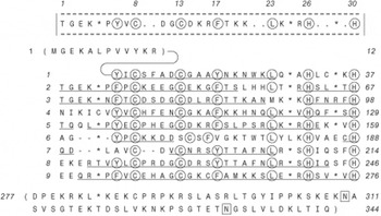
This novel idea of small Zn-stabilized domains was strengthened by the timely publication by Roeder's group (Ginsberg et al. Reference Ginsberg, King and Roeder1984) of the amino acid sequence of TFIIIA derived from a cDNA clone. Upon inspection of the sequence, the large number of cysteines and histidines present in the protein appeared to occur in a more or less regular pattern. A rigorous computer analysis by McLachlan showed that, of the 344 amino acids of the TFIIIA sequence residues, numbers 13–276 form a continuous run of nine tandemly repeated, similar units of about 30 amino acids, each containing two invariant pairs of histidines and cysteines (Miller et al. Reference Miller, Mclachlan and Klug1985) (Fig. 1).
Amino acid sequence (Ginsberg et al. Reference Ginsberg, King and Roeder1984) of TFIIIA from X. laevis oocytes aligned to show the repeating units (Miller et al. Reference Miller, Mclachlan and Klug1985). The best conserved residues are ringed. The boxed-in consensus sequence is shown at the top, indexed on a repeat unit of 30 amino acids. The structural units or finger modules (Fig. 2) begin at amino acid number 6, separated by a five-amino-acid linker.

3. A repeating structure for TFIIIA
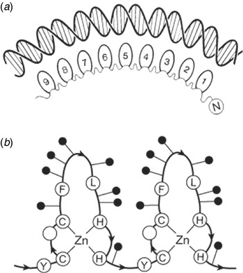
On the basis of the three different lines of evidence described above, namely (a) a 30-amino-acid repeat in the sequence, which (b) corresponds in size to the observed periodic intermediates and the limit-digest product of 3 kDa, and (c) the measured Zn content of 7–11 atoms, we proposed (Miller et al. Reference Miller, Mclachlan and Klug1985) that the 30-kDa region of the TFIIIA protein has a repeating structure consisting of nine 30 amino acid units (Fig. 2 a). Twenty-five of the 30 amino acids in the repeat fold around a Zn ion to form a small independent structural domain or module, the ‘finger’, and the five intervening amino acids provide the linkers between consecutive fingers (Fig. 2 b). The Zn ion forms the basis of the folding by being tetrahedrally coordinated to the two invariant pairs of cysteines and histidines. In addition to this unique conserved pattern of Cys-Cys … His-His, each repeat also contains three other conserved amino acids, namely Tyr6 (or Phe6), Phe17 and Leu23, all of which are large hydrophobic residues (Fig. 1). I therefore suggested that these might interact to form a hydrophobic cluster stabilizing the compact finger module. The 30-amino-acid repeat is rich in basic and polar residues, but the largest number is found concentrated in the region between the second cysteine and the first histidine, implicating this region, in particular, in nucleic acid binding. This was later found to be the case (Pavletich & Pabo, Reference Pavletich and Pabo1991).
(a) Interpretation (Miller et al. Reference Miller, Mclachlan and Klug1985) of the main structural feature of the protein TFIIIA and its interactions with 50 bp of DNA, showing combinatorial recognition by the modular design. (b) Folding scheme for a linear arrangement of repeating structural units (‘zinc finger modules’), each centred on a tetrahedral arrangement of zinc ligands, Cys2 His2. Also shown are the three hydrophilic groups that were proposed to form a structural core, as confirmed later (Fig. 5 below).

Formally, when indexed on a 30-amino-acid repeat, the repeating structure could be written as

where h represents a conserved (large) hydrophobic residue. The proposal that each 25-amino-acid module formed an independently folded, Zn-stabilized domain soon gained support from two lines of research. First, a study using extended X-ray absorption fine structure confirmed that the Zn ligands are two cysteines and two histidines (Vincent et al. Reference Vincent, Colot and Rosbash1985). Second, Tso et al. (Rosenberg et al. Reference Rosenberg, Schroder, Preiss, Kienlin, Cote, Riede and Jackle1986) found that, in the DNA sequence of the gene for TFIIIA, the position of the intron–exon boundaries marks most of the proposed finger module domains.
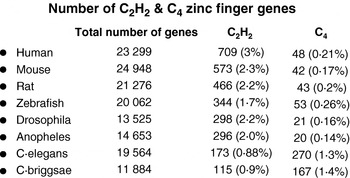
In evolutionary terms, the multi-fingered TFIIIA may have arisen by gene duplication of an ancestral domain comprising about 30 amino acids. Because one such self-contained small domain would have had the ability to bind to nucleic acids, and could be passed on by exon shuffling, we suggested (Miller et al. Reference Miller, Mclachlan and Klug1985) that these domains might occur more widely in gene control proteins than in just this case of TFIIIA. The extent to which this prediction has been borne out (3% of the genes of the human genome, at the latest count; Fig. 3) is astonishing. Indeed, within months of the paper's publication, we received word of sequences homologous to the zinc finger motif of TFIIIA. The first two were from Drosophila, the serendipity gene from Roshbash's group (Vincent et al. Reference Elrod-Erickson, Benson and Pabo1985) and the Kruppel gene from Jackle's group (Berg, Reference Berg1988).
4. A new principle of DNA recognition
The two key points that emerged from our paper (Miller et al. Reference Miller, Mclachlan and Klug1985) were a new protein fold for nucleic acid binding and a novel principle of DNA recognition (Fig. 2). The overall design for specific DNA recognition was distinctly different from that of the helix–turn–helix motif, found in the first DNA-binding proteins to be described. The latter binds to DNA as a symmetric dimer to a palindromic sequence on the DNA, thus making use of both the 2-fold symmetry of the DNA helix backbones and also the nucleotide sequence. Heterodimeric variations of this and other 2-fold symmetric designs were found later, but they still make use of the double helix symmetry.
In contrast, the zinc finger is a DNA-binding module that can be linked tandemly in a linear, polar fashion to recognize DNA (or RNA) sequences of different lengths. Each finger domain has a similar structural framework, but can achieve chemical distinctiveness through variations in a number of key amino acid residues. This modular design thus offers a large number of combinational possibilities for the specific recognition of DNA (or RNA). It is not surprising that it is found widespread throughout so many different types of organisms (Fig. 3).
Number of Cys2 His2 and ‘Cys4’ genes in the genomes of various organisms. The Cys4 genes refer to the steroid and thyroid hormone family of nuclear receptors, which were also later named zinc fingers because of a similarity in the amino acid sequences suggesting the presence of zinc tetrahedrally ligated to four cysteines. The structures are, however, quite different from the classical C2H2 finger. Note how the proportion of C2H2 genes increases with greater complexity on the evolutionary scale. Data taken from Bateman et al. (Reference Bateman, Birney, Cerruti, Durbin, Etwiller, Eddy, Griffiths-Jones, Howe, Marshall and Sonnhammer2002).

5. The structure of the zinc finger and its interaction with DNA
Miller et al. (Reference Miller, Mclachlan and Klug1985) noted that, in addition to the characteristic arrangement of conserved cysteines and histidines that are fundamental in the folding of the finger by the coordinating Zn, there are three other conserved amino acids, notably Tyr6, Phe17 and Leu23, and suggested that they were likely to form a hydrophobic structural core in the folded structure. In other words, the seven conserved amino acids in each unit would provide the framework of tertiary folding, whereas some of the variable residues determined the specificity of each domain. Berg (Klug et al. Reference Klug, Jack, Viswamitra, Kennard, Shakked and Steitz1979) built on these original observations by fitting known structural motifs from other metallo proteins to the consensus sequence of the TFIIIA finger motifs. His proposed model consisted of an antiparallel β-sheet, which contains a loop formed by the two cysteines, and an α-helix containing the His-His loop. The two structural units are held together by the Zn ion. In analogy with the way in which the bacterial helix–turn–helix motif binds DNA, DNA recognition was postulated to reside mostly in the helical region of the protein structure.
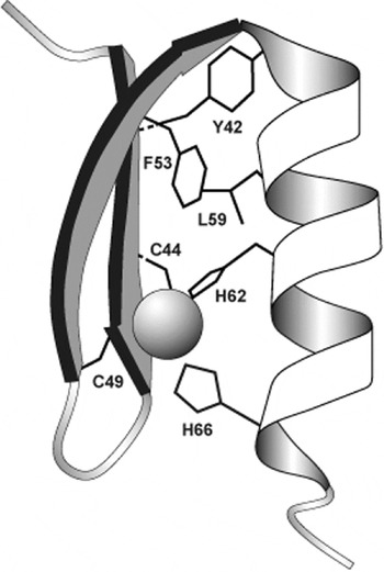
Berg's model was confirmed in outline by the nuclear magnetic resonance (NMR) studies of Wright's group (Lee et al. Reference Lee, Gippert, Soman, Case and Wright1989) on a single zinc finger in solution, and by Neuhaus in the MRC Laboratory (Neuhaus et al. Reference Neuhaus, Nakaseko, Schwabe and Klug1992) on a two-finger peptide (Fig. 4). Neuhaus' work took longer to solve the structure, but it had the merit of showing that adjacent zinc fingers are structurally independent in solution because they are joined by flexible linkers.
The structure of a zinc finger from a two-dimensional (2D) NMR study of a two-finger peptide in solution (Neuhaus et al. Reference Neuhaus, Nakaseko, Schwabe and Klug1992). The same study showed that the linker between the two modules is highly flexible.

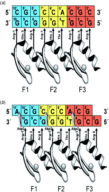
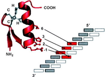
The precise pattern of amino acid interactions of zinc fingers with DNA was revealed in 1991 when Pavletich & Pabo (Reference Pavletich and Pabo1991) solved the crystal structure of a complex of a DNA oligonucleotide specifically bound to the three-finger DNA-binding domain (DBD) of the mouse transcription factor Zif268, an early response gene. The primary contacts are made by the α-helix that binds in the DNA major groove through specific hydrogen-bond interactions from amino acids at helical positions −1, 3 and 6 to three successive bases (a triplet) on one strand of the DNA (Fig. 5 a). Later, the second zinc finger–DNA complex to be solved, by Fairall et al. (Reference Fairall, Schwabe, Chapman, Finch and Rhodes1993) in the MRC Laboratory, revealed an important secondary interaction from helical position 2 to the other strand (Figs. 5 b and 6). This is the canonical docking arrangement, but there are, however, some wide variations from this arrangement in the family of zinc finger–DNA complexes now known (Elrod-Erickson et al. Reference Elrod-Erickson, Benson and Pabo1998). There are also, of course, still other interactions such as with the phosphates of the DNA backbones, but these do not play a direct role in specific recognition.
(a) Schematic diagram (Jamieson et al. Reference Jamieson, Miller and Pabo2003) of the first model of modular recognition of DNA by a three-zinc-finger peptide, illustrating the results of the first crystal structure determination of the complex between the DBD of the transcription factor Zif268 and an optimized DNA-binding site (Choo & Klug, Reference Choo and Klug1994b). Each finger interacts with a 3-bp subsite on one strand of the DNA, using amino acid residues in helical positions −1, 3 and 6. (b) Refined model of DNA recognition (Jamieson et al. Reference Jamieson, Miller and Pabo2003; Reynolds et al. Reference Reynolds, Ullman, Moore, Isalan, West, Clapham, Klug and Choo2003). View of the potential hydrogen bonds to the second strand of the DNA (‘cross-strand interactions’) emanating from position 2 on the recognition helix. This is based on the crystal structure of the tramtrack–DNA complex (Choo & Klug, Reference Choo and Klug1994a), the mutagenesis (Jamieson et al. Reference Jamieson, Miller and Pabo2003) and phage display selection studies of Isalan and co-workers (Reynolds et al. Reference Reynolds, Ullman, Moore, Isalan, West, Clapham, Klug and Choo2003) and the refined structure of the Zif268–DNA complex, and of variants by Pabo and his colleagues (Townsend et al. Reference Townsend, Wright, Winfrey, Fu, Maeder, Joung and Voytas2009). The fingers ideally bind 4-bp overlapping subsites, so that adjacent fingers are functionally synergistic though structurally independent.

Another view of the refined model. Contacts with the DNA are made by amino acids at helical positions −1, 3 and 6 to the coding strand and from position 2 to the non-coding strand. Thus, the binding site for a finger is not simply a triplet of three successive bases, but a 4-bp site, overlapping with that of the preceding finger.

6. Zinc finger peptides for the regulation of gene expression
The mode of DNA recognition by a finger is thus principally a one-to-one interaction between individual amino acids from the recognition helix to individual DNA bases (Pavletich & Pabo, Reference Pavletich and Pabo1991). This is quite unlike the case of other DNA-binding proteins, where one amino acid may contact two bases and vice versa. Moreover, because the fingers function as independent modules, fingers with different triplet specificities can be linked to give specific recognition of longer DNA sequences. For this reason, the zinc finger motifs are ideal natural building blocks for the de novo design of proteins for recognizing any given sequence of DNA. Indeed the first experiments by Berg, Reference Berg1988 and others, using site-directed mutagenesis, showed that it is possible to rationally alter the DNA-binding characteristics of individual zinc fingers when one or more of the amino acids in the α-helical positions are varied. As a small collection of these mutants accumulated, it became possible to find some regularities or ‘rules’ relating amino acids on the recognition α-helix to corresponding bases in the bound DNA sequence.
In the MRC Laboratory, we adopted a different approach. The reason was that these ‘rules’ did not take into account the fact that real DNA structures are not fixed in the canonical B form but, as I had shown earlier, there are wide departures, depending on the DNA sequence (Klug et al. Reference Klug, Jack, Viswamitra, Kennard, Shakked and Steitz1979; Rhodes & Klug, Reference Rhodes and Klug1981). This was further supported by the structure of the zinc finger–tramtrack DNA complex (Fairall et al. Reference Fairall, Schwabe, Chapman, Finch and Rhodes1993). Here, the helical position used for the primary contact by the first finger with the 3′-most base of the cognate triplet (thymine) is not the canonical −1, but 2. The reason is that the DNA helix is deformed from the B form, with a thymine followed by an adenine at a helical rotation angle of 39°, rather than the canonical 36°, and preceded by another adenine at an angle of 33°. The interaction with the finger thus occurs at an ATA sequence that has unusual flexibility, as I had noted long ago (Klug et al. Reference Klug, Jack, Viswamitra, Kennard, Shakked and Steitz1979). DNA is not a rigid, passive participant in its interaction with proteins.
7. Affinity selection from a library of zinc fingers by phage display
The alternative to this rational but biased design of proteins with new specificities was the isolation of desirable variants from a large pool or library. A powerful method, namely phage display, of selecting such proteins is the cloning of peptides (Smith, Reference Smith1985) or protein domains (McCafferty et al. Reference McCafferty, Griffiths, Winter and Chiswell1990) as fusions to the minor coat protein (pIII) of bacteriophage fd, which leads to their expression on the tip of the capsid. Phage displaying the peptides of interest can be affinity purified by binding to the target and then amplified in bacteria for use in further rounds of selection and for DNA sequencing of the cloned genes.
Phage display libraries comprising about 107 variants of the middle finger from the DBD of Zif268 were created by Choo in the MRC Laboratory (Choo & Klug, Reference Choo and Klug1994a, Reference Choo and Klugb). A DNA oligonucleotide of fixed sequence was used to bind and hence purify phage from this library over several rounds of selection, returning a number of different but related zinc fingers that bind the given DNA. By comparing similarities in the amino acid sequence of functionally equivalent fingers, the likely mode of interaction of these fingers with DNA was deduced (Choo & Klug, Reference Choo and Klug1994b). Remarkably, most base contacts were found to occur from three primary positions on the α-helix of the zinc finger, correlating well with the implications of the crystal structure of Zif268 bound to DNA (Pavletich & Pabo, Reference Pavletich and Pabo1991).
This demonstrated ability to select zinc fingers with desired specificity meant that, as the data from the selections accumulated, some wider general rules could be devised for a recognition code (Choo & Klug, Reference Choo and Klug1994a), and hence DNA-binding peptides could be made to measure using the combinatorial strategy exemplified by TFIIIA. In other words, these general rules could be used for the ‘rational design’ of a zinc finger peptide to recognize a short run of DNA sequence by ‘mixing and matching’ individual specific fingers. Where the general rules for finger specificity led to an ambiguity, as in the case of closely related triplets, e.g. GCG and GTG, Choo & Klug (Reference Choo and Klug1994a) showed that zinc finger modules could nevertheless be selected to discriminate between them.
8. Use of engineered zinc finger peptides to repress gene expression in a mouse cell line
One interesting possibility for the use of such zinc finger peptides is to target selectively genetic differences in pathogens or transformed cells. In December 1994, Choo et al. (Reference Choo, Sanchez-Garcia and Klug1994) reported the first such application, which built a protein that recognized a specific DNA sequence both in vitro and in vivo. This was a crucial test of our understanding of the mechanism of zinc finger DNA recognition.
In summary, a three-finger peptide that was able to bind site-specifically to a unique 9-bp region of the p190 bcr-abl cDNA was created: this is a transforming oncogene that arises by translocation between the tips of chromosomes 9 and 22, of which one product is the Philadelphia chromosome. Chromosome 22 contains a novel DNA sequence at the junction of two exons, one each from the two genomic parent bcr and abl genes. Our engineered peptide discriminated in vitro against like regions of the parent bcr and c-abl genes, which differ in only a single base out of the 9-bp target, by factors greater than one order of magnitude.
This peptide also contained a nuclear localization signal (NLS) fused to the zinc finger domain so that the peptide could accumulate in the nucleus. Consequently, stably transformed mouse cells, made interleukin-3 (IL-3) independent by the action of the oncogene, were found to revert to IL-3 dependence on transient transfection with a vector expressing the peptide. This construct was also engineered to contain a c-myc epitope, which enabled investigators to follow by immunofluorescence the localization of the peptide to the nuclei of the transfected cells. When IL-3 is subsequently withdrawn from cell culture, over 90% of the transfected p190 cells become apoptotic (i.e. showing chromosome degradation) within 24 h. These experiments were repeated on cells transformed by another related, but different, oncogene p210 bcr-abl, which served as a control. All transfected p210 cells maintained their IL-3 dependence, and remained intact on entry of the engineered peptide (Choo et al. Reference Choo, Sanchez-Garcia and Klug1994).
The proof of principle led us, and later others, to devote future studies on zinc fingers to potential applications in gene regulation for research purposes or for therapeutic correction. It also stimulated the creation of the first biotech companies, Sangamo Biosciences in Richmond California and later Gendaq in Cambridge, to exploit the new technology.
9. Promoter-specific activation by a chimaeric zinc finger peptide
These experiments showed that a zinc finger peptide could be engineered to switch off gene expression in vivo. The same paper (Choo et al. Reference Choo, Sanchez-Garcia and Klug1994) also described other experiments on a different cell system (cultured mouse fibroblasts) to show that a gene could also be switched on by a zinc finger construct. The same 9-bp sequence was used, but this time as a promoter for a CAT reporter gene contained in a plasmid. The peptide, which recognized the promoter sequence, was fused to a VP16 activation domain from herpes simplex virus (HSV) and, on transient transfection, stimulated expression of the reporter gene by a factor of 30-fold above controls.
10. Improving zinc finger specificity by protein engineering
First, having shown proof of the principle that engineered zinc finger peptides could be used to target DNA, improving the specificity of recognition became the focus of our subsequent work. Although the main source of specificity lies in the amino acids at positions −1, 3 and 6 of the recognition α-helix of a zinc finger for successive bases lying on one strand of a DNA triplet, Isalan et al. (Reference Isalan, Choo and Klug1997) found that the ‘cross-strand’ interaction described above, from helical position 2 to the neighbouring base pair on the adjacent triplet (Fig. 5 b), can significantly influence the specificity. Therefore, it has been necessary to revise the simple model that zinc fingers are essentially independent modules that bind 3-bp subsites to a model that considers functional synergy at the interface between adjacent independently folded zinc fingers. In this refined model, Zif268-like zinc fingers potentially bind 4-bp overlapping subsites (Isalan et al. Reference Isalan, Klug and Choo1998) (Figs. 5 b and 6).
Consequently, our colleague Mark Isalan (Isalan et al. Reference Isalan, Klug and Choo2001) redesigned our method of phage library construction to allow for the optimization of the interaction that a finger makes with the DNA-binding site of the adjacent N-terminal finger. Isalan adopted a bipartite selection strategy in which two halves of a three-finger DBD are selected separately and then recombined in vitro to make a complete three-finger domain that binds 9-bp and automatically allows the interface synergy between its constituent three fingers. These two separate (non-overlapping) libraries are then used to perform the bipartite selections. Because all the steps are carried out in vitro, the method is rapid and easily adapted to a high-throughput automated format. This was applied commercially by our MRC spin-off company Gendaq for up to 2×45=2048 binding sites to create a large archive of zinc finger peptides that recognize a vast number of nine DNA base pair sequences. This archive was later acquired by Sangamo Biosciences, and used in their zinc finger constructs.
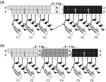
Second, an important step forward was to increase the length of the DNA sequence targeted and hence its degree of rarity; at the same time, an increase was expected in the binding affinity of the longer zinc finger construct. Three zinc fingers recognize a 9-bp sequence, which would occur randomly many thousands of times in a large genome. Therefore, six fingers linked together would recognize a DNA sequence 18-bp in length, which is sufficiently long to constitute a rare address in the human genome. However, one cannot simply go on adding fingers with the conventional linkers, because the periodicity of the packed fingers does not quite match the DNA periodicity. They thus tend to get out of register and are strained in doing so, leading to only a small increase in affinity. We have, therefore, learned how to engineer longer runs of zinc fingers that can target longer DNA sequences and have affinities a thousand times greater than three finger peptides (Liu et al. Reference Liu, Rebar, Zhang, Liu, Jamieson, Liang, Qi, Li, Chen, Mendel, Zhong, Lee, Eisenberg, Spratt, Case and Wolffe2001; Rebar et al. Reference Rebar, Huang, Hickey, Nath, Meoli, Nath, Chen, Xu, Liang, Jamieson, Zhang, Spratt, Case, Wolffe and Giordano2002) (Fig. 7).
Two modes of generating six ZFPs for specific recognition of 18-bp sequences (Liu et al. Reference Liu, Rebar, Zhang, Liu, Jamieson, Liang, Qi, Li, Chen, Mendel, Zhong, Lee, Eisenberg, Spratt, Case and Wolffe2001; Rebar et al. Reference Rebar, Huang, Hickey, Nath, Meoli, Nath, Chen, Xu, Liang, Jamieson, Zhang, Spratt, Case, Wolffe and Giordano2002). Top: Two three-finger peptides fused together using an extended canonical linker (2×3F scheme). Bottom: Three two-finger peptides linked using canonical linkers extended by an insertion of either a glycine residue or a glycine-serine-glycine sequence in the canonical linkers between fingers 2 and 3 and fingers 4 and 5, respectively.

An early design by Kim & Pabo (Reference Kim and Pabo1998) was to use a longer more flexible linker between two different pre-existing three-finger domains to form a six-finger peptide, but this has not been used much in practical applications. The method of choice in current use is that developed in Cambridge by Moore et al. (Reference Moore, Klug and Choo2001) (and later transferred to Sangamo via Gendaq). This uses two-finger binding domains, which can be obtained from the archive described above. Three of these are assembled into six-finger domains, using longer variants of the conserved six amino acid linker TGEKP linkers (Fig. 7 b, bottom). The linkers contain an extra glycine residue or a glycine-serine-glycine tripeptide between the constituent two-finger modules. Such six-finger domains (denoted 3×2F) bind their 18-bp targets with picomolar affinity, as also do six fingers made by linking two three-finger domains (2×3F) made with an extended linker (Moore et al. Reference Moore, Klug and Choo2001). However, the advantage of the 3×2F over the 2×3F strategy is that it is much more sensitive to a mutation or an insertion in the target sequence, with a loss of affinity of up to a 100-fold. Thus, the 3×2F peptide discriminates more strongly than the 2×3F peptide between closely related DNA target sequences. The logic behind our 3×2F design was that the strain in binding longer DNA targets is more evenly distributed than in a 2×3F construct, and it indeed turned out to have the advantage expected. These six-finger peptides not only have picomolar affinities for their 18-bp targets but also give virtually single gene recognition (Tan et al. Reference Tan, Guschin, Davalos, Lee, Snowden, Jouvenot, Zhang, Howes, Mcnamara, Lai, Ullman, Reynolds, Moore, Isalan, Berg, Campos, Qi, Spratt, Case, Pabo, Campisi and Gregory2003) when tested on DNA microarrays displaying 20,000 different sequences. As mentioned above, a library of two-finger peptides begun in the MRC Laboratory, Cambridge, was transferred via Gendaq to Sangamo.
11. Some early applications of engineered zinc finger proteins (ZFPs)
ZFPs can be engineered with a variety of effector domains fused to poly-zinc finger peptides that can recognize virtually any desired DNA sequence with high affinity and specificity. They thus formed the basis of a novel technology that has increasing uses in research and medicine. A summary of the first such applications was given by Pabo and his colleagues (Jamieson et al. Reference Jamieson, Miller and Pabo2003). Applications in the MRC Laboratory, Sangamo, and the Scripps Research Institute include the following:
(i) Inhibition of HIV expression (Reynolds et al. Reference Reynolds, Ullman, Moore, Isalan, West, Clapham, Klug and Choo2003). It was shown that ZFPs targeted to the HIV promoter long terminal repeat activated by the tat protein effectively repressed expression, and preliminary experiments in a cellular infection assay gave a 3-fold drop in infectivity.
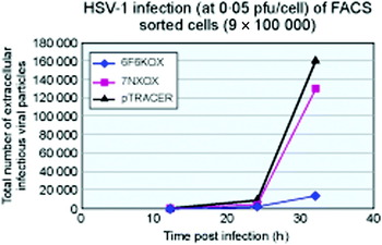
(ii) Disruption of the effective cycle of infection of HSV (Papworth et al. Reference Papworth, Moore, Isalan, Minczuk, Choo and Klug2003). A ZFP transcription factor designed to repress the promoter of a viral gene that is normally activated first in the replication cycle produced a 10-fold reduction in the virus titre in an infected cell line (Fig. 8). This was a good result, considering that there are five other ‘immediate early’ genes that contribute to the infection. Several more of these would have to be repressed to reduce the titre further.
(iii) Activating the expression of vascular endothelial growth factor-A (VEGF-A) in a human cell line (Liu et al. Reference Liu, Rebar, Zhang, Liu, Jamieson, Liang, Qi, Li, Chen, Mendel, Zhong, Lee, Eisenberg, Spratt, Case and Wolffe2001) and in an animal model (Rebar et al. Reference Rebar, Huang, Hickey, Nath, Meoli, Nath, Chen, Xu, Liang, Jamieson, Zhang, Spratt, Case, Wolffe and Giordano2002): these experiments have led to a therapeutic application (see below).
(iv) Regulation of the level of zinc finger expression by a small molecule (Beerli et al. Reference Beerli, Schopfer, Dreier and Barbas2000). This can be used in controlling the dose and/or timing for a therapeutic application. An efficient way of achieving this is by fusing the ZFP to the ligand binding domain of a steroid hormone nuclear receptor. In the absence of hormone, the ZFP transcription factor is retained in the cytoplasm, but after ligand binding the ZFP translocates to the cell nucleus in active form.
12. Stimulation of new vasculature by engineered ZFPs
Following the work at Sangamo on the activation of the transcription factor VEGF by ZFPs in mouse and human cell lines (Liu et al. Reference Liu, Rebar, Zhang, Liu, Jamieson, Liang, Qi, Li, Chen, Mendel, Zhong, Lee, Eisenberg, Spratt, Case and Wolffe2001), experiments showed that new blood vessels could be formed in a mouse ear (Rebar et al. Reference Rebar, Huang, Hickey, Nath, Meoli, Nath, Chen, Xu, Liang, Jamieson, Zhang, Spratt, Case, Wolffe and Giordano2002). These did not leak, unlike the results that had been obtained by earlier workers who had been delivering various cDNA spliced isoforms of the gene. The reason for the success with ZFP activation is that the latter acts on the promoter of the VEGF gene, and hence all spliced isoforms are naturally produced when the gene is induced to promote angiogenesis.
Repression of HSV infection (at 0·05 pfu/cell) of FACS sorted HeLa cells by zinc finger peptides fused to a KOX1 repression domain (Bibikova et al. Reference Bibikova, Beumer, Trautman and Carroll2003). The peptides are targeted to the viral gene IE175 K, the first of the six immediate early genes to be expressed by the virus. A six-finger recognition peptide 6F6KOX reduces the virus titre 10-fold, while a three-finger peptide produces only a 3-fold reduction.

Subsequent work by Frank Giordano at Yale and by Brian Annex at Duke University showed increased blood flow in the hind limbs of ischaemic rabbits, with the ZFP delivered by a retrovirus or simply by injecting the DNA. Several years ago Sangamo began clinical trials to evaluate the ability of an appropriate ZFP to stimulate the natural growth of normal blood vessels in treating claudication, a symptom of peripheral arterial obstructive disease that causes poor blood flow in the legs. The phase 2 trials have shown great improvements, and indeed also in the more serious condition of critical limb ischaemia, in which the arteries are blocked.
13. Gene correction by homologous recombination (HR) using sequence-specific zinc finger nucleases (ZFNs)
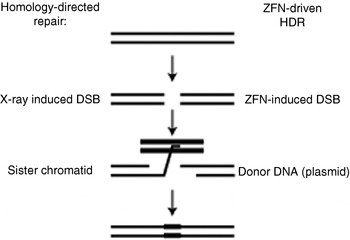
Gene correction is the process by which sequence alterations in defective or deleterious genes can be changed or ‘corrected’ by HR-mediated gene conversion between the target locus and a donor construct encoding the corrective sequence (Fig. 9). Monogenic disorders such as X-linked severe combined immune deficiency (SCID), sickle-cell anaemia (SCA), haemophilia and Gaucher's disease are caused by the inheritance of defective alleles of a single gene. The ability to replace this gene sequence via HR-mediated gene correction has the potential of fully restoring the gene function and providing a permanent cure for patients with these disorders. However, this process is highly inefficient in that the frequency of unaided HR at a specific locus occurs in only about 1 in 105 cells (Capecchi, Reference Capecchi1989). This is far below a level that would be considered therapeutic. However, a DSB has been demonstrated by Jasin (Reference Jasin1996) to potentiate HR at a specific genetic locus by ~5000-fold. Therefore, the introduction of a corrective donor sequence together with a site-specific nuclease that would produce a DSB at or near the location of the mutation could stimulate gene correction to levels that would provide a therapeutic impact.
Gene modification or correction using HR (Elrod-Erickson et al. Reference Elrod-Erickson, Benson and Pabo1998) via ‘short-path gene conversion’ stimulated by a DSB (Perez et al. Reference Perez, Wang, Miller, Jouvenot, Kim, Liu, Wang, Lee, Bartsevich, Lee, Guschin, Rupniewski, Waite, Carpenito, Carroll, Orange, Urnov, Rebar, Ando, Gregory, Riley, Holmes and June2008). The left side schematically depicts the repair of a random X-ray-induced DSB by HR using the sister chromatid as the repair donor. As shown on the right, a site-specific DSB is created by ZFP nucleases (Fig. 10 below).

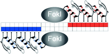
Jasin's demonstration was based on artificially introducing into an endogenous gene of a human cell line an 18-bp DNA sequence, which was the specific binding site for the homing endonuclease Sce1 and had only a small probability of occurring naturally elsewhere in the genome. However, to carry out gene correction in native cells requires the specific targeting of the mutated sequence, and a zinc finger peptide fused to a nuclease domain is the natural choice (Fig. 9). A nuclease of this type was developed by Chandrasegaran and co-workers (Kim et al. Reference Kim, Cha and Chandrasegaran1996) using an engineered ZFP fused to the non-specific cleavage domain of the Fok1 type II restriction enzyme. This type of ZFN was used by Carroll and co-workers (Bibikova et al. Reference Bibikova, Beumer, Trautman and Carroll2003) to produce mutants in Drosophila by gene ‘correction’ and by Porteus & Baltimore (Reference Porteus and Baltimore2003) in a green fluorescent protein model system to study gene modification in a human cell line. Following Chandrasegaran, the three-finger ZFP nucleases were introduced as pairs with tandem binding sites engineered in opposite orientations (Fig. 10), with a 6-bp spacing separating the two half sites (Kim et al. Reference Kim, Cha and Chandrasegaran1996). In both cases the efficiency of the targeting is not very high, but this was not crucial to these studies. Indeed some of the Drosophila mutants are lethal, but they are normally selected on the phenotype. Both studies showed that a ZFN-produced DSB can markedly increase the rate of HR between a donor DNA construct and a reporter gene in their two different systems.
Gene correction using a pair of three ZFNs (Urnov et al. Reference Urnov, Miller, Lee, Beausejour, Rock, Augustus, Jamieson, Porteus, Gregory and Holmes2005) to produce a double-stranded DNA break. The zinc finger peptides are linked to the non-specific catalytic domain of the Fok1 endonuclease by a short amino acid linker. In the 3D structure, the two catalytic domains form a dimeric association.

The results of Carroll's and of Porteus' laboratories (Bibikova et al. Reference Bibikova, Beumer, Trautman and Carroll2003; Porteus & Baltimore, Reference Porteus and Baltimore2003) stimulated work at Sangamo to apply the gene correction method to tackle monogenic disorders of the human genome. The workers set out to determine whether ZFNs could create a comparable increase in HR frequency at an endogenous human gene. They focused their efforts on the IL2Rγ gene in which loss-of-function point mutations cause X-linked severe combined immunodeficiency disease (SCID). In the absence of bone marrow transplantation or gene therapy, this malady leads to death in early childhood. Treatment for the disease by gene therapy (Cavazzana-Calvo et al. Reference Cavazzana-Calvo, Hacein-Bey, De Saint Basile, Gross, Yvon, Nusbaum, Selz, Hue, Certain, Casanova, Bousso, Deist and Fischer2000) had been performed by others by inserting one or more copies of the normal gene in the chromosomes of a number of affected children, that is, by gene addition rather than gene correction. After successful treatment of the majority of the patients, two of them died some years later of leukaemia, probably because the random insertion of the gene led to its coming under inappropriate control.
The middle exon of IL2Rγ gene contains SCID mutation hotspots. Since maximal homology-driven recombination occurs when a DSB is evoked at or close to the mutated site, the Sangamo workers engineered a pair of ZFNs specific for the exact location of the mutation on the X chromosome. Two DBDs were assembled from the zinc finger archive described above, each containing four highly specific zinc finger motifs, and thus simultaneously recognizing two different 12-bp sites, separated by a fixed distance between them. The chance of this particular pattern existing elsewhere in the genome is negligible. Having confirmed in vitro that the proteins bind as intended, they next improved them further by single amino acid substitutions in the zinc finger recognition helices, which gave a further 5-fold increase in potency. The results showed an 18–20% rate of gene correction in the target cells, which was stable after one month in culture in the target cells (Urnov et al. Reference Urnov, Miller, Lee, Beausejour, Rock, Augustus, Jamieson, Porteus, Gregory and Holmes2005).
This accomplishment was dramatic, with an increase by many orders of magnitude over anything achieved in the past by ‘gene targeting’, particularly as no selection has been used. Moreover, measurements of both the mRNA and protein levels expressed by the corrected IL2Rγ gene showed that the mutation had been efficiently and stably corrected. The next steps in curing SCID disease are to isolate CD34 progenitor cells from the patient's bone marrow, and correct them ex vivo. After this gene correction, the cells would be allowed to expand and then re-introduced into the bone marrow to repopulate it with corrected cells. Of course an extensive study of ZFN safety (e.g. verifying that no other DSBs are created outside the target and checking immunogenicity) must first be undertaken, but there is every prospect of gene correction eventually becoming a reality for SCID and other monogenic diseases.
14. Specificity of zinc finger constructs
Like any conventional small-molecule drug, a ZFN can produce secondary effects, but in this case it is possible to assess this by measuring the binding to off-target sites on the DNA. This was done by Sangamo in their experiments on establishing HIV resistance in CD4 T-cells by genome editing (Perez et al. Reference Perez, Wang, Miller, Jouvenot, Kim, Liu, Wang, Lee, Bartsevich, Lee, Guschin, Rupniewski, Waite, Carpenito, Carroll, Orange, Urnov, Rebar, Ando, Gregory, Riley, Holmes and June2008). Here, the target was the HIV receptor CCR5, which was disrupted at 36% efficiency, and effects at the top 15 DNA sequence-related sites were read by 454 DNA pyrosequencing technology. The largest off-target effect (5·4%) was at the biologically related CCR2 site, and of the other 13 sites, one was at an intron of a gene AβLIMZ on chromosome 4, which showed a very low frequency of mutations in the two 38 000 sequences examined. Taken together with the ‘surveyer’ Cel-1 nuclease data and the preservation of the biological properties of the cell, all the results support the conclusion that the ZFNs used in the CCR5 work are highly specific.
15. Targeted gene knockout
Gene knockout is the most powerful tool for determining the function of a gene and also for permanently modifying the phenotype of a cell. Recent methods use HR (Capecchi, Reference Capecchi1989) where recognition of the target is by the homology of the extrachromosomal donor DNA (see above). The low efficiency of this process can necessitate screening thousands of clones. Rapid gene knockout can be achieved by simply using a ZFN to create a DSB at the target and, in the absence of a DNA donor, allowing it to be repaired by the natural process of non-homologous end-joining (NHEJ). This is an imperfect repair process and usually results in changes to the DNA sequence at the site of the break, and hence to mutant (null) alleles of the protein products. In this way, in a collaboration with Sangamo we produced disruption of the dihydrofolate reductase gene in CHO cells at frequencies greater than 1%, thus obviating the need for selection markers. This established a new method for gene knockouts (Santiago et al. Reference Santiago, Chan, Liu, Orlando, Zhang, Urnov, Holmes, Guschin, Waite, Miller, Rebar, Gregory, Klug and Collingwood2008), with applications to reverse genetics and recombinant protein productions in mammalian cells in serum-free media.
As a reviewer remarked, it is ironic that the NHEJ pathway, responsible for the annoying background of random integrants that has plagued gene targeting (Capecchi, Reference Capecchi1989) for so long, is the basis of a simple and efficient method for knockouts. Two spectacular demonstrations of the power of NHEJ targeted knockout have appeared recently. Meng et al. (Reference Meng, Noyes, Zhu, Lawson and Wolfe2008) and Doyon et al. (Reference Doyon, Mccammon, Miller, Faraji, Ngo, Katibah, Amora, Hocking, Zhang, Rebar, Gregory, Urnov and Amacher2008) have both shown that heritable mutations can be produced in zebrafish by generating mRNAs encoding ZFNs for the locus of interest and injecting them into embryos at the one-cell stage. Both groups also measured off-target effects: Meng et al. found a rate of ~1% but Doyon et al. detected no off-target cleavage. The most likely (and comforting) explanation for the difference is that the Doyon et al. used four-finger ZFNs, which were more specific than the three-finger ZFNs used by Meng et al.
16. Other applications of engineered ZFPs
The Sangamo ZFP development programmes for obstructive limb disease, diabetic neuropathy and HIV/AIDs are the most advanced, but other programmes underway include amyotrophic lateral sclerosis (ALS) (a Motor neurone disease) and nerve regeneration. These can be followed on the company's website at www.sangamo.com.
It should also be mentioned that customized zinc finger constructs for given DNA sequences can now be obtained commercially from Sigma-Aldrich, as laboratory reagents under the name Compo-Zr Technology.
Another application of zinc finger technology outside ‘the therapeutic space’ is in the strategic partnership Sangamo has with Dow Agricultural Sciences for use of zinc fingers in the breeding of enhanced plant crops (Shukla et al. Reference Shukla, Doyon, Miller, Dekelver, Moehle, Worden, Mitchell, Arnold, Gopalan, Meng, Choi, Rock, Wu, Katibah, Zhifang, Mccaskill, Simpson, Blakeslee, Greenwalt, Butler, Hinkley, Zhang, Rebar, Gregory and Urnov2009). One example that has been announced is the layering of traits (quantitative trait loci) in which particular alleles of a set of genes can be introduced to give a particular variant of a crop.
Acknowledgements
I thank my former colleagues, Yen Choo, Mark Isalan and Michael Moore at the MRC in Cambridge, and at our spin-off company Gendaq, for helping to develop the promise of zinc fingers into a robust technology.
I also thank Edward Lanphier and Philip Gregory for creating and leading Sangamo Biosciences, for their innovations in applying the technology to medical therapeutics and, in collaboration with Dow Agricultural Sciences (DAS), extending it to plant crop breeding. The latter has been described by Dow Agricultural Sciences as ‘game changing’. See also Townsend et al. (Reference Townsend, Wright, Winfrey, Fu, Maeder, Joung and Voytas2009) for another group working on plants.
I should also state that I have drawn heavily on the illustrations in my earlier review in Annual Reviews of Biochemistry 2010, online 2009.
17. Appendix: highlights from Sangamo Biosciences Inc., December 2009
17.1. Clinical
1. Diabetic neuropathy (DN)
• Data on neuroregeneration demonstrate proof of concept and define a drug responsive population for a phase 2b study (SB-509-901) that began in January 2010 with $3.0M support from the juvenile Diabetes Research Foundation.
2. Motoneurone disease, ALS
• Presentation of preliminary data from the phase 2 study of SB-509 in subjects with ALS.
3. HIV/AIDS
• Initiation of two phase 1 safety trials of a ZFN-based T-cell product (SB-728-T) in which the CCR5 gene, coding for the HIV receptor on T-cells, is disrupted in subjects with HIV/AIDS.
• Presentation of preliminary data from a single subject treated with SB-728-T demonstrating that ZFN-modified cells were well tolerated and stably maintained at up to 20 weeks and total CD4+ T-cells were stable during this time.
17.2. Further applications
• Expansion of licence with Sigma-Aldrich to include the use of ZFNs in cell line engineering and transgenic animal applications.
• Launch of Sigma Advanced Genetic Engineering (SAGE) facility for custom and off-the-shelf transgenic rat production.
17.3. Preclinical/Research
• Sangamo scientists and collaborators receive multiple awards to advance research and preclinical stage programmes (CIRM, Doris Duke Foundation and Bill and Melinda Gates Foundation).