INTRODUCTION
Lophelia pertusa (L., 1758) is a scleractinian framework-forming cold-water coral. It is a cosmopolitan species that can develop reef complexes in the Atlantic Ocean providing important habitat and making these reefs significant centres of biodiversity in deep water shelf settings (Roberts et al., Reference Roberts, Wheeler and Freiwald2006).
‘It has long been recognized that corals contain regular growth patterns in their skeleton’ (Buddemeier & Kinzie III, Reference Buddemeier and Kinzie1976). Such patterns as annual density bands in massive tropical corals provide a chronology to estimate growth rates and are vital for examining geochemical parameters in relation to palaeo-temperatures, long-term climate events and pollution (Swart, Reference Swart1983; Shen & Boyle, Reference Shen and Boyle1987; Beck et al., Reference Beck, Edwards, Ito, Taylor, Recy, Rougerie, Joannot and Henin1992; Tudhope et al., Reference Tudhope, Chilcott, McCulloch, Cook, Chappell, Ellam, Lea, Lough and Shimmield2001; Fallon et al., Reference Fallon, White and McCulloch2002). Cold-water corals are found across a wide range of latitudes and at great depths. This broad range compared to tropical corals means that cold-water species have tremendous potential as new palaeo-proxies over a wide portion of the Earth's oceans. The expansion of palaeoclimate research to cold-water corals has revealed that their skeletal growth differs significantly from that of the well studied massive shallow corals. In studying two deep-water scleractinian corals for future palaeoclimate studies, Adkins et al. (Reference Adkins, Henderson, Wang, O'Shea and Mokadem2004) found a radial growth rate of 0.07 mm yr−1, a minimum vertical extension rate of 5 mm yr−1 and clear but non-annual banding. Vertical extension rates in the cup coral Desmophyllum cristagalli ranged from 0.5 mm yr−1 to 2 mm yr−1 (Adkins et al., Reference Adkins, Henderson, Wang, O'Shea and Mokadem2004). Chronologies from other cold-water scleractinians, including the branching ahermatypic L. pertusa, are proving more difficult to interpret because they are obscured by complex and inconsistent skeletal banding patterns (Freiwald et al., Reference Freiwald, Henrich, Patzold, James and Clarke1997; Mortensen & Rapp, Reference Mortensen and Rapp1998; Lazier et al., Reference Lazier, Smith, Risk and Schwarcz1999; Risk et al., Reference Risk, Hall-Spencer, Williams, Freiwald and Roberts2005). Because L. pertusa is the most commonly studied and widespread cold-water coral, recent research has attempted to use trace elements as proxies for palaeoclimate reconstructions (Lutringer et al., Reference Lutringer, Blamart, Frank, Labeyrie, Freiwald and Roberts2005; Risk et al., Reference Risk, Hall-Spencer, Williams, Freiwald and Roberts2005; Cohen et al., Reference Cohen, Gaetani, Lundalv, Corliss and George2006). The current study investigated skeletal growth of L. pertusa sampled from oil platform structures in a productive shelf regime, the North Sea. Skeletal characteristics were examined from sequential polyps along branches to identify possible patterns that might reveal a chronology for further palaeoclimate and pollution record studies.
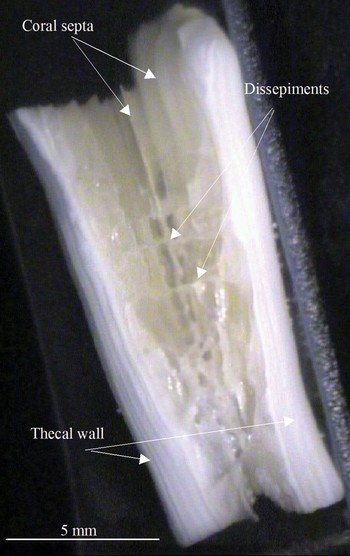
Lophelia pertusa colonies from North Sea oil platforms form a characteristic dome-shape with densely packed polyps (Gass & Roberts, Reference Gass and Roberts2006). Corallites are cylindrical to conical with septa arranged in 3 cycles (Mortensen & Rapp, Reference Mortensen and Rapp1998) and dissepiments visible across the coral cavity. Branches extend by asexual intratentacular polyp budding. Longitudinal dark and light growth bands in the thecal wall and skeletal patterns along the growth direction in septa have been previously documented (Wainwright, Reference Wainwright1964; Freiwald et al., Reference Freiwald, Henrich, Patzold, James and Clarke1997; Mortensen & Rapp, Reference Mortensen and Rapp1998; Blamart et al., Reference Blamart, Rollion-Bard, Cuif, Juillet-Leclerc, Lutringer, van Weering, Henriet, Freiwald and Roberts2005; Risk et al., Reference Risk, Hall-Spencer, Williams, Freiwald and Roberts2005) (Figure 1).
A longitudinal section from a coral polyp under reflected light. The septa and dissepiments are visible in the inner cavity. Grey and white bands are visible running along the thecal wall.

MATERIALS AND METHODS
Skeletal morphology
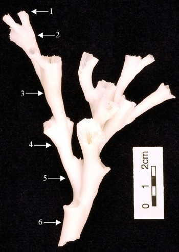
Eight colonies of Lophelia pertusa were sampled from two offshore oil platforms (North Cormorant and Thistle Alpha) in June 2003 from the northern North Sea located between 60° and 61°N and 0° and 2°E where water depths reach between 146 and 160 m. Corals were sampled using ‘work class’ oil industry remotely operated vehicles from depths ranging between 48 and 109 m and from those parts of the platforms with the least influence from drilling discharges (Gass, Reference Gass2006). Branches consisting of three to four sequential living polyps were selected from each colony. Coral tissue was dissolved in dilute bleach (<5% sodium hypochlorite), the skeletons rinsed and dried at 60°C. Polyps were assigned a number starting from one for the outermost polyp and counting back along the branch towards the centre and older portion of the colony (Figure 2). Using this approach, Polyp 1 always corresponded to the youngest generation. Digital calipers were used to measure: calyx diameter from outer edges at the top and bottom of the polyp; polyp height from its budding point to the top of the corallite, and thickness of the thecal wall at the rim of the calyx.
A branch of Lophelia pertusa from a North Sea coral. Arrows indicate polyp number along a branch.

Datasets for polyp numbers and skeletal characters were not all normally distributed and skeletal characteristics were therefore compared between polyp generations using the non-parametric Kruskal–Wallis test, followed by Mann–Whitney U-tests.
Microscopy
Three to five sequential polyps from two branches from two colonies sampled from North Cormorant were sectioned and examined under reflected light. The number of dark and light growth bands was counted. Corals were sectioned longitudinally and etched in weak nitric acid (1%) for 30 seconds, rinsed in deionized water and dried at 60°C. The samples were sputtered with gold under high vacuum and viewed using a Cambridge S90 scanning electron microscope (SEM).
RESULTS AND DISCUSSION
Corallites were trumpet-shaped with a distinctly narrow base and wider top. Mean polyp length excluding the newest generation of corallites, which will not yet have grown for a full year, was 27.4 ± 5 mm (mean ± SD). This falls within the 26 ± 5 mm (mean ± SD) annual linear extension based on video images of 15 Lophelia pertusa colonies from the Tern platform (also northern North Sea) (Gass & Roberts, Reference Gass and Roberts2006). These combined results suggest that colonies of L. pertusa growing on platforms in the North Sea bud and extend the full length of one polyp per year; hence Polyp 1 is in its first year of growth, Polyp 2 is two years old, Poly p 3 is three years old and so forth. These results are unique because this is the only study which provides a known timeline of several years for in situ growth of L. pertusa. The results are based on videos taken of the same coral colonies through time which was only made possible by the opportunities of working with Shell Expro Ltd and being able to revisit the same colony on a platform. In a natural deep reef environment this would pose vast challenges. Our results also suggest that the growth rate of 26 mm yr−1 for L. pertusa applies to both the growth of the colony as a whole (Gass & Roberts, Reference Gass and Roberts2006) and to the first year of polyp growth. This makes an important distinction between average growth rates of a polyp over its lifetime, which is not consistent through time but is fastest in its first year and dramatically slower thereafter (see further results below). Future studies examining L. pertusa as a proxy for palaeoclimate or historical pollution events must not assume average growth rates over the full length of the polyp but must take into consideration this initial fast growth.
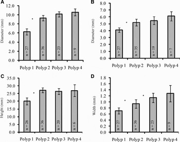
The width of the upper calyx was 9.7 ± 2 mm (mean ± SD) and the width of the base of coral polyps was 5.5 ± 1 mm. Thecal widths varied between colonies (1.05 ± 0.5 mm). Figure 3a & b show the calyx diameter at the top and bottom of the corallite increasing in size from Polyp 1 to Polyp 3, illustrating significant changes between Polyps 1 and 2. Figure 3c shows a significant increase in polyp height between Polyps 1 and 2 and no increase in size thereafter for Polyps 2, 3 and 4. Finally, thecal width (Figure 3c) showed consistent growth through time and significantly widened at an even rate from Polyp 1 through to Polyp 3, but slowing thereafter.
Coral colonies were sampled in late spring and the average height of Polyp 1 is approximately three-quarters of the full grown height of subsequent polyps. We speculate that budding events may follow the spring and autumn plankton blooms in the North Sea. As a result, one would expect average height of new polyps from colonies sampled in June to be almost fully grown as the data suggest in the present study.
The results from skeletal measurements (a) calyx diameter across the top of the polyp, (b) calyx diameter at the base of the polyp, (c) polyp height, and (d) thecal width as measured at the top of the calyx. *, indicates significant difference between polyp numbers (P < 0.05); N = number of polyps.

The results show that the majority of polyp growth occurs in its first year as the polyp reaches its maximum height and creates its characteristic trumpet shape. Faster growth in younger polyps of L. pertusa has also been documented by Maier et al. (Reference Maier, Hegemen, Weinbauer and Gattuso2009) using 45Ca labelling in aquaria. It has also been inferred based on skeletal growth lines (but without a known timeline as in the current study) for L. pertusa from a Norwegian fjord where polyps reached a third of their height in their first year of growth (Mortensen & Rapp, Reference Mortensen and Rapp1998), but did not show annual budding. In comparison, the rate of coral skeletal growth in the North Sea may reflect the locally more productive environment. Emiliani et al. (Reference Emiliani, Hudson and Shinn1978) also found that growth in the solitary scleractinian coral Bathypsammia tintinnabulum decreased from the base upwards, stopping altogether at maturation.
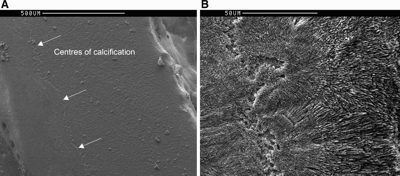
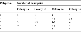
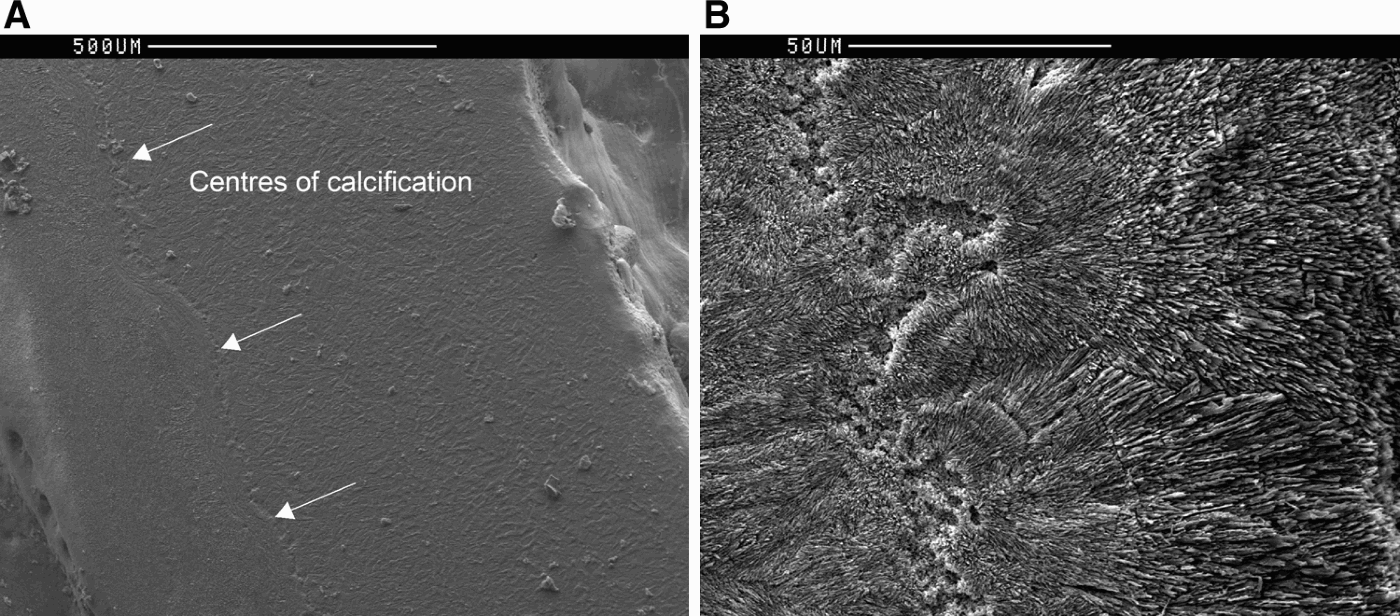
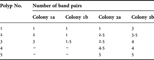
Longitudinal polyp sections revealed growth bands running down thecal walls. However, the innermost, dark band was the only band that clearly ran the full length of polyps in all cases. The number and clarity of banding varied between colonies, and even between branches from the same colony (Table 1) (Figure 1 shows an example of banding from a Polyp 4). However, there was some indication that the band pairs might be annual in some cases, particularly Colony 1a where each new polyp generation showed an additional set of band pairs, suggesting one set of bands per year. Inconsistencies between branches may indicate that banding does not correspond with time but is a more random event influenced by the possible retreating and advancing of the coral's outer tissue layer (Lazier et al., Reference Lazier, Smith, Risk and Schwarcz1999). Polyps examined under SEM revealed clear centres of calcification running along the inner half of the theca and measuring up to 50 µm wide (Figure 4).
Longitudinal cross-sections of theca. (a) One line of centres of calcification is visible; (b) centres of calcification where aragonite crystals can be seen growing away from the centre.
Number of band pairs visible in thecal walls of two Lophelia pertusa colonies from the North Cormorant oil platform.
Thecal growth banding in L. pertusa is well-documented from corals from the north-east Atlantic (Freiwald et al., Reference Freiwald, Henrich, Patzold, James and Clarke1997; Mortensen & Rapp, Reference Mortensen and Rapp1998; Risk et al., Reference Risk, Hall-Spencer, Williams, Freiwald and Roberts2005), but its timing is unknown, although Risk et al. (Reference Risk, Hall-Spencer, Williams, Freiwald and Roberts2005) suggested that they are annual. Our results show variation between the number of dark and light growth bands and the corresponding age of the polyp as well as variations among and within colonies; hence banding was not annual in all cases. Zibrowius (Reference Zibrowius1984) also documented morphological variations in L. pertusa. Despite this, the results demonstrate an annual chronology as new polyps bud along a branch. However, to obtain the maximum resolution for palaeoclimate studies, the question remains of how to distinguish the skeletal material formed during the first year of the polyp's life from that formed during subsequent thecal thickening and the influence of outer tissue retreat on this process. Centres of calcification indicate early skeletal formation (Cuif & Dauphin, Reference Cuif and Dauphin1998). All polyps in the current study showed a clear innermost dark band and a primary line of centres of calcification, both located in the inner theca. Centres of calcification have been reported as darker and optically dense growth bands regions in thecal walls of L. pertusa and Desmophyllum cristagalli (Lazier et al., Reference Lazier, Smith, Risk and Schwarcz1999; Adkins et al., Reference Adkins, Boyle, Curry and Lutringer2003; Blamart et al., Reference Blamart, Rollion-Bard, Cuif, Juillet-Leclerc, Lutringer, van Weering, Henriet, Freiwald and Roberts2005; Lutringer et al., Reference Lutringer, Blamart, Frank, Labeyrie, Freiwald and Roberts2005). The line of centres of calcification observed in the current study reflects the primary nucleation zone as described by Cohen et al. (Reference Cohen, Gaetani, Lundalv, Corliss and George2006). However, contrary to other studies (Wainwright, Reference Wainwright1964; Cohen et al., Reference Cohen, Gaetani, Lundalv, Corliss and George2006), additional dark bands did correspond to further nucleation zones and the corals did not appear to have secondary, tertiary, etc., nucleation zones. Therefore, it is likely that the centres of calcification and the adjacent skeletal material would represent the first skeletal material laid down in the polyp's first year. Initial dense skeletal growth might provide young polyps with more stability to endure the high currents often associated with environments favoured by L. pertusa. Therefore, by isolating skeletal material adjacent to the primary line of centres of calcification it may be possible to obtain a year long skeletal chronology for each polyp along a branch, at least in the case of North Sea corals. This would offer a relatively high sampling resolution considering the growth rate of the polyp. The next steps will be to test this model with environmental proxies such as Sr/Ca or Mg/Ca ratios previously documented in coral skeletons, and to investigate how this model corresponds to colonies of L. pertusa living in other areas.
ACKNOWLEDGEMENTS
We acknowledge BP, Lundin Britain Ltd, and Shell Expro Ltd for assistance with sampling. We acknowledge the Atlantic Frontier Environmental Network for funding (AFEN PhD grant). J.M.R. received additional support from the European Commission Marie Curie international fellowship ‘Trans-Atlantic Coral Ecosystem Studies’ (Contract No. MOIF-CT-2006-040018) under the programme ‘Structuring the European Research Area’ and the HERMES project (Contract No. GOCE-CT-2005-511234) through the European Commission's Sixth Framework Programme under the priority ‘Sustainable Development, Global Change and Ecosystems’.