Introduction
Foreign body ingestion amongst children is a common presentation to the emergency department. It requires prompt recognition and management to prevent potentially life-threatening complications, including oesophageal perforation, mediastinitis and tracheoesophageal fistula formation.Reference Gregori, Scarinzi, Morra, Salerni, Berchialla and Snidero1
The narrowest point of the oesophagus is at the level of the cricopharyngeus muscle, and as a result this is the most common site for foreign body impaction.Reference Stack and Munter2 Depending on the nature of the foreign body and the skills of the attending medical teams, a range of approaches can be employed for removal of upper oesophageal foreign bodies. Flexible upper gastrointestinal endoscopy can be undertaken with a host of retrieval devices (e.g. grasping forceps, snares baskets), but alternatives include rigid oesophagoscopy or balloon extraction under fluoroscopic guidance. Hollow foreign bodies present a unique challenge, as they can be difficult to visualise and grasp if the lumen is large, and difficult to deliver past oesophageal sphincters or the adjacent inflammation or granulation tissue.
It is current practice at our centre to undertake rigid oesophagoscopy in the first instance for suspected upper oesophageal foreign body impaction. We present a novel technique using oesophageal balloon dilatation with fluoroscopic guidance, which we propose can be employed following unsuccessful rigid and flexible oesophagoscopy with the usual retrieval devices.
Technical description
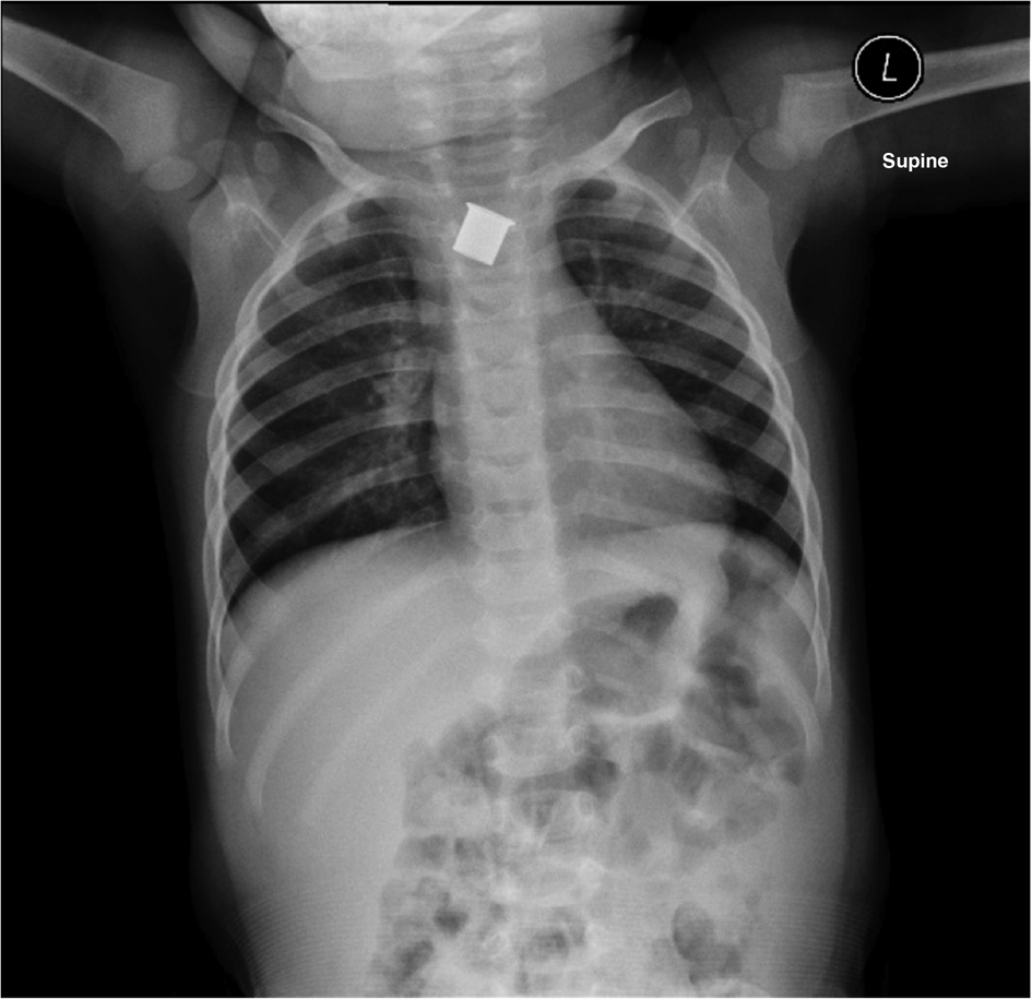
A 13-month-old child presented with an oesophageal foreign body. Under general anaesthetic, with an endotracheal tube in situ, the upper margin of a metallic, cylindrical foreign body (Figure 1) could be only partially visualised with a rigid oesophagoscope. The upper rim of the object was covered in granulation tissue. Direct removal was deemed too risky as the condition of the surrounding tissues could not be determined.
Plain anteroposterior radiograph showing position of the foreign body. L = left

An XP260 paediatric flexible gastroscope (Olympus, Tokyo, Japan) was passed into the oesophagus. Under visualisation, it was possible to pass a 0.035-inch (0.889 mm) guide wire (Terumo, Leuven, Belgium) between the foreign body and the oesophageal wall, into the stomach. Attempts to pass a nasogastric tube over this guide wire were unsuccessful, as the foreign body was tightly impacted against the oesophageal wall.
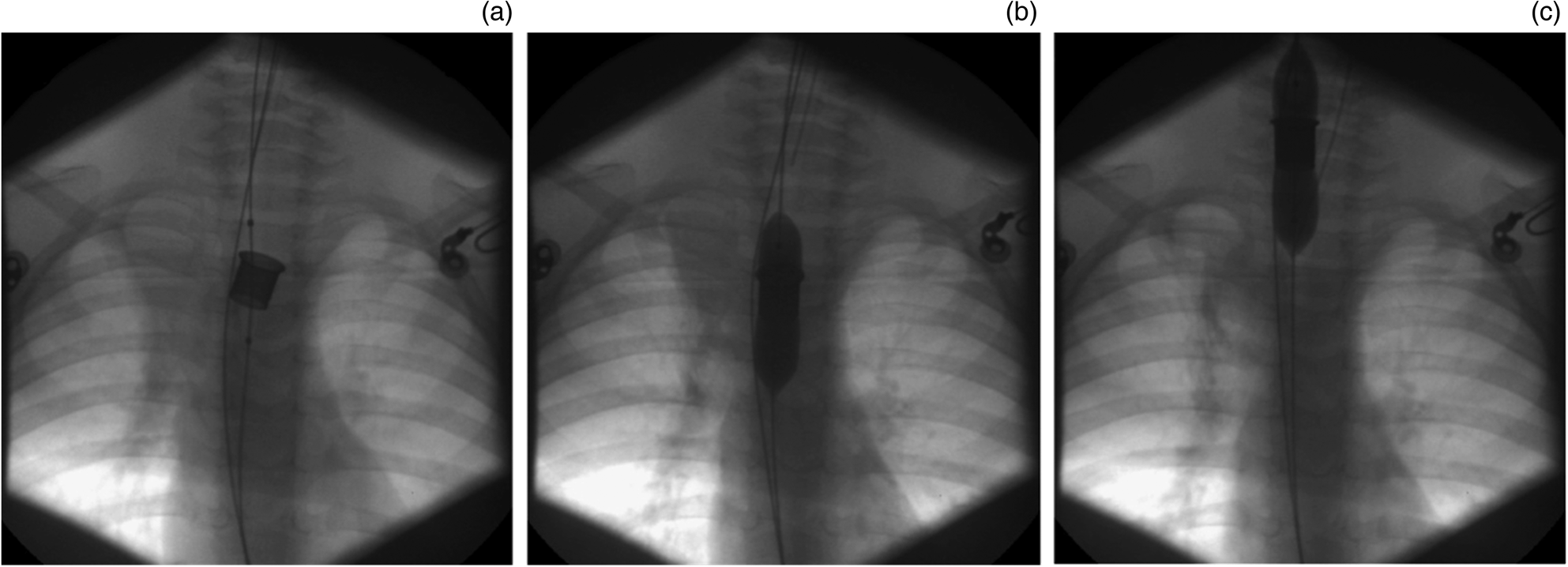
Under fluoroscopic guidance, a second guide wire was passed through the lumen of the object (Figure 2a), and an oesophageal dilatation balloon (Wanda, 12 mm × 40 mm × 80 cm; Boston Scientific, Marlborough, Massachusetts, USA) was passed over the guide wire, with radio-opaque markers used to approximate position. The balloon was inflated to 4 bar pressure (Figure 2b). With gentle traction, the foreign body was removed safely, along with the balloon, under fluoroscopic screening (Figure 2c). A nasogastric tube was advanced over the original guide wire and secured in place for post-operative enteral feeding.
(a–c) Fluoroscopy images showing oesophageal dilatation balloon extraction.

Discussion
Flexible and rigid oesophagoscopy are successful in retrieving upper oesophageal foreign bodies in the vast majority of instances on the first attempt.Reference Russell, Lucas, Johnson, Yannam, Griffin and Beierle3 The most commonly retrieved items in the paediatric population are coins and food boluses, although the prevalence of button battery ingestion is increasing.Reference Haddad, Wilson, Fard and Levi4 Tissue necrosis and formation of granulation tissue can complicate button battery ingestion, making extraction challenging, similar to the case described. Conventional methods can usually be employed, even with unusual foreign bodies, but there is a paucity of literature addressing those that cannot be adequately visualised directly or endoscopically.
For many years, Foley catheter balloon inflation under fluoroscopic guidance had been employed as a less invasive means of removing upper oesophageal foreign bodies. However, it has largely been superseded by oesophagoscopy because of: the inability to examine surrounding mucosa, the possibility of missing radiolucent objects and a high failure rate.Reference Hawkins5
Creative solutions are required intra-operatively during foreign body extraction, as soft tissue abnormalities and radiolucent objects are often missed on plain radiographs. We present a reproducible method for retrieving foreign bodies associated with significant inflammation and granulation, using equipment readily available in most tertiary paediatric hospital operating theatres. Expertise in operating under fluoroscopy is required, and the involvement of interventional radiology or paediatric surgery staff may be needed for this technique. The Foley catheter technique was not felt appropriate in this case given the overlapping granulation tissue proximal to the foreign body. The use of an oesophageal dilatation balloon, with the help of a guide wire, enabled dilatation of the oesophageal lumen proximal and distal to the impacted object, thereby potentially increasing the efficacy of the extraction.Reference Towbin, Lederman, Dunbar, Ball and Strife6
Although there are no published studies or reports discussing the retrieval of hollow foreign bodies from the oesophagus, Munoz and colleaguesReference Munoz, Habashi, Corregidor, Bass, Alizadeh and Gupta7 described a small case series of hollow objects successfully removed from the stomach using an anchoring oesophageal dilatation balloon: this was used to mount the object onto a flexible endoscope, with subsequent removal without fluoroscopic guidance.
Conclusion
While the majority of impacted objects (e.g. coins and food boluses) can be definitively managed in the first instance with a low incidence of complications, hollow oesophageal foreign bodies present a unique challenge, with no published literature to inform surgeons. We report a novel technique for removing impacted objects using an oesophageal dilatation balloon under fluoroscopic guidance that may avoid the need for an open procedure.
Competing interests
None declared