Introduction
One hundred and eighty species of monogenoids have been reported to infest characiform fish from Brazil (Cohen et al., Reference Cohen, Justo and Kohn2013). However, among these, only ten species have been reported to be members of Erythrinidae (Cohen et al., Reference Cohen, Justo and Kohn2013; Moreira et al., Reference Moreira, Scholz and Luque2015; Santos-Neto et al., Reference Santos-Neto, Rodrigues and Domingues2015): six species of monogenoids are known to parasitize the gills and body surfaces of Hoplias malabaricus (Bloch) (trahira), two species were collected from the gills of Hoplias aimara (Valenciennes) (trahira) and the two remaining species were from the gills of Hoplerythrinus unitaeniatus (Agassiz) (trahira pixuna, jeju). Additionally, two undescribed species of dactylogyrids were found on the gills of H. malabaricus from the Paraná Basin (table 1).
List of host species, parasite species, site, localities and references. G, Gills; BS, body surface; 1, Solimões, Negro, Branco; 2, Amazonas, Madeira, Guaporá; 3, Paraná, Paranapanema; 4, Macaé, São João, others; 5, Paraguay, São Lourenço; 6, Amazonas, Xingú, Iriri, Paru; 7, Meruu, Acará, Guamá; 8, Gurupi, Tiriaçu.

* Suriano (Reference Suriano1997) reported U. eremitus from Chascomus Lake, Argentina; however, information about the Basin and Sub-basin of this locality is not available at the Agência Nacional de Águas, Ministério do Meio Ambiente, Brazil (http://hidroweb.ana.gov.br/).
During a survey of the parasites infecting H. malabaricus, which inhabit the streams of the coastal drainage ecosystem of the State of Pará, Brazil, one new species of Urocleidoides and two new species of a new genus of dactylogyrid were encountered on the gills of H. malabaricus. Descriptions of the new species and the proposal of the genus are presented herein. Urocleidoides brasiliensis Rosim, Mendoza-Franco & Luque, 2011, U. cuiabai Rosim, Mendoza-Franco and Luque, 2011, U. eremitus Kritsky, Thatcher & Boeger, 1986 and U. malabaricusi Rosim, Mendoza-Franco & Luque, 2011 are reported for the first time parasitizing H. malabaricus from the coastal rivers of the Oriental Amazon Basin. Urocleidoides eremitus reported by Graça et al. (Reference Graça, Ueda, Oda and Takemoto2013) from the Upper Paraná River floodplain is now considered by the authors as a new species of Urocleidoides. We also address the importance of host distributional range, especially in ‘species complex’ cases (e.g. H. malabaricus) in order to understand the patterns of morphological variation in parasites and their species delimitation.
Materials and methods
Host sample collection
Fish hosts were collected by trammel net, and line and hook from the Caeté River (North/North-east Atlantic Basin; Gurupi, Turiaçu Sub-basin), Municipality of Bragança, Pará State, Brazil (1°3′54.82″S, 46°41′37.60″W) in April 2013, October 2013 and August 2014; Itabocal River (North/North-east Atlantic Basin; Meruu, Acará, Guamá Sub-basin), Municipality of Irituia, Pará State, Brazil (1°51′59.82″S, 47°24′17.15″W) in November 2013 and July 2014; Maracanã River (North/North-east Atlantic Basin; Gurupi, Turiaçu Sub-basin), Municipality of Nova Timboteua, Pará State, Brazil (1°7′46.32″S, 47°21′11.64″W) in July 2013; Maparanim River (North/North-east Atlantic Basin; Gurupi, Turiaçu Sub-basin), Municipality of Terra Alta, Pará State, Brazil (1°5′0.10″S, 47°55′43.98″W) in July 2013; Gurupi River (North/North-east Atlantic Basin; Gurupi, Turiaçu Sub-basin), Municipality of Viseu, Pará State, Brazil (1°17′37.6″S, 46°11′0.49″W) in May 2014; and Piriá River (North/North-east Atlantic Basin; Gurupi, Turiaçu Sub-basin), Municipality of Viseu, Pará State, Brazil (1°12′44.65″S, 46°17′36.72″W) in March 2014. Host scientific names were validated according to Oyakawa (Reference Oyakawa, Reis, Kullander and Ferraris2003) and Oyakawa & Mattox (Reference Oyakawa and Mattox2009). The nomenclature of basins and sub-basins follows the Agência Nacional de Águas, Ministério do Meio Ambiente, Brazil (http://hidroweb.ana.gov.br/).
Parasitological procedures
Gill arches were removed and placed in vials containing heated water (~65°C). Each vial was shaken vigorously and formalin was added to obtain a 5% solution. In the laboratory, the contents of each vial were examined under a dissecting microscope (Leica S6D; Leica Microsystems, Wetzlar, Germany) and helminths were removed from the gills or sediment using small probes. Some specimens were stained with Gomori's trichrome (Humason, Reference Humason1979; Boeger & Vianna, Reference Boeger, Vianna and Thatcher2006) and mounted in Damar Gum or Canada balsam to determine internal soft structures, and others were mounted in Hoyer's medium or Gray & Wess medium (Humason, Reference Humason1979; Boeger & Vianna, Reference Boeger, Vianna and Thatcher2006) for the study of sclerotized structures. The measurements, all in micrometres, were obtained according to the procedures of Mizelle & Klucka (Reference Mizelle and Klucka1953). Dimensions of organs and other structures represent the greatest measurement in dorso-ventral view; lengths of curved or bent structures (anchors, bars and accessory piece) represent the straight-line distances between extreme ends; total lengths of the male copulatory organ were carried out using ImageJ (Rasband, Reference Rasband1997–2016) on drawing-tube images. Each average measurement is followed by the range and the number (n) of specimens measured in parentheses. Illustrations were prepared with the aid of a drawing tube on a Leica DM 2500 microscope with differential interference contrast and phase contrast optics. Illustrations of soft structures were prepared using pen and ink; illustrations of hard structures were scanned and redrawn on a digitizing tablet using Adobe Illustrator and Corel Draw software. Plates were prepared using PhotoPaint software. Definitions of prevalence and mean intensity followed Bush et al. (Reference Bush, Lafferty, Lotz and Shostak1997). Type specimens and vouchers were deposited in the following collections: Helminthological Collection of the Instituto Oswaldo Cruz (CHIOC), Rio de Janeiro, RJ, Brazil; Invertebrate Collection of the Instituto de Pesquisas da Amazônia (INPA), Manaus, AM, Brazil; Invertebrate Collection of the Museu Paraense Emílio Goeldi (MPEG), Belém, PA, Brazil. The following museum specimens were examined: 20 specimens of U. eremitus (INPA PA260 1–3, CHIOC 37471), 2 specimens of U. malabaricusi (CHIOC 37467a–b), 4 specimens of U. cuiabai (CHIOC 37469b–e) and 5 specimens of U. brasiliensis (CHIOC 37470b–f). Historical review of species containing relevant taxonomic contributions, such as description (descr.), redescription (redes.), citation (citat.) and figure (fig.), are included after valid species' names.
Results
Class Monogenoidea Bychowsky, 1937
Subclass Polyonchoinea Bychowsky, 1937
Order Dactylogyridea Bychowsky, 1937
Dactylogyridae Bychowsky, 1933
Urocleidoides Mizelle & Price, 1964
Urocleidoides bulbophallus n. sp.
Description
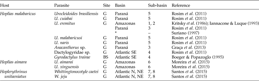
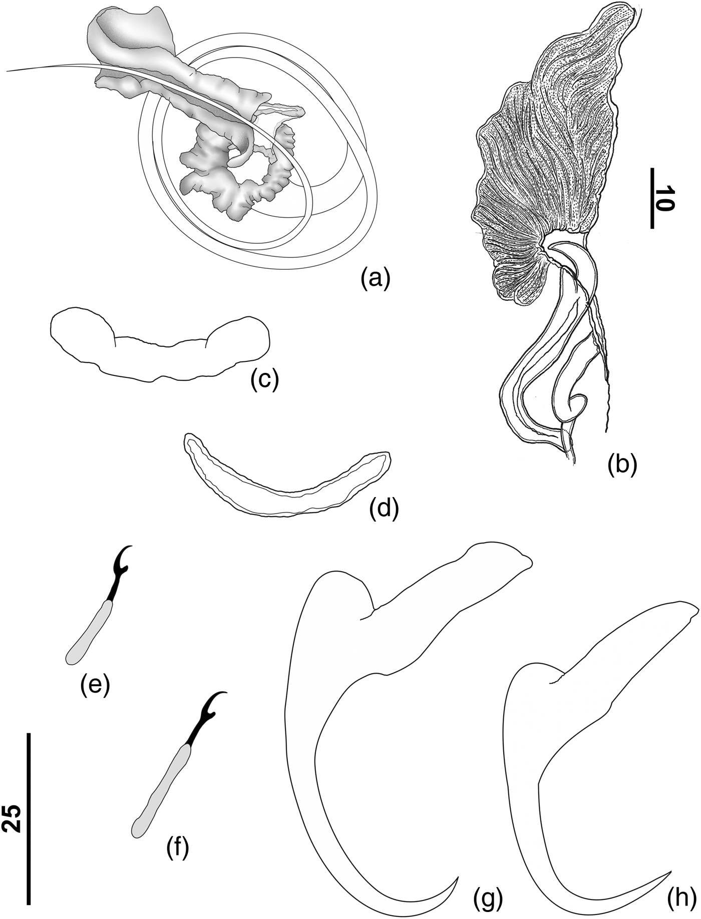
Based on eight specimens (fig. 1); one mounted in Gomori's trichrome, seven mounted in Gray & Wess medium. Body fusiform, total length excluding haptor 268 (225–275; n = 7) long, 101 (72–130; n = 8) wide at level of germarium. Tegument smooth. Cephalic margin tapered; moderately developed terminal lobes; three bilateral pairs of head organs with rod-shaped secretion; cephalic glands unicellular, posterolateral to pharynx. Four eyes, posterior pair larger than anterior pair; accessory granules present in cephalic area, elliptical. Mouth subterminal, midventral; pharynx muscular, glandular; oesophagus short. Two intestinal caeca, confluent posteriorly to gonads, lacking diverticula. Genital pore opening midventral; genital atrium muscular. Testis, vas deferens, prostatic reservoir not observed. Copulatory complex comprising male copulatory organ (MCO), accessory piece; MCO sclerotized, coiled, counterclockwise, with approximately 1½ rings, 75 (75; n = 3) long, base with sclerotized cap; circular sclerotized tandem brim associated with the base of the male copulatory organ; proximal portion of the MCO slightly expanded, bulb-shaped, distal aperture acute (fig. 1b). Accessory piece sclerotized, non-articulated with the MCO, comprising a bent sheath, ‘e’ shape. Germarium, seminal receptacle, Mehlis' glands, ootype not observed. Vagina single, muscular; vaginal aperture sinistro-ventral, marginal; vagina comprising vaginal vestibule with soft-tissue canal, elongated, sigmoid, slightly sclerotized. Vaginal sclerite 31 (28–35; n = 6) long, robust, with longitudinal superficial groove, distally hooked (fig. 1a). Vitellaria dense throughout trunk, except in region of reproductive organs. Eggs not observed. Peduncle short. Haptor subtriangular, 52 (47–54; n = 3) long, 67 (54–77; n = 3) wide. Anchors similar; each with well-developed superficial root, short deep root, evenly curved shaft and point; point acute, extending just past level of tip of superficial root. Ventral anchor (fig. 1g), base 18 (17–20; n = 4) long, superficial root with small sclerotized cap, 35 (33–38; n = 7) long; shaft and point, forming angle of approximately 115°. Dorsal anchor more delicate than ventral anchor (fig. 1h) 19 (17–22; n = 8) in length, base 10 (9–12; n = 4) long; shaft and point, forming angle of approximately 110°. Ventral bar (fig. 1c) 35 (30–40; n = 5) long, slightly curved or straight rod with small terminal enlargements, ends curved in anterior direction. Dorsal bar (fig. 1d) 22 (18–25; n = 4) long, narrow, broadly U-shaped, slightly curved in posterior direction. Hooks similar in shape (fig. 1e, f), shank with inflation, erected thumb, lightly curved long shaft, delicate point, filamentous hook, loop of hook extending to union of shank subunits; hook pair 1, 12 (11–13; n = 6) long; pairs 2–4 and 6–7, 21 (18–25; n = 8) long; hook pair 5 not observed.
Urocleidoides bulbophallus n. sp. (a) Vaginal sclerite; (b) copulatory complex; (c) ventral bar; (d) dorsal bar; (e, f) hooks; (g) ventral anchor; (h) dorsal anchor. Scale bars in μm.

Taxonomic summary
Type host
Hoplias malabaricus (Bloch).
Site of infection
Gill filaments.
Type locality
Caeté River, Municipality of Bragança, Pará State, Brazil (1°3′54.82″S, 46°41′37.60″W).
Other records
Hoplias malabaricus, Itabocal River, Municipality of Irituia, Pará State, Brazil (1°51′59.82″S, 47°24′17.15″W); Piriá River, Municipality of Viseu, Pará State, Brazil (1°12′44.65″S, 46°17′36.72″W); Maparanim River, Municipality of Terra Alta, Pará State, Brazil (1°5′0.10″S, 47°55′43.98″W); Gurupi River, Municipality of Viseu, Pará State, Brazil (1°17′37.6″S, 46°11′0.49″W).
Specimens deposited
Holotype: CHIOC no. 38621a. Six paratypes: CHIOC nos 38620a–b, 38621b; INPA no. 706; MPEG nos 0081–0082. Thirteen vouchers: CHIOC nos 38616–38619, 38635a–b, 38639; INPA nos 707–708; MPEG nos 0083–0085.
Etymology
The specific name refers to the presence of a bulb in the proximal portion of the male copulatory organ of this species.
Remarks
Most specimens of U. bulbophallus n. sp. were mounted in Gray & Wess medium. Measurements and description of internal organs are therefore limited. The new species resembles U. visiofortatus Mendoza-Franco & Reina 2008 mainly by the dissimilarity in the size of anchors (ventral anchors twice as large as dorsal anchors) and U. neotropicalis Mendoza-Franco & Reina 2008 by having a male copulatory organ with bulbous base. It differs from U. visiofortatus mainly by possessing a male copulatory organ with bulbous base proximal (bulbous base absent in U. visiofortatus) and a vaginal aperture marginal (midventral position in U. visiofortatus). It is easily distinguished from U. neotropicalis by the comparative size of anchors and shape of the accessory piece. In U. neotropicalis, the anchors are approximately similar in size (ventral anchors twice as large as dorsal anchors in U. bulbophallus n. sp.), and the accessory piece comprising a variable sheath along the distal portion of male copulatory organ (a bent sheath, ‘e’ shape in U. bulbophallus n. sp.).
Urocleidoides paranae n. sp.
Description
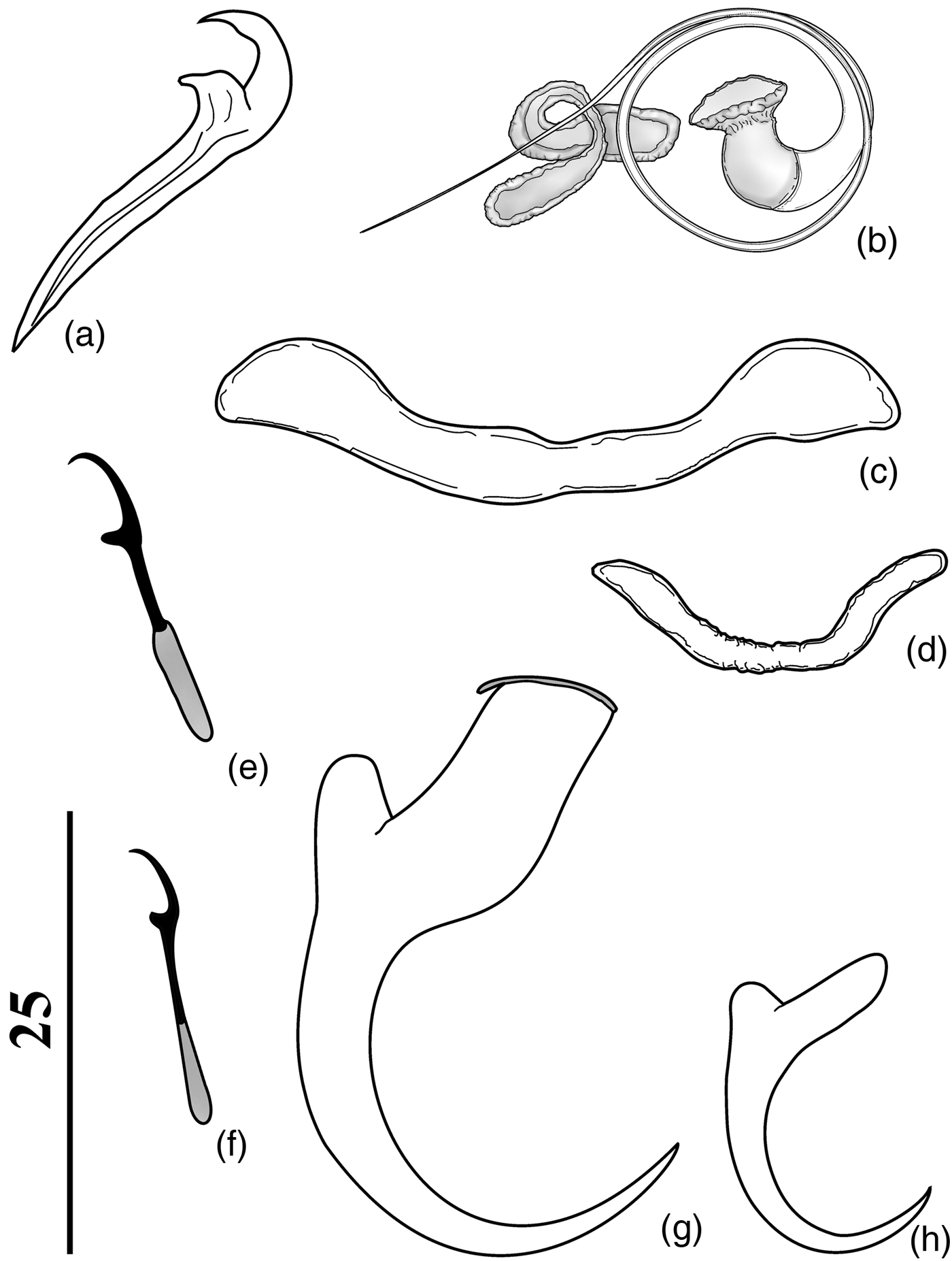
Based on two specimens mounted in Hoyer's medium (fig. 2). Body fusiform, total length excluding haptor 361 (346–377; n = 2) long, 156 (155–158; n = 2) wide at level of germarium. Tegument smooth. Cephalic margin tapered; moderately developed terminal lobes; three bilateral pairs of head organs with rod-shaped secretion; cephalic glands unicellular, posterolateral to pharynx. Four eyes, posterior pair larger than anterior pair; accessory granules present in cephalic area, elliptical. Mouth subterminal, midventral; pharynx muscular, glandular 29 (n = 2) in diameter; oesophagus, intestinal caeca not observed. Genital pore, gonads, ootype, uterus, egg, seminal receptacle not observed. Copulatory complex comprising MCO, accessory piece; MCO sclerotized, coiled, counterclockwise, with approximately 2½ rings, 108 (105–110; n = 2) long, base with sclerotized cap; circular sclerotized tandem brim associated with the base of the MCO present; distal aperture acute (fig. 2a). Accessory piece sclerotized, non-articulated with the MCO, comprising variably flattened sheath along distal shaft of MCO. Vagina single; vaginal aperture sinistro-ventral, marginal; vagina comprising vaginal vestibule with slightly sclerotized funnel; vaginal canal heavily sclerotized at proximal portion with a dilatation at middle portion, distal portion an elongate tube slightly sclerotized (fig. 2b). Vaginal sclerite 29 (n = 1) long, robust, with longitudinal superficial groove, distally hooked. Vitellaria dense throughout trunk, except in region of reproductive organs. Peduncle short. Haptor subtriangular, 95 (85–105; n = 2) long, 107 (104–111; n = 2) wide. Anchors similar. Ventral anchor (fig. 2h) 47.5 (47– 48; n = 2) in length, base 22.5 (22–23; n = 2) long with depressed, moderately short superficial root, non-existent deep root, elongate shaft and short point, forming angle of approximately 98°. Dorsal anchor (fig. 2i) 39 (n = 1) in length, base 16 (n = 1) long with elongate superficial root, poorly developed deep root, evenly curved shaft and point, forming angle of approximately 95°. Ventral and dorsal bars slightly curved rods with enlarged ends; ventral bar (fig. 2d) 46 (45–47; n = 2) long; dorsal bar (fig. 2e) 41.5 (41–42; n = 2) long. Hooks similar in shape (fig. 2f, g), shank with inflation, erected thumb, lightly curved long shaft, delicate point; hook pairs 1 and 5, 17 (16–18; n = 2) long; pairs 2–4 and 6–7, 28 (n = 2) long.
Urocleidoides paranae n. sp. (a) Copulatory complex; (b) vagina; (c) vaginal sclerite; (d) ventral bar; (e) dorsal bar; (f, g) hooks; (h) ventral anchor; (i) dorsal anchor. Scale bars in μm.

Taxonomic summary
Type host
Hoplias malabaricus (Bloch).
Type locality
Upper Paraná River floodplain (Paraná River Basin; Paraná, Paranapanema Sub-basin), Paraná and Mato Grosso do Sul State, Brazil (22°43′00″S, 53°10′00″W).
Specimens studied
Holotype: CHIOC no. 37789. One paratype: CHIOC no. 37790.
Etymology
The specific name refers to the Paraná River, from which the type host was collected.
Remarks
Examination of the vouchers of U. eremitus collected from H. malabaricus from the Upper Paraná River floodplain (see Graça et al., Reference Graça, Ueda, Oda and Takemoto2013) indicates that these specimens are members of a new species of Urocleidoides, described above as U. paranae n. sp. The new species could be confused with U. eremitus by having similar hooks, bars and dorsal anchors. However, examination of the type series of U. eremitus (INPA PA260 1–3) allowed us to differentiate both species. The new species differs from U. eremitus by possessing a ventral anchor with a depressed, moderately short superficial root and non-existent deep root, whereas the latter species has a well-developed superficial root and small deep root in the ventral anchor. Also, the accessory piece in U. paranae n. sp. is a variable sheath along the distal shaft of MCO (fig. 2a), whereas in U. eremitus, the accessory piece is represented by an elongate proximal portion and expanded distal portion, ventrally bent (see figs 3a and 4a).
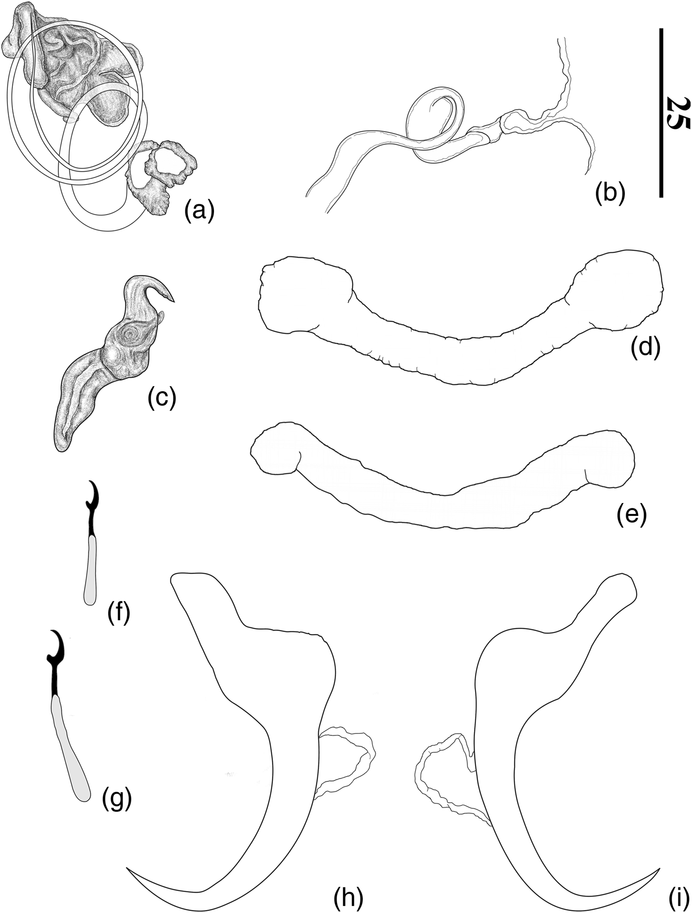
Urocleidoides eremitus Kritsky, Thatcher & Boeger, 1986, holotype (INPA 141). (a) Copulatory complex; (b) vaginal region showing vaginal sclerite, vaginal vestibule and vaginal canal; (c) hook pair 1; (d) hook pair 7; (e) ventral bar; (f) dorsal bar; (g) ventral anchor; (h) dorsal anchor. Scale bars in μm.

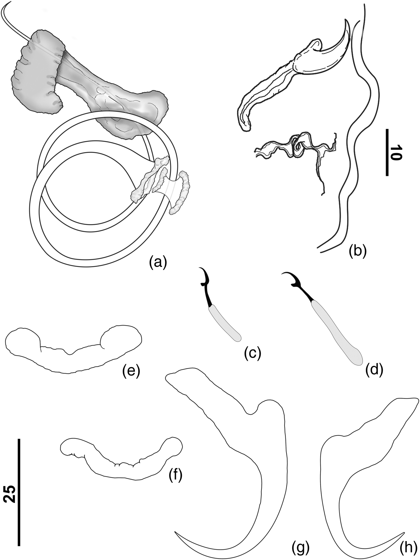
Urocleidoides eremitus Kritsky, Thatcher & Boeger, 1986, voucher (CHIOC 37471). (a) Copulatory complex; (b) vaginal region showing vaginal sclerite, vaginal canal and muscular pad; (c) ventral bar; (d) dorsal bar; (e) hook pair 1; (f) hook pair 7; (g) ventral anchor; (h) dorsal anchor. Scale bars in μm.

Urocleidoides brasiliensis Rosim, Mendoza-Franco & Luque, 2011
Urocleidoides brasiliensis. Rosim et al. (Reference Rosim, Mendoza-Franco and Luque2011): 410–411, figs 40–49 (descr.); Cohen et al. (Reference Cohen, Justo and Kohn2013): 67, 126, fig. 331 (citat.); Graça et al. (Reference Graça, Ueda, Oda and Takemoto2013): 1486 (citat.).
Taxonomic summary
Type host
Hoplias malabaricus (Bloch).
Site of infection
Gill filaments.
Type locality
Baía das Pedras, Cuiabá River (Paraná River Basin; Paraguay, São Loureço Sub-basin), Mato Grosso State, Brazil (16°58′S, 56°25′W).
Other records
Hoplias malabaricus, Guandú River (East Atlantic Basin; Macaé, São João Sub-basin), Municipality of Rio de Janeiro, Rio de Janeiro State, Brazil (22°48′N, 43°37′W); Upper Paraná River floodplain, Paraná and Mato Grosso do Sul State, Brazil (22°43′00″S, 53°10′00″W); Cristalino River (Tocantins River Basin; Araguaia, Mortes Javaés Sub-basin), Mato Grosso State, Brazil (13°22′00″S, 50°52′00″W); Caeté River, Municipality of Bragança, Pará State, Brazil (1°3′54.82″S, 46°41′37.60″W); Maracanã River, Municipality of Nova Timboteua, Pará State, Brazil (1°7′46.32″S, 47°21′11.64″W); Piriá River, Municipality of Viseu, Pará State, Brazil (1°12′44.65″S, 46°17′36.72″W); Gurupi River, Municipality of Viseu, Pará State, Brazil (1°17′37.6″S, 46°11′0.49″W); Maparanim River, Municipality of Terra Alta, Pará State, Brazil (1°5′0.10″S, 47°55′43.98″W); Itabocal River, Municipality of Irituia, Pará State, Brazil (1°51′59.82″S, 47°24′17.15″W).
Specimens studied
Five paratypes: CHIOC nos 37470b–f. Fourteen vouchers: CHIOC nos 38612–38615, 38648a–c, 38654; INPA nos 721a–b; MPEG nos 0096–0099.
Comparative measurements
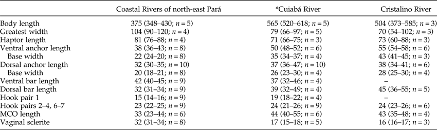
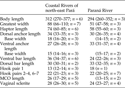
The comparative measurements of specimens of U. brasiliensis from three localities are listed in table 2.
Comparative measurements (in μm) of specimens of Urocleidoides brasiliensis Rosim, Mendoza-Franco & Luque, 2011 from three localities.

* Type locality; MCO, male copulatory organ.
Remarks
The comparative analysis of the type material of U. brasiliensis (CHIOC 37470) and specimens of Urocleidoides parasitizing the gills of H. malabaricus from the streams of the coastal drainages of the State of Pará indicated that they are conspecific, mainly by sharing the morphology of anchors and vagina. The specimens studied here differ morphometrically from specimens from the type locality (Cuiabá River) and voucher specimens from Cristalino River.
Urocleidoides cuiabai Rosim, Mendoza-Franco & Luque, 2011
Urocleidoides cuiabai. Rosim et al. (Reference Rosim, Mendoza-Franco and Luque2011): 409–410, figs 3, 21–39 (descr.); Cohen et al. (Reference Cohen, Justo and Kohn2013): 67–68, fig. 334 (citat.); Graça et al. (Reference Graça, Ueda, Oda and Takemoto2013): 1485–1486 (citat.).
Taxonomic summary
Type host
Hoplias malabaricus (Bloch).
Site of infestation
Gill filaments.
Type locality
Baía das Pedras Cuiabá River, Mato Grosso State, Brazil (16°58′S, 56°25′W).
Other records
Hoplias malabaricus, Maracanã River, Municipality of Nova Timboteua, state of Pará, Brazil (1°7′46.32″S, 47°21′11.64″W); Gurupi River, Municipality of Viseu, Pará State, Brazil (1°17′37.6″S, 46°11′0.49″W); Piriá River, Municipality of Viseu, Pará State, Brazil (1°12′44.65″S, 46°17′36.72″W).
Specimens studied
Four paratypes: CHIOC nos 37469b–e. Eleven vouchers: CHIOC nos 38622–38624, 38645, 38647; INPA nos 720a–b; MPEG nos 0100–0103.
Comparative measurements
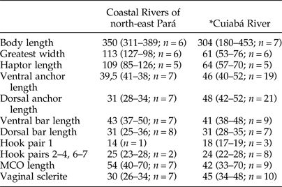
The comparative measurements of specimens of U. cuiabai from two localities are listed in table 3.
Comparative measurements (in μm) of specimens of Urocleidoides cuiabai Rosim, Mendoza-Franco & Luque, 2011 from two localities. MCO = Male copulatory organ.

* Type locality; MCO, male copulatory organ.
Remarks
The analysis of specimens of Urocleidoides from H. malabaricus found in the coastal rivers of Pará with type material of U. cuiabai from Cuiabá River (CHIOC 37469b–e) indicated that they are conspecific based on the morphology of the male copulatory organ and bars. These specimens have a male copulatory organ with 2–3 rings, and a dorsal bar with U-shaped with bifurcated ends. The specimens studied here differ morphometrically from the other locality where this species was previously reported.
Urocleidoides eremitus Kritsky, Thatcher & Boeger, 1986
Urocleidoides eremitus (figs 3, 4). Kritsky et al. (Reference Kritsky, Thatcher and Boeger1986): 5, figs 1–9 (descr.); Rosim et al. (Reference Rosim, Mendoza-Franco and Luque2011): 411, figs 54–61, 64–65 (redes.); Cohen et al. (Reference Cohen, Justo and Kohn2013): 68, fig. 335 (citat.); Graça et al. (Reference Graça, Ueda, Oda and Takemoto2013): 1485–1486 (citat.).
Taxonomic summary
Type host
Hoplias malabaricus (Bloch).
Site of infestation
Gill filaments.
Type locality
Janauacá Lake (Amazon River Basin; Solimões, Negro, Branco Sub-basin) near Manaus, Amazonas State, Brazil.
Other records
Hoplias malabaricus, Maracanã River, Municipality of Nova Timboteua, Pará State, Brazil (1°7′46.32″S, 47°21′11.64″W); Piriá River, Municipality of Viseu, Pará State, Brazil (1°12′44.65″S, 46°17′36.72″W); Caeté River, Municipality of Bragança, Pará State, Brazil (1°3′54.82″S, 46°41′37.60″W).
Specimens studied
Holotype: INPA no. 141. Two paratypes: INPA nos 142a–b. Thirty-two vouchers: CHIOC nos 37471a–q, 38625a–d, 38636, 38644, 38651; INPA nos 723a–c; MPEG nos 0104–0108.
Comparative measurements
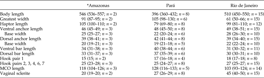
The comparative measurements of specimens of U. eremitus from three localities are listed in table 4.
Comparative measurements (in μm) of specimens of Urocleidoides eremitus Kritsky, Thatcher & Boeger, 1986 from three localities.

* Type locality; MCO, male copulatory organ.
Remarks
Recently, Rosim et al. (Reference Rosim, Mendoza-Franco and Luque2011) reported U. eremitus for specimens collected from H. malabaricus from the East Atlantic Basin (Guandú River) and Paraná River Basin (Cuiabá, Jaguari-Mirim and Machado rivers). These authors recognized some morphological differences among those specimens when compared with specimens from the type series (i.e. four paratypes, USNPC 78764). Most significantly, these authors reported the presence of a conspicuous muscular pad on the left side of the body midline, at the level of the vaginal sclerite. Most of the available voucher specimens deposited in the CHIOC (37471a–q) by Rosim et al. (Reference Rosim, Mendoza-Franco and Luque2011) were, in general, in very poor condition (all specimens stained in Gomori's trichrome). However, the presence of the muscular pad was confirmed for some specimens (fig. 4b). Nonetheless, examination of the type specimens, particularly the holotype (INPA 141) (stained in Gomori's trichrome) (fig. 3b) and specimens of U. eremitus collected from coastal rivers of the Oriental Amazon Basin, demonstrated that the studied specimens do not possess the muscular pad. Although this feature can be relevant taxonomically, the comparative analysis of studied specimens (type and voucher specimens) allowed us to confirm that they are conspecific by sharing the similar morphology of the copulatory complex and haptoral structures (see figs 3a, c–h and 4a, c–h).
Rosim et al. (Reference Rosim, Mendoza-Franco and Luque2011) recognized morphometrical differences in the length of the male copulatory organ among specimens collected in their study and the type material of U. malabaricusi (17–30 vs. 136, respectively). These authors considered this feature, besides the presence of the muscular pad, an important characteristic in distinguishing the difference between both morphotypes. However, an examination of Rosim et al.’s (Reference Rosim, Mendoza-Franco and Luque2011) specimens of U. eremitus during the present study demonstrated that the authors apparently measured the length of the male copulatory organ incorrectly. The measurements conducted herein demonstrate that those specimens did not differ morphometrically from the type specimens from other localities (table 4).
Iannacone & Luque (Reference Iannacone and Luque1993) reported the same species from H. malabaricus captured in the Tambopata River, Madre de Dios, Perú. Urocleidoides eremitus was also found parasitizing the same host in Chascomus Lake, Argentina by Suriano (Reference Suriano1997). Examination of specimens from both localities will be necessary to determine the identity of the specimens (with or without muscular pad) and whether or not all specimens are conspecific with U. eremitus.
The presence of a muscular pad for some specimens assigned as U. eremitus from the East Atlantic Basin and Paraná River Basin, for instance, does not seem to be sufficient enough evidence to propose a new species. We agree with Rosim et al. (Reference Rosim, Mendoza-Franco and Luque2011) that in order to prevent future synonyms, those specimens with a muscular pad should be provisionally accepted as conspecific with U. eremitus until the impact of a representative sampling of the geographic distribution (East Atlantic and Paraná River Basins vs. Amazon River Basin) on colonization/speciation events within this group of parasites is better understood.
Urocleidoides malabaricusi Rosim, Mendoza-Franco & Luque, 2011
Urocleidoides malabaricusi. Rosim et al. (Reference Rosim, Mendoza-Franco and Luque2011): 407, figs 1, 4–12 (descr.); Cohen et al. (Reference Cohen, Justo and Kohn2013): 68–69, fig. 338 (citat.); Graça et al. (Reference Graça, Ueda, Oda and Takemoto2013): 1485–1486 (citat.).
Taxonomic summary
Type host
Hoplias malabaricus (Bloch).
Site of infestation
Gill filaments.
Type locality
Baía das Pedras, Cuiabá River, Mato Grosso State, Brazil (16°58′S, 56°25′W).
Other records
Hoplias malabaricus, Caeté River, Municipality of Bragança, Pará State, Brazil (1°3′54.82″S, 46°41′37.60″W); Gurupi River, Municipality of Viseu, Pará State, Brazil (1°17′37.6″S, 46°11′0.49″W); Maracanã River, Municipality of Nova Timboteua, Pará State, Brazil (1°7′46.32″S, 47°21′11.64″W); Maparanim River, Municipality of Terra Alta, Pará State, Brazil (1°5′0.10″S, 47°55′43.98″W); Piriá River, Municipality of Viseu, Pará State, Brazil (1°12′44.65″S, 46°17′36.72″W); Itabocal River, Municipality of Irituia, Pará State, Brazil (1°51′59.82″S, 47°24′17.15″W).
Specimens studied
Two paratypes: CHIOC nos 37467a–b. Thirteen vouchers: CHIOC nos 38626–38629, 38641; INPA nos 722a–c; MPEG nos 0109–0113.
Comparative measurements
The comparative measurements of specimens of U. malabaricusi from two localities are listed in table 5.
Comparative measurements (in μm) of specimens of Urocleidoides malabaricusi Rosim, Mendoza-Franco & Luque, 2011 from two localities.

MCO, Male copulatory organ.
Remarks
A comparative analysis of the type material of U. malabaricusi (CHIOC 37467) and specimens of Urocleidoides from H. malabaricus of the coastal rivers of Pará indicated that they are conspecific, mainly because they both share the presence of a pad surrounding the copulatory complex.
Constrictoanchoratus n. gen.
Diagnosis
Body divisible into cephalic region, trunk, haptor. Tegument thin, smooth. Cephalic region with terminal ventral cephalic lobe poorly developed or absent. Bilateral pairs of head organs opening laterally to cephalic region; cephalic glands lateral or posterolateral to pharynx. Eyes present (2 pairs); granules elongate. Mouth subterminal, midventral; pharynx muscular, glandular; oesophagus short. Two intestinal caeca, confluent posteriorly to gonads, lacking diverticula. Genital pore midventral near level of caecal bifurcation. Genital atrium muscular. Gonads tandem or testis post-germarial; testis dorsal to germarium. Vas deferens looping left intestinal caecum; seminal vesicle a dilatation of vas deferens, sigmoid, looping dorso-ventrally before entering into the MCO. Copulatory complex comprising MCO, accessory piece; MCO sclerotized, coiled, clockwise, with conical base surrounded by sclerotized cap; circular sclerotized tandem brim associated with the base of the MCO present or absent; accessory piece sclerotized, non-articulated with the MCO. Vagina single; vaginal aperture sinistro-ventral, marginal, opening anteriorly or at mid-level of the trunk; vaginal vestibule muscular or heavily sclerotized at distal portion; vaginal canal muscular or heavily sclerotized, straight. Seminal receptacle present, anterior to germarium. Vitellaria well developed, coextensive with caeca. Haptor armed with, 14 hooks (7 pairs) with ancyrocephaline distribution; hook comprising shank of two subunits. Pair of ventral, dorsal anchors; anchors with elongate superficial root, inconspicuous deep root; constriction present at the intersection between shaft and point. Ventral, dorsal bar present. Parasites of gills of Erythrinidae (Characiformes).
Taxonomic summary
Type species
Constrictoanchoratus ptilonophallus n. gen. n. sp. from Hoplias malabaricus (Bloch).
Site of infestation
Gill filaments.
Type locality
Maracanã River, Municipality of Nova Timboteua, Pará State, Brazil (1° 7′ 46.32″ S 47° 21′ 11.64″ W).
Other species
Constrictoanchoratus lemmyi n. gen. n. sp.
Other records
Constrictoanchoratus ptilonophallus n. gen. n. sp. from Hoplias malabaricus, Caeté River, Municipality of Bragança, Pará State, Brazil (1°3′54.82″S, 46°41′37.60″W); Itabocal River, Municipality of Irituia, Pará State, Brasil (1°51′59. 82″S, 47°24′17.15″W); Piriá River, Municipality of Viseu, Pará State, Brasil (1°2′44.65″S, 46°17′36.72″W); Marapanim River, Municipality of Terra Alta, Pará State, Brazil (1°5′0.10″S, 47°55′43.98″W); and Gurupi River, Municipality of Viseu, Pará State, Brazil (1°17′37.6″S, 46°11′0.49″W). Constrictoanchoratus lemmyi n. gen. n. sp. from Hoplias malabaricus, Itabocal River, Municipality of Irituia, Pará State, Brasil (1°51′59. 82″S, 47°24′17.15″W).
Etymology
The generic name is from the Latin (constrict = constriction) and refers to the morphology of anchors.
Remarks
Features that distinguish Constrictoanchoratus n. gen. from other dactylogyrid genera that occur in erythrinid hosts include the presence of a male copulatory organ coiled with clockwise rings; ventral and dorsal anchors with elongate superficial root and inconspicuous deep root, a constriction at the intersection between the shaft and the point; and hook with inflated shank.
The presence of a constriction at the intersection between the shaft and point in ventral and dorsal anchors in the haptor is an unusual feature in Neotropical dactylogyrids. The character also occurs in the monotypic, Rhinonastes pseudocapsaloideum Kritsky, Thatcher & Boeger, 1988. However, R. pseudocapsaloideum possesses a single ventral anchor–bar complex, and 1 ventral, 6 marginal pairs of hooks located in the disc-shaped haptor, whereas species of Constrictoanchoratus n. gen. have two pairs of anchor–bar complexes (1 ventral, 1 dorsal) and hooks with ancyrocephaline distribution (Mizelle, Reference Mizelle1936). Also, R. pseudocapsaloideum was encountered in the nasal cavities of characiform fish from Prochilodontidae, while species of Constrictoanchoratus n. gen. were encountered on the gills of Erythrinidae.
Constrictoanchoratus ptilonophallus n. sp.
Description
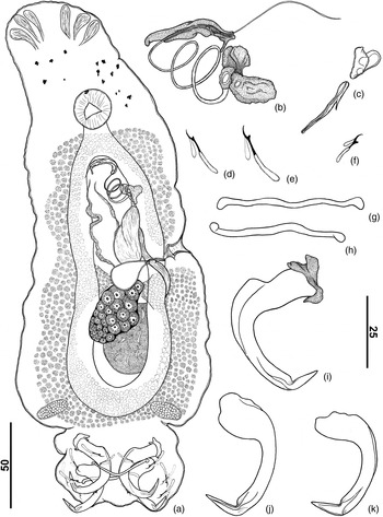
Based on 35 specimens (fig. 5); 13 mounted in Gomori's trichrome, 22 mounted in Hoyer's medium. Body fusiform (fig. 5a), total length excluding haptor 386 (315–538; n = 20) long, 104 (81–160; n = 18) wide at level of germarium. Tegument smooth. Cephalic margin tapered; poorly developed terminal lobes; three bilateral pairs of head organs with rod-shaped secretion; cephalic glands unicellular, posterolateral to pharynx. Four eyes, posterior pair larger and slightly farther apart than anterior pair; accessory granules present in cephalic area, spherical. Mouth subterminal, midventral; pharynx subspherical, 28 (24–32; n = 11) long, 23 (20–27; n = 11) wide. Genital pore opening midventral; genital atrium muscular. Gonads overlapping; testis dorsal to germarium. Testis saccate, 54 (48–58; n = 3) long, 18 (16–21; n = 4) wide. Prostatic reservoir not observed. MCO, with approximately 2½ rings, 171 (160–180; n = 11) long, base with a unilateral expanded sclerotized cap, distal aperture acute; circular sclerotized tandem brim associated with the base of the MCO with bilateral expanded sclerotized projections, wing shaped (fig. 5b). Accessory piece comprising an elongated sheath with a groove, which serves as a guide to MCO; proximal portion rounded, distal portion with one small elongate projection. Germarium 54 (48–58; n = 3) long, 18 (16–21; n = 4) wide, elongated. Vagina comprising vaginal vestibule with soft tissue at proximal portion, heavily sclerotized at distal portion, cup-shape vaginal canal sclerotized, elongated, straight with spines at midpoint (fig. 5c). Seminal receptacle pyriform; Mehlis' glands, ootype not observed. Vitellaria dense throughout trunk, except in region of reproductive organs. Eggs not observed. Peduncle short. Haptor subhexagonal, 58 (55–75; n = 10) long, 86 (79–98; n = 11) wide. Anchors dissimilar. Ventral anchor (fig. 5i), base 38 (36–40; n = 8) long, with elongate superficial root 25 (24–28; n = 5) long, inconspicuous deep root, tip of superficial root covered with sclerotized cap; evenly curved shaft and point, forming angle of approximately 110°; distal portion of shaft, intersection shaft and point with longitudinal superficial groove; external shaft with expansion keel shaped; short point, robust; point extending at the level of tip of superficial root. Dorsal anchor (fig. 5j, k) 32 (30–34; n = 6) long, base 15 (14–15; n = 6) long, robust, with inconspicuous roots, evenly curved shaft, point; forming angle of approximately 110°; distal portion of shaft, intersection shaft and point with longitudinal superficial groove; short point, robust; point extending well past level of tip of inner base. Ventral bar (fig. 5h) 45 (41–50; n = 7) long, narrow, broadly V-shaped, with slightly enlarged ends. Dorsal bar (fig. 5g) 38 (38–45; n = 7) long, narrow, rod-shaped. Hooks similar in shape (fig. 5d–f), shank with inflation, erected thumb, lightly curved long shaft, delicate point, filamentous hook, loop of hook extending to union of shank subunits; hook pair 1, 18 (17–19; n = 7) long; pair 5, 15 (14–16; n = 3) long; pairs 2–4 and 6–7, 23 (22–23; n = 7) long.
Constrictoanchoratus ptilonophallus n. gen. n. sp. (a) Holotype, whole-mount (ventral); (b) copulatory complex; (c) vagina; (d) hook 1; (e) hooks 2–4, 6 and 7; (f) hook pair 5; (g) dorsal bar; (h) ventral bar; (i) ventral anchor; (j, k) dorsal anchor. Scale bars in μm.

Taxonomic summary
Type host
Hoplias malabaricus (Bloch).
Site of infestation
Gill filaments.
Type locality
Maracanã River, Municipality of Nova Timboteua, Pará State, Brazil (1°7′46.32″S, 47°21′11.64″W).
Other records
Hoplias malabaricus, Caeté River, Municipality of Bragança, Pará State, Brazil (1°3′54.82″S, 46°41′37.60″W); Itabocal River, Municipality of Irituia, Pará State, Brazil (1°51′59.82″S, 47°24′17.15″W); Maparanim River, Municipality of Terra Alta, Pará State, Brazil (1°5′0.10″S, 47°55′43.98″W); Piriá River, Municipality of Viseu, Pará State, Brazil (1°12′44.65″S, 46°17′36.72″W).
Specimens deposited
Holotype: CHIOC no. 38630a. Fourteen paratypes: CHIOC nos 38630b, 38631a–e, 38653; INPA no. 709; MPEG nos 0086–0090. Nineteen vouchers: CHIOC nos 38632a–i, 38637; INPA no. 710; MPEG nos 0091–0095.
Etymology
The specific name (a noun) is from Greek (ptilon = wing + phallos = penis) and refers to the circular, sclerotized, tandem brim associated with the base of the male copulatory organ, with wing-shaped, bilateral, expanded, sclerotized projections.
Remarks
Constrictoanchoratus ptilonophallus n. gen. n. sp. is the type species of the genus. The new species is characterized by having: (1) vaginal vestibule and vaginal canal heavily sclerotized; (2) male copulatory organ comprising a coil of about 2½ rings, base surrounded by two circular, sclerotized, tandem brims, proximal brim expanded and wing-like; (3) accessory piece sheath-like with small appendage on the distal portion.
Constrictoanchoratus lemmyi n. sp.
Description
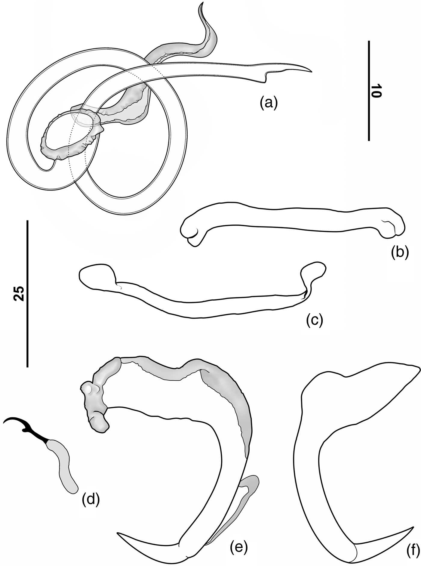
Based on six specimens (fig. 6); one mounted in Gomori's trichrome, five mounted in Gray & Wess medium. Body fusiform, may be constricted near midlength, total length excluding haptor 415 (380–465; n = 4) long, 131(120–140; n = 4) wide at level of germarium. Tegument smooth. Cephalic margin tapered; poorly developed terminal lobes; three bilateral pairs of head organs with rod-shaped secretion; cephalic glands unicellular, posterolateral to pharynx. Four eyes, posterior pair larger than anterior pair; accessory granules present in cephalic area, elliptical. Mouth subterminal, midventral; pharynx spherical, 131 (120–140; n = 4) in diameter. Genital pore opening midventral; genital atrium muscular. Gonads overlapping; testis dorsal to germarium. Testis, prostatic reservoir not observed. MCO with approximately 1½ rings, 83 (80–85; n = 3) long base with sclerotized cap; distal aperture subterminal, hook shaped (fig. 6a). Accessory piece comprising an elongated sheath. Germarium 94 (87–100; n = 2) long, 40 (34–45; n = 2) wide, elongated. Vagina comprising vaginal vestibule, vaginal canal with soft tissue. Seminal receptacle pyriform; Mehlis' glands, ootype not observed. Vitellaria dense throughout trunk, except in region of reproductive organs. Eggs not observed. Peduncle short. Haptor subhexagonal, 62 long, 103 wide. Anchors dissimilar. Ventral anchor (fig. 6e), base 16 (15–16; n = 3) long, with elongate superficial root, 38 (37–39; n = 3) long, inconspicuous deep root, anterior portion of superficial root covered with sclerotized cap; evenly curved shaft and point, forming angle of approximately 110°; short point, robust, extending at the level of tip of superficial root. Dorsal anchor (fig. 6f) base 18 (18; n = 2) long, robust, with elongate superficial root, subtriangular, 34 (33–35; n = 3) long, inconspicuous deep roots, evenly curved shaft, point; forming angle of approximately 100°, short point, robust; point extending well past level of tip of inner base. Ventral bar (fig. 6c) 54 (50–58; n = 2) long, slightly curved or straight rod with small terminal enlargements at ends, curved in anterior direction. Dorsal bar (fig. 6b) 40 (36–44; n = 2) long, narrow, rod-shaped, with bifurcated ends, slightly curved in posterior direction. Hooks similar in shape (fig. 6d), 18 (18–19; n = 4) long, shank with inflation, erected thumb, lightly curved long shaft, delicate point, filamentous hook, loop of hook extending to union of shank subunits.
Constrictoanchoratus lemmyi n. gen. n. sp. (a) Copulatory complex; (b) dorsal bar; (c) ventral bar; (d) hook; (e) ventral anchor; (f) dorsal anchor. Scale bars in μm.

Taxonomic summary
Type host
Hoplias malabaricus (Bloch).
Site of infestation
Gill filaments.
Type locality
Caeté River, Municipality of Bragança, Pará State, Brazil (1°3′54.82″S, 46°41′37.60″W).
Other records
Hoplias malabaricus, Itabocal River, Municipality of Irituia, Pará State, Brazil (1°51′59.82″S, 47°24′17.15″W).
Specimens deposited
Holotype: CHIOC no. 38634. Four paratypes: CHIOC nos 38638, 38642, 38646, 38650. One voucher: CHIOC no. 38633.
Etymology
The specific name is in honor of ‘Lemmy’ Kilmister (1945–2015), leader of the heavy-metal band Motorhead, of whom the senior author is a big fan.
Remarks
This species differs from C. ptilonophallus n. gen. n. sp. mainly by having a male copulatory organ comprising a coil of about 1½ rings (2½ rings in C. ptilonophallus), distal aperture subterminal, hook-shaped (aperture terminal, acute in C. ptilonophallus); base with sclerotized margin, without sclerotized brims (present in C. ptilonophallus); vaginal vestibule and canal with soft tissue (heavily sclerotized in C. ptilonophallus); and dorsal bar with bifurcated ends (not bifurcated in C. ptilonophallus).
A key to the Dactyologyridea species from Erythrinidae is given below.
Key to Dactyologyridae species from Erythrinidae
1 Prostatic reservoir simple; male copulatory organ (MCO) a coiled tube; circular sclerotized tandem brim associated with the base of the MCO present or absent 2
– Prostatic reservoir separated into two/three zones; MCO a spiral tube (corkscrew like); circular sclerotized tandem brim associated with the base of the MCO present or absent 11 (Whittingtonocotyle)
2(1) Vaginal sclerite present; anchors with evenly curved shaft and point 3 (Urocleidoides)
– Vaginal sclerite absent; anchors with constriction at the intersection between shaft and point 12 (Constrictoanchoratus)
3(2) MCO with two rings or fewer than two rings 4
– MCO with more than two rings 7
4(3) Anchors with superficial root at least three times larger than deep root; elongate shaft and point 5
– Anchors with superficial root twice as large as the deep root; short shaft and point; vaginal vestibule slightly sclerotized, bulb-shaped U. aimarai
5(4) Accessory piece delicate sheath, uniform; vaginal vestibule muscular or slightly sclerotized 6
– Accessory piece distally bifurcated; vaginal vestibule muscular; parasites of nasal cavities U. naris
6(5) Vaginal vestibule slightly sclerotized; anchors similar in size; dorsal bar slightly U-shaped with rounded ends; muscular pad surrounding the copulatory complex present U. malabaricusi
– Vaginal vestibule muscular; ventral anchor twice as large as the dorsal anchor; dorsal bar slightly U-shaped with bifurcated ends; MCO with proximal bulb U. bulbophallus sp. n.
7(3) Anchors with inconspicuous deep roots (at least ⅛ the size of superficial root) 8
– Anchors with developed deep roots (no more than ¼ the size of superficial root) 9
8(7) Dorsal bar slightly U-shaped with rounded ends; vaginal canal heavily sclerotized at proximal portion with a dilatation at middle portion, distal portion an elongate tube slightly sclerotized U. paranae sp. n.
– Dorsal bar slightly U-shaped with bifurcated ends; vaginal canal slightly sclerotized, expanded as a corrugated bag U. cuiabai
9(7) Eyespots present; vaginal aperture marginal; vaginal sclerite with grooves 10
– Eyespots absent; vaginal aperture ventral; vaginal sclerite without grooves U. xinguensis
10(9) Vaginal canal, heavily sclerotized, an undulated tube with a proximal looping U. eremitus
– Vaginal canal slightly sclerotized, a corrugated bag U. brasiliensis
11(1) Prostatic reservoir separated into two zones; vaginal canal convolute; dorsal bar with long anteromedial process W. caetei
– Prostatic reservoir separated in three zones; vaginal canal sigmoid; dorsal bar with short anteromedial process W. jeju
12(2) Vaginal vestibule and vaginal canal heavily sclerotized; MCO with 2½ rings; circular sclerotized tandem brim associated with the base of the MCO present C. ptilonophallus n. gen. n. sp.
– Vaginal vestibule and vaginal canal with soft tissue; MCO with 1½ rings C. lemmyi n. gen. n. sp.
Discussion
From the eight valid species of Monogenoidea known to parasitize the gills of species of Hoplias in Brazil, Argentina and Peru, only five species were reported for H. malabaricus. Urocleidoides eremitus was the first species of monogenoid described from this host species, which was captured in the rivers of the Occidental Brazilian Amazon Basin by Kritsky et al. (Reference Kritsky, Thatcher and Boeger1986). Later, four other species of this genus (U. brasiliensis Rosim, Mendoza-Franco & Luque, 2011, U. cuiabai Rosim, Mendoza-Franco & Luque, 2011, U. malabaricusi Rosim, Mendoza-Franco & Luque, 2011 and Urocleidoides naris Rosim, Mendoza-Franco & Luque, 2011) were described and/or reported from other Brazilian regions (south, south-east and mid-west regions of Brazil) (Rosim et al., Reference Rosim, Mendoza-Franco and Luque2011).
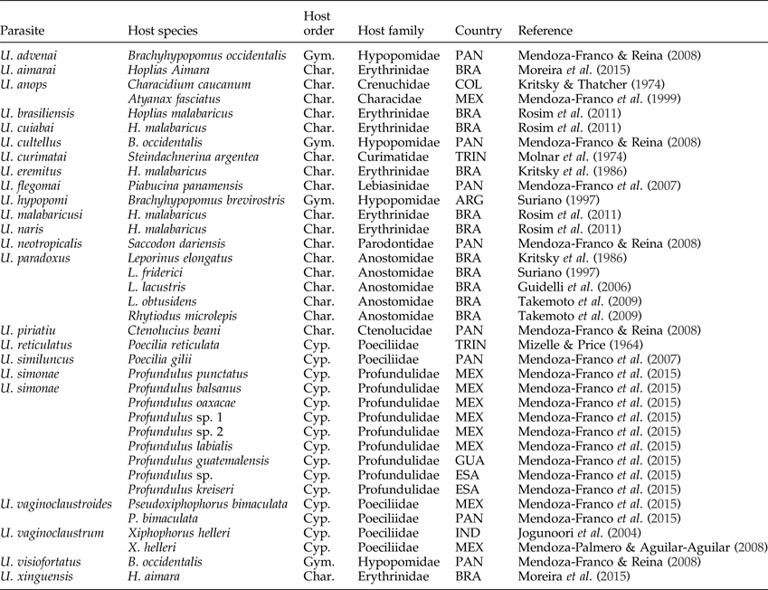
Mizelle & Price (Reference Mizelle and Price1964) proposed Urocleidoides Mizelle & Price 1964 for their new species, U. reticulatus Mizelle & Price, 1964. The new species was found parasitizing the gills of Poecilia reticulata Peters (Poeciliidae) collected in the Capitol Aquarium, Sacramento, California, USA. After the revision of the genus proposed by Kritsky et al. (Reference Kritsky, Thatcher and Boeger1986), Urocleidoides was restricted to species possessing a sinistral vaginal sclerite, overlapping or tandem gonads, a male copulatory organ with counterclockwise rings, and the morphology of haptoral structures. Actually, the genus contains 20 valid species (Kritsky et al., Reference Kritsky, Thatcher and Boeger1986; Mendoza-Franco et al., Reference Mendoza-Franco, Scholz, Vivas-Rodrigues and Vargas-Vazquez1999, Reference Mendoza-Franco, Aguirre-Macedo and Vidal-Martínez2007; Jogunoori et al., Reference Jogunoori, Kritsky and Venkatanarasaiah2004; Mendoza-Franco & Reina, Reference Mendoza-Franco and Reina2008; Moreira et al., Reference Moreira, Scholz and Luque2015) from fish hosts representing two ostariophysian teleost orders (Characiformes and Gymnotiformes) and Cyprinodontiformes from South America, Central America and Mexico (table 6).
List of species of Urocleidoides sensu stricto. Orders of fishes: Gym., Gymnotiformes; Char., Characiformes; Cyp, Cyprinodontiformes. Countries: ARG, Argentina; BRA, Brazil; COL, Colombia; ESA, El Salvador; GUA, Guatemala; IND, India; MEX, Mexico; PAN, Panama; TRI, Trinidad.

Mendoza-Franco & Reina (Reference Mendoza-Franco and Reina2008) described Urocleidoides advenai Mendoza-Franco & Reina, 2008 taken from the gills of Brachyhypopomus occidentalis (Regan) (Gymnotiformes) in Central America. This species shares the morphology of the copulatory complex with other species of the Urocleidoides; however, it is also characterized by the absence of the vaginal sclerite. Mendoza-Franco & Reina (Reference Mendoza-Franco and Reina2008) considered that the main limitation in determining the diagnostic limits of Urocleidoides was the lack of a cladistic analysis for this genus.
For taxonomy purposes, we opted to follow Kritsky et al. (Reference Kritsky, Thatcher and Boeger1986) in their taxonomic diagnosis of Urocleidoides; therefore, we consider U. advenai as belonging to Urocleidoides sensu lato. Besides U. advenai, nine other species of Urocleidoides are currently considered as incertae sedis and remain to be re-assigned to appropriate genera in the Neotropics: U. astyanacis Gioia, da Silva Cordeiro & de Toledo Artigas 1988, U. strictus Mizelle, Kritsky & Crane 1968, U. trinidadensis Molnar, Hanek & Fernando 1974 from Characiformes; U. carapus Mizelle, Kritsky & Crane 1968, U. gymnotus Mizelle, Kritsky & Crane 1968 and U. virescens from Gymnotiformes; and U. amazonensis Mizelle & Kritsky 1969, U. catus Mizelle & Kritsky 1969 and U. megorchis Mizelle & Kritsky 1969 from Siluriformes. We believe that future phylogenetic studies using morphological and/or molecular characters with appropriate taxa sampling will help us to define the real taxonomic status of these ten species.
Urocleidoides brasiliensis Rosim, Mendoza-Franco & Luque, 2011, U. cuiabai Rosim, Mendoza-Franco and Luque, 2011, U. eremitus Kritsky, Thatcher & Boeger, 1986 and U. malabaricusi Rosim, Mendoza-Franco & Luque, 2011 are reported here for the first time to be parasitizing the gills of H. malabaricus from streams located in the Oriental Amazon Basin. The analysis of specimens of U. eremitus from different localities revealed that they are morphologically similar (figs 3 and 4). The only exception was U. eremitus from the Upper Paraná River floodplain, which represents a new species of Urocleidoides, described here as U. paranae sp. n. We detected that the specimens of U. brasiliensis, U. cuiabai and U. malabaricusi reported in the present work, differ morphometrically from those presented by Rosim et al. (Reference Rosim, Mendoza-Franco and Luque2011). These authors also detected variations in the shape and size of haptoral structures of U. brasiliensis, U. cuiabai and U. eremitus from H. malabaricus captured in different locations in Brazil and considered them to be intraspecific variations. This is probably due to the geographic distance or even due to the results of phenotypic plasticity of parasites or host-induced morphological change, as suggested by León-Règagnon et al. (Reference León-Règagnon, Guillén-Hernández and Arizmendi-Espinosa2005).
Domingues & Marques (Reference Domingues and Marques2011) also considered that the morphometric differences observed in some species of monogenoids from the genus Potamotrygonocotyle (Monocotylidae), which parasitize the gills of freshwater stingrays (Potamotrygonidae), could not be considered as evidence of interspecific variation. These authors conducted a cladistic analysis based on morphological characters for the species of Potamotrygonocotyle. The results suggested that there was no autapomorphic character that would sustain the maintenance of some nominal species as valid. Fehlauer-Ale & Littlewood (Reference Fehlauer-Ale and Littlewood2011) conducted a molecular cladistic analysis on some species of Potamotrygonocotyle and discovered the existence of cryptic lineages, suggesting that the diversity of the genus may be underestimated.
Gasques et al. (Reference Gasques, Graça, Prioli, Takemoto and Prioli2015) proposed the first molecular characterization of Urocleioides cuiabai and U. malabaricusi from Hoplias aff. malabaricus captured in the Upper Paraná River floodplain (Brazil) based on a fragment of the cytochrome c oxidase subunit 1 (COI) gene. These authors suggested that U. malabaricusi could represent a cryptic lineage, based on the magnitude of genetic divergence. Although U. malabaricusi is characterized mainly by the possession of a muscular pad surrounding the copulatory complex, this feature was also reported for U. eremitus from the East Atlantic Basin and Paraná River Basin (Rosim et al., Reference Rosim, Mendoza-Franco and Luque2011) (see also comments under the Remarks on U. eremitus), which challenges the taxonomy of the species when based on morphology or molecular data alone. Therefore, it raises the question: whether Gasques et al. (Reference Gasques, Graça, Prioli, Takemoto and Prioli2015) were dealing with a cryptic species of ‘U. malabaricusi complex’ or if there was a misidentification of a congeneric species, such as U. eremitus. We suggest that a detailed taxonomic/morphological characterization be undertaken, and we also recommend that more than two species be included to propose an optimal phylogenetic tree for this group.
Parasitic organisms have been used as biological markers to discriminate fish stocks and to determine migration routes (Mackenzie, Reference Mackenzie1987, Reference Mackenzie2002), as well as to evaluate the phylogenetic relationships of their hosts (Brooks & Glen, Reference Brooks and Glen1982; Brooks et al., Reference Brooks, Bandoni, MacDonald and O'Grady1989; Brooks & McLennan, Reference Brooks and McLennan1991, Reference Brooks and McLennan1993; Hoberg, Reference Hoberg1992; Klassen, Reference Klassen1992). Among the parasite groups in fish hosts, monogenoidean parasites represent an excellent biological marker (Tirard et al., Reference Tirard, Berrebi, Raibaut and Frenaud1992; Lambert & El Gharbi, Reference Lambert and El Gharbi1995) and an excellent evolutionary model (Boeger & Kritsky, Reference Boeger and Kristky1989, Reference Boeger and Kristky1997, Reference Boeger and Kristky2003; Domingues & Boeger, Reference Domingues and Boeger2005), mainly because they possess a direct life cycle (monoxenic) and an exceptional host specificity (Bychowsky, Reference Bychowsky1957). In addition to morphological, genetic and molecular assessment, other features, such as parasite infestation, could be a valuable source of information and could potentially be used for host species recognition.
Morphological, cytogenetic and molecular evidence indicates that H. malabaricus is a species complex (Bertollo et al., Reference Bertollo, Born, Dergam, Fenocchio and Moreira-Filho2000; Oyakawa, Reference Oyakawa, Reis, Kullander and Ferraris2003; Santos et al., Reference Santos, Volcker, Belei, Cioffi, Bertollo, Paiva and Dergam2009). Santos et al. (Reference Santos, Volcker, Belei, Cioffi, Bertollo, Paiva and Dergam2009) reported that some lineages of erythrinid fish recognized as H. malabaricus belong to a different species when comparing cytogenetic and molecular characters, and at least some karyotypic strains are related to H. malabaricus. Recent studies indicate that a single karyomorph of H. malabaricus can harbour more than one species of Hoplias (Marques et al., Reference Marques, Santos, Silva, Sampaio and Rodrigues2013). Among parasites, Urocleidoides sensu stricto has the largest range, parasitizing nine families of three orders. On the host–parasite network proposed by Braga et al. (Reference Braga, Araújo and Boeger2014), Urocleidoides is indicated as a provincial hub with many interactions, and most of them are modular, being influenced by spatial structure and phylogenetic relatedness of species.
The occurrence of monogenoidean parasites infesting H. malabaricus from different Brazilian river basins provides evidence that the diversity of monogenoids from this host requires further study. Nadler & Pérez-Ponce de Léon (Reference Nadler and Pérez-Ponce de Léon2011) suggested that parasitological studies should include broader aspects of comparative biology, such as systematics, evolution, ecology and biogeography/phylogeography.
Finally, it is an open question whether U. paranae n. sp. should be restricted only to the Paraná River, in the same way as U. bulbophallus n. sp. and species of Constrictoanchoratus are restricted only to the coastal drainage ecosystem of the Oriental Amazon rivers. The taxonomic status of species of Urocleidoides infecting H. malabaricus collected from other hydrographic basins should be studied for a more refined analysis, especially with the verification of molecular data and appropriate taxa sampling.
Acknowledgements
The authors thank the following individuals for supporting this study: Marcelo Knoff and Daniela Lopes, Coleção Helmintológica do Instituto Oswaldo Cruz, Rio de Janeiro, RJ (Brazil), and Célio Magalhães, Coleção de Invertebrados do Instituto Nacional de Pesquisas da Amazônia, Manaus, AM (Brazil) allowed access to specimens under their care; Bárbara Branches, Daniel Ribeiro, Geusivam Soares, João Santos-Neto, Laís Araújo, Nayna Costa, Raquel Nascimento and Renan Reis for assistance during the collecting trips; George Mattox (Universidade Federal de São Carlos, Campus Sorocaba, Brazil) for host identification.
Financial support
This work was supported partially by Programa de Pós-Graduação em Biologia Ambiental da Universidade Federal do Pará by granting an MSc Scholarship and collecting trip grant to K.D.C.F.; research grants from the Conselho Nacional de Desenvolvimento Científico e Tecnológico (CNPq) (PROTAX no. 001/2015–440526/2015-9) to M.V.D; and Fundação Amazônia de Amparo a Estudos e Pesquisa (FAPESPA) (ICAAF no. 41/2011) to J.-M. C.
Conflict of interest
None.
Ethical standards
Specimens were collected under the license for collection of biological material (43381–1) granted by the Instituto Chico Mendes de Conservação da Biodiversidade (ICMBio).