Coronary artery fistula is a rare congenital anomaly in which a coronary artery connects to any of the four chambers of the heart or any of its great vessels. Even when they are asymptomatic, coronary fistulae can be associated with the ultimate development of severe complications; therefore, surgical correction or catheter-based interventions are often indicated. Of all types of coronary fistulae, those that connect the left main coronary artery to the right atrium are uncommon,Reference Brussee and Gasser 1 and very few surgically corrected cases have been reported previously.
Clinical summary
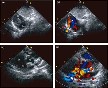
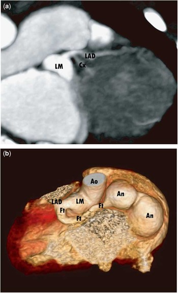
A 2-year-old boy was admitted to our institute after a heart murmur was appreciated during a routine physical examination. Upon auscultation, a continuous heart murmur was best heard in the left second intercostal space. The transthoracic echocardiogram revealed a “cyst-like” structure within the right atrium (Fig 1a). Color Doppler demonstrated a continuous systolic–diastolic flow inside the structure and a high-velocity systolic–diastolic signal directed from the inside of the structure towards the right atrium (Fig 1b). After careful examination, the systolic–diastolic signal was revealed to originate from the left coronary artery orifice and to course behind the aorta until it reached the superior portion of the right atrium (Fig 1c and d). A coronary arteriovenous fistula was suspected and the patient was referred for cardiac CT. The left main coronary artery origin was dilated with a maximum diameter of 7 mm. The left main coronary artery bifurcated into the left anterior descending coronary artery and a small circumflex coronary artery (Fig 2a). The arteriovenous fistula originated before the bifurcation of the left main coronary artery and coursed tortuously, posterior to aorta, before finally draining into the superior portion of the right atrium (Fig 2b). In addition, two aneurysm-like enlargements were evident on the CT.
( a ) Subcostal two-dimensional view demonstrated a “cyst-like” structure that is the aneurysm-like fistula (An) draining into the right atrium (RA). ( b ) Color Doppler of the same image shows the blood flow in the fistula. ( c ) Aortic valve short-axis view demonstrated the enlargement of the origin of the fistula from the enlarged left coronary artery and its course behind the aorta (AO) before it drained into the RA. ( d ) Color Doppler of the same image shows the blood flow in the fistula. LA=left atrium; LM=left main coronary artery; RVOT=right ventricular outflow tract.

( a ) CT scan showed the enlargement of the section of origin of the left main coronary artery (LM) and the normal left anterior descending artery (LAD) and circumflex artery (Cx). ( b ) Three-dimensional reconstruction of the CT scan demonstrated the origin, course, and two aneurysm-like enlargement (An) of the fistula (Ft) before it drained into the right atrium. Ao=aorta.

The patient underwent surgical repair using cardiopulmonary bypass. The proximal and distal portions of the fistula were divided, decompressed, sutured close, and left in situ. At the conclusion of the case, there was no flow in the fistula, as confirmed by postoperative transthoracic echocardiogram. The patient had an uneventful recovery and was discharged home on postoperative day 7.
Discussion
A coronary artery fistula is an abnormal connection between a coronary artery and any of the four chambers of the heart or any of its great vessels.Reference Sercelik, Mavi and Ayalp 2 Of all types of coronary fistulae, the connection between the left main coronary artery and the right atrium is uncommon, and only a few cases have been reported in the literature.Reference Brussee and Gasser 1 Despite remaining asymptomatic in the majority of children, coronary fistulae are associated with left-to-right shunting, myocardial ischaemia secondary to coronary steal, and the potential for the development of mural thrombosis. Proximal coronary fistulae with aneusymal dilatation left untreated are also associated with potential rupture and death.Reference Angelini 3 Percutaneous transcatheter closure techniques can be used for the closure of the fistula.Reference Wisniewska-Szmyt, Swiatkiewicz and Chojnicki 4 In the present case, the complex anatomy of the fistula precluded the use of transcatheter techniques and surgical repair was therefore indicated. Coronary angiography is a commonly used modality for the diagnosis and tracing of the anatomical course of coronary fistulae. In the present study, we demonstrated that transthoracic echocardiography and contrast CT are sufficient for defining the complex anatomy of the coronary fistula and its relationship to adjacent structures in anticipation of surgical intervention.
Acknowledgements
None.
Financial Support
This work was supported by the National Natural Science Foundation of China (G.T., Grant No. 81500183), (Z.S., Grant No. 81400366).
Conflicts of Interest
None.