




No CrossRef data available.
Article contents
Paleohaemoproteus burmacis gen. n., sp. n. (Haemospororida: Plasmodiidae) from an Early Cretaceous biting midge (Diptera: Ceratopogonidae)
Published online by Cambridge University Press: 24 March 2005
Abstract
Paleohaemoproteus burmacis gen. n., sp. n. (Haemospororida: Plasmodiidae) is described from the abdominal cavity of a female biting midge (Diptera: Ceratopogonidae) preserved in 100 million year old amber from Myanmar (Burma). The description is based on the developmental stages of oocysts and sporozoites. The fossil species differs from extant species of Haemoproteus by its wide range of oocyst sizes, small sporozoites and occurrence in an extinct species of biting midge. Numerous sporozoites in the abdominal cavity suggest that the biting midge was an effective vector of this malarial parasite. Characters of the biting midge suggest that the host was a large, cold-blooded vertebrate. This is the earliest record of a malaria parasite and first indication that Early Cretaceous reptiles were infected with haemosporidial parasites.
Information
- Type
- Research Article
- Information
- Copyright
- © 2005 Cambridge University Press
INTRODUCTION
Biting midges (Diptera: Ceratopogonidae) are ancient haematophagous insects, extending back to at least the Lower Cretaceous (Poinar & Milki, 2001). Extant biting midges are known to carry a number of vertebrate pathogens (Fallis & Bennett, 1961b; Linley, 1985). During a survey of haematophagous insects in amber, a female biting midge was observed to harbour oocysts and sporozoites of a malarial parasite. The present study identifies these parasites as a Haemoproteus-like malarial pathogen, and provides a description based on characters of the sporogonic cycle.
MATERIALS AND METHODS
The amber piece containing the fossil biting midge was re-cut and polished in order to better view the specimen. The final piece is rectangular in outline, measuring 6 mm long, 2 mm wide and 2 mm deep. Observations, drawings and photographs were made with a Nikon SMZ-10 R stereoscopic microscope and Nikon Optiphot compound microscope with magnifications up to 1050X.
Amber from Myanmar (Burma) occurs in lignitic seams in sandstone-limestone deposits in the Hukawng Valley. Palynomorphs obtained from the amber beds have been assigned to the Upper Albian (~100–105 mya) (Cruickshank & Ko, 2003). Nuclear magnetic resonance (NMR) spectra of amber samples taken from the same locality as the fossil indicated an araucarian (possibly Agathis) plant source (Lambert and Wu, personal communication).
RESULTS
Most of the insect's tissues and cells in the abdomenal cavity of the fossil biting midge had been completely cleared, leaving only an outer cuticular border and some tissue at the posterior end (Fig. 1A). Most of the clearing probably resulted from bacterial activity before the insect was completely covered by the resin. After complete entombment, chemicals from the resin could have further cleared the abdomen. The cleared abdomen allowed an unobstructed view of a number of developing oocysts and sporozoites of a malarial parasite. The head of the biting midge was complete (mouthparts, antennae, palps) as are all legs except the right foreleg. Identification of the biting midge was difficult since, except for the basal portion of one, the wings were missing. However, the remaining basal portion of the wing had the R and M veins exiting from the basal arculus, which is typical for ceratopogonids. The absence of macrotrichia on the existing portion of the wing, empodia absent, tarsal claws equal, hind-femora not noticeably larger than fore- and mid-femora, a 13-segmented flagellum with the terminal 5 segments longer than the preceding, the palpus with 2 segments beyond palpal segment 3, the hind-leg claws moderately developed, scattered setae on the first tarsomere of the hind-leg and a hind-leg/foreleg tarsal ratio of 1·1, align the fossil biting midge with the genus Protoculicoides (Boesel, 1937; Borkent, 1995).

Fig. 1. (A) Lateral view of the fossil biting midge in Burmese amber. Note the cleared abdominal cavity, showing the numerous oocysts as dark dots. (B) Developing oocysts of Paleohaemoproteus burmacis in Burmese amber. Lower two oocysts (arrows) contain numerous nucleated bodies and upper one shows short, curved sporozoites differentiating from the nucleated bodies. (C) Section of the midgut region of the biting midge with a number of oocysts of P. burmacis at various stages of development. Note empty oocyst with faint wall (arrow) and associated residual body (arrowhead).
Systematics
Phylum Apicomplexa Levine, 1970; Class Aconoidasida Mehlhorn, Peters & Haberkorn, 1980; Order Haemospororida Danilewsky, 1885; Family Plasmodiidae Mesnil, 1903.
Paleohaemoproteus n. gen.
This genus is established as a collective genus for fossil members of the genus Haemoproteus sensu lato associated with biting midges. There are no type species associated with collective genera and such genera have no taxonomic standing.
Paleohaemoproteus burmacis n.sp. (Figs 1 and 2)
Oocysts
Some 35 oocysts were observed in the abdomen of the fossil biting midge. It is presumed that the oocysts had been associated with the epithelial cells of the gut wall (Fig. 1A,C). The oocysts ranged from 6 to 31 μm in greatest diameter. A distinct wall, 0·8 μm in thickness, could be observed on some of them (Fig. 2D,E). Within the oocysts in the early stages of development were spherical cells ranging from 3·2 to 4·6 μm in greatest length. The nuclei within these cells ranged from 1·7 to 2·3 μm in diameter.

Fig. 2. Oocyst and sporozoite development in Paleohaemoproteus burmacis. (A) Differentiating oocyst in early stage of development showing subcapsular vacuolation with a large central vacuole (arrow). (B) Differentiating oocyst with nucleated bodies indicating division of the chromatin and cytoplasm. Some sporozoites (arrows) have begun to form. (C) Spherical oocyst containing sporozoites. Note central residual body (arrow) with pigment granules. (D) Oval oocyst filled with sporozoites. Pointed end was probably attached to the insect's midgut epithelium. Note oocyst wall (arrow). (E) Mature oocyst containing sporozoites preparing to exit. Note oocyst wall (arrow). (F) Released from their oocysts, sporozoites cluster in the anterior portion of the abdominal haemocoel of the biting midge.
Sporozoites
Developing sporozoites within the oocysts varied from 2·3 to 3·5 μm in length and from 0·3 to 0·4 μm in width. Released sporozoites ranged from 0·4 to 0·6 μm in length.
Comments
The fossil species differs from extant species of Haemoproteus by having a greater range of oocyst size and smaller sporozoites (Table 1). It is further characterized by its presence in a biting midge entombed in 100 million-year-old Burmese amber.

Oocysts in the early stages of development showed subcapsular vacuolation (Fig. 2A), which has been noted on extant haemosporidians (Fallis & Bennett, 1961a). A further stage of development was subdivision of the cytoplasm into separate units, each with a nucleus and surrounding cytoplasm (Figs 1B,C and 2A–C), similar to that observed in the development of extant haemosporidians (Fallis & Bennett, 1961a). Still later development involved the formation of immature sporozoites within the oocysts (Figs 1B and 2B). At this stage, it was possible to see the residual body (often with dark granules) within the oocyst (Fig. 2C). Other oocysts contained mature elongate sporozoites (Fig. 2D,E). Empty oocysts could be identified by their thin wall and associated residual body (Fig. 1C). Large numbers (N=254) of free sporozoites were clustered together in the anterior portion of the abdominal cavity (Fig. 2F).
Type specimen
Developing oocysts with sporozoites in the body cavity of a female biting midge deposited in the Poinar amber collection (accession no. B-D-32) maintained at Oregon State University.
Host
An extinct species of biting midge, probably belonging to the genus Protoculicoides (Ceratopogonidae: Diptera) in Burmese amber.
Type locality
Amber mine in the Hukawng Valley, southwest of Maingkhwan in the state of Kachin (26°20′N, 96°36′E), northern Myanmar (Burma).
DISCUSSION
There are several genera of Plasmodiidae vectored by insects that resemble the present fossil (Telford, 1994; Perkins et al. 2000), namely Hepatocystis Levaditi and Schoen, 1932, Plasmodium Marchiafava & Celli, 1885, Leucocytozoon Sambon, 1908, Saurocytozoon Lainson & Shaw, 1969 and Haemoproteus Kruse, 1890. Species in these genera all have similar life-cycles involving a vertebrate and insect host (Atkinson & van Riper, 1991; Telford, 1994; Perkins et al. 2000). The biting insect serves as the vector and acquires gametocytes of the parasite from infected vertebrate blood cells during feeding. The gametocytes are liberated in the stomach of the biting insect, where sexual reproduction occurs, thus beginning the sporogonic stage of the life-cycle. The zygote differentiates into a motile ookinete that penetrates into or between the epithelial cells of the insect's midgut and becomes an oocyst. The oocysts form discrete nucleated bodies that mature into sporozoites. The motile sporozoites leave the oocysts and migrate into the body cavity and eventually into the salivary glands where they are transferred back to the vertebrate at the next bloodmeal.
Members of the genus Hepatocystis occur in mammals and are vectored by Culicoides biting midges. However, the oocysts occur in the head and thorax of the vector (Perkins et al. 2000). Members of the genus Plasmodium have large oocysts that develop in mosquitoes and phlebotomine flies. The genus Leucocytozoon has small (from 10 to 25 μm in diameter), non-expanding oocysts, occurs in birds and is vectored mainly by simuliid blackflies (one species is transmited by a Culicoides biting midge) (Desser & Bennett, 1993; Perkins et al. 2000). In Leucocytozoon, separate nucleated bodies are not formed in the oocyst (only multinucleated bodies with the sporozoites budding off a central mass) (Desser & Bennett, 1993). The genus Saurocytozoon infects reptiles and has culicine mosquitoes as experimental vectors. It forms oocysts larger than 20 μm (up to 62 μm in diameter), each containing several hundred long, thin sporozoites (Landau et al. 1973). Species of Haemoproteus infect birds and reptiles and are vectored by hippoboscid flies, Culicoides biting midges and Chrysops deerflies. They produce expanding oocysts that have discrete nucleated bodies which give rise to sporozoites (Desser & Bennett, 1993) and form up to several hundred elongate thin sporozoites (Garnham, 1966). On the basis of the position of the oocysts in the biting midge, the number and size of the oocysts, the number of sporozoites produced per oocyst, the size of the sporozoites and the systematic placement of the vector, the fossil protozoa most closely resemble extant members of the genus Haemoproteus. Variability in oocyst and sporozoite size and numbers of sporozoites per oocyst appears to be common in species of Haemoproteus (Adie, 1924; Sterling & Deguisti, 1974).
The oocysts and sporozoites described in the fossil biting midge have features in common with the developmental stages of Haemoproteus canachites Fallis & Bennett in Culicoides sphagnumensis Williams (Fallis & Bennett, 1960) and H. nettionis (Johnson & Cleland) in Culicoides sp. (Fallis & Bennett, 1961a). These authors reported developing oocysts ranging from 5 to 11 μm in diameter, which compares with the smaller ones found in the fossil biting fly.
In the early stages of sporozoite formation, spherical-oval bodies, similar to those observed in the fossil oocysts, are formed in Haemoproteus species (Fallis & Bennett, 1960; Desser & Bennett, 1993). Sporozoites of Haemoproteus may be released though a rupture of the oocyst wall or leave the oocyst gradually (Khan & Fallis, 1971; Desser & Bennett, 1993) and it appears that timed, gradual release occurs with P. burmacis since some oocysts contained mature sporozoites as well as oval nucleated bodies. This condition is also apparent in H. nettionis Fallis & Bennett (1961a, Fig. 33) where nucleated pre-sporozoite stages occur in the oocyst along with exiting sporozoites.
Today, the haemoproteids are one of the most commonly occurring blood parasites of birds (Fallis & Wood, 1957; Bennett & Peirce, 1988; Atkinson & van Riper, 1991) and reptiles, especially Old and New World turtles and African cobras (Telford, 1984). Most members of Haemoproteus are transmitted by biting midges and the wide distribution of these pathogens can be explained by the nearly cosmopolitan occurrence of Culicoides, the genus most commonly incriminated in the transmission of Haemoproteus species (Linley, 1985; Desser & Bennett, 1993).
Numerous sporozoites in the abdominal cavity of the fossil suggest that the biting midge was an effective vector of P. burmacis. While the vertebrate host cannot be definitely determined, some indication can be ascertained from morphological features of the biting fly. A long, slender third palpal segment with a small number (6–8) of capitate sensilla, as occurs on the fossil, is indicative of species that feed on large vertebrates (Borkent, 1995). An additional character related to the vertebrate host is the number and disposition of sensilla coeloconica (small glands) on the antennal flagellomeres (Borkent, 1995). The fossil biting midge appears to lack flagellar sensilla coeloconica, which indicates that the host is likely to have been cold-blooded. Possible candidates in the Burmese amber forest would have been large lizards, crocodilians and dinosaurs. Dinosaurs were suggested as possible hosts for Upper Cretaceous biting midges (Borkent, 1995). The idea that biting midges could have transmitted pathogens to dinosaurs in the Lower Cretaceous was proposed by Poinar & Milki (2001). The present study indicates that this latter scenario could have been possible. This report is the earliest record of a malarial pathogen and the first evidence that Early Cretaceous reptiles could have been infected by haemosporidial parasites.
We thank Roberta Poinar for editorial comments.
References
REFERENCES
ADIE, H. (
1924).
The sporogony of Haemoproteus columbae.
Bulletin de la Société de Pathologie Exotique
17,
605–613.Google Scholar
ATKINSON, C. T. &
VAN RIPER Iii, C. (
1991).
Pathogenicity and epizootiology of avian Haematozoa: Plasmodium, Leucocytoozoon, and Haemoproteus. In
Bird-Parasite Interactions: Ecology, Evolution, and Behaviour
(ed. Loye, J. E. & Zuk, M.), pp.
19–48.
Oxford University Press,
New York.
BENNETT, G. F. &
PEIRCE, M. A. (
1988).
Morphological form in the avian Haemoproteidae and an annotated checklist of the genus Haemoproteus Kruse, 1890.
Journal of Natural History
22,
1683–1696.CrossRefGoogle Scholar
BOESEL, M. W. (
1937).
Order Diptera. In
Insects and Arachnids from Canadian Amber, vol. 4 (ed. Carpenter, F. M., Folsom, E. D., Essig, A. C., Kinsey, A. C., Brues, C. T., Boesel, M. W. & Ewing, H. E.), pp.
44–55.
University of Toronto Studies,
Geological Series.
BORKENT, A. (
1995).
Biting Midges in the Cretaceous Amber of North America (Diptera: Ceratopogonidae).
Backhuys Publishers,
Leiden.
CRUICKSHANK, R. D. &
KO, K. (
2003).
Geology of an amber locality in the Hukawng Valley, northern Myanmar.
Journal of Asian Earth Sciences
21,
441–445.CrossRefGoogle Scholar
DESSER, S. S. &
BENNETT, G. F. (
1993).
The genera Leucocytozoon, Haemoproteus, and
Hepatocystis. In
Parasitic Protozoa, 2ndEdn, vol. 4,(ed.KREIER, J. P.),
pp.273–307.
Academic Press,
San Diego.
FALLIS, A. M. &
BENNETT, G. F. (
1960).
Description of Haemoproteus canachites n. sp. (Sporozoa: Haemoproteidae) and sporogony in Culicoides (Diptera: Ceratopogonidae).
Canadian Journal of Zoology
38,
455–464.Google Scholar
FALLIS, A. M. &
BENNETT, G. F. (
1961 a).
Sporogony of Leucocytozoon and Haemoproteus in simuliids and ceratopogonids and a revised classification of the Haemosporidiida.
Canadian Journal of Zoology
39,
215–228.Google Scholar
FALLIS, A. M. &
BENNETT, G. F. (
1961 b).
Ceratopogonidae as intermediate hosts for Haemoproteus and other parasites.
Mosquito News
21,
21–28.Google Scholar
FALLIS, A. M. &
WOOD, D. M. (
1957).
Biting midges (Diptera: Ceratopogonidae) as intermediate hosts for Haemoproteus of ducks.
Canadian Journal of Zoology
35,
425–435.CrossRefGoogle Scholar
GARNHAM, P. C. C. (
1966).
Malaria Parasites and other Haemosporidia.
Blackwell Scientific Publications,
Oxford.
KHAN, R. A. &
FALLIS, A. M. (
1971).
A note on the sporogony of Parahaemoproteus velans (=Haemoproteus velans Coatney and Roudabush) (Haemosporidia: Haemoproteidae) in species of Culicoides.
Canadian Journal of Zoology
49,
420–421.CrossRefGoogle Scholar
LANDAU, I.,
LAINSON, R.,
BOULARD, Y.,
MICHEL, Y. &
SHAW, J. J. (
1973).
Dévelopment chez Culex pipiens de Saurocytozoon tupinambi (Sporozaire, Leucocytozoidae), parasite de lézards brésiliens.
Comptes Rendus Hebdomadaires des Seances de l'Academie des Sciences
276,
2449–2452.Google Scholar
LINLEY, J. R. (
1985).
Biting midges (Diptera: Ceratopogonidae) as vectors of nonviral animal pathogens.
Journal of Medical Entomology
22,
589–599.CrossRefGoogle Scholar
PERKINS, F. O.,
BARTA, J. R.,
CLOPTON, R. E.,
PIERCE, M. A. &
UPTON, S. J. (
2000).
Phylum Apicomplexa Levine, 1970. In
An Illustrated Guide to the Protozoa, 2nd Edn, vol. 1
(ed. Lee, J. J., Leedale, G. F. & Bradbury, P.), pp.
190–369.
Society of Protozoologists,
Lawrence, Kansas.
POINAR, G. O. Jr. &
MILKI, R. (
2001).
Lebanese Amber.
Oregon State University Press,
Corvallis,
USA.
STERLING, C. R. &
DE GUISTI, D. L. (
1974).
Fine structure of differentiating oocysts and mature sporozoites of Haemoproteus metchnikovi in its intermediate host Chrysops callidus.
Journal of Protozoology
21,
276–283.CrossRefGoogle Scholar
TELFORD, S. R. Jr., (
1984).
Haemoparasites of reptiles. In
Diseases of Amphibians and Reptiles
(ed. Hoff, G. L., Frye, F. L. & Jacobson, E. R.), pp.
385–517.
Plenum Press,
New York.CrossRef
TELFORD, S. R. Jr. (
1994).
Plasmodia of reptiles. In
Parasitic Protozoa, 2nd Edn, vol. 7
(ed. Kreier, J. P.), pp.
1–71.
Academic Press,
San Diego.
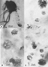
Fig. 1. (A) Lateral view of the fossil biting midge in Burmese amber. Note the cleared abdominal cavity, showing the numerous oocysts as dark dots. (B) Developing oocysts of Paleohaemoproteus burmacis in Burmese amber. Lower two oocysts (arrows) contain numerous nucleated bodies and upper one shows short, curved sporozoites differentiating from the nucleated bodies. (C) Section of the midgut region of the biting midge with a number of oocysts of P. burmacis at various stages of development. Note empty oocyst with faint wall (arrow) and associated residual body (arrowhead).
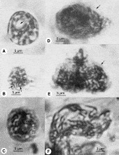
Fig. 2. Oocyst and sporozoite development in Paleohaemoproteus burmacis. (A) Differentiating oocyst in early stage of development showing subcapsular vacuolation with a large central vacuole (arrow). (B) Differentiating oocyst with nucleated bodies indicating division of the chromatin and cytoplasm. Some sporozoites (arrows) have begun to form. (C) Spherical oocyst containing sporozoites. Note central residual body (arrow) with pigment granules. (D) Oval oocyst filled with sporozoites. Pointed end was probably attached to the insect's midgut epithelium. Note oocyst wall (arrow). (E) Mature oocyst containing sporozoites preparing to exit. Note oocyst wall (arrow). (F) Released from their oocysts, sporozoites cluster in the anterior portion of the abdominal haemocoel of the biting midge.

Table 1. Comparison of the oocysts and sporozoites of Paleohaemoproteus burmacis with extant Haemoproteus spp.
 You have
Access
- 33
- Cited by