Introduction
Adults of the genus Anisakis Dujardin, 1845 are gastric parasites of marine mammals, especially cetaceans. The definitive hosts acquire the infection by preying on crustacean intermediate hosts or fish and squid paratenic hosts infected with the third-stage larvae of the nematode. In the past 20 years, the systematics and the taxonomic status of the species belonging to this genus have been largely clarified using genetic/molecular and morphological approaches. As a result, two main clades encompassing nine taxa are currently recognized in the genus Anisakis (Mattiucci & Nascetti, Reference Mattiucci and Nascetti2006; Valentini et al., Reference Valentini, Mattiucci, Bondanelli, Webb, Mignucci-Giannone, Colom-Llavina and Nascetti2006). A first clade includes the three sibling species in the Anisakis simplex complex (i.e. A. simplex s.s., A. pegreffii and A. simplex C), A. typica and A. ziphidarum. A sixth taxon, identified by allozyme markers and mitochondrial cox2 sequences and named provisionally Anisakis sp., is also included in this clade. The second clade encompasses the species A. physeteris, A. brevispiculata and A. paggiae, all of them parasites of sperm whales. Of the six taxa recognized in the first clade, only two species, A. ziphidarum and Anisakis sp., have been found parasitizing the stomach of beaked whales (family Ziphiidae) (Paggi et al., Reference Paggi, Nascetti, Webb, Mattiucci, Cianchi and Bullini1998; Valentini et al., Reference Valentini, Mattiucci, Bondanelli, Webb, Mignucci-Giannone, Colom-Llavina and Nascetti2006). However, the adult stage of Anisakis sp. has never been described.
In addition to these nine taxa, recent polymerase chain reaction-restriction fragment length polymorphism (PCR-RFLP) studies carried out with third-stage larval anisakids recovered from marine fish caught in Madeiran waters have revealed the existence of an undescribed taxon, which has been provisionally named Anisakis sp. A (Pontes et al., Reference Pontes, D'Amelio, Costa and Paggi2005). As in the case of Anisakis sp., these type I larvae are closely related to A. ziphidarum suggesting that it too could complete its life cycle in beaked whales. Herein we present molecular evidence that confirms that Anisakis sp. A and Anisakis sp. are in fact the same species. In addition, the adult stage of this species is described for the first time.
Materials and methods
The materials studied were 25 ethanol-fixed nematodes (1 male, 22 females and 2 fourth-stage larvae) collected from the stomach of a male Blainville's beaked whale, Mesoplodon densirostris (de Blainville, 1817), which stranded in Arteixo (Galicia, NW Spain; 43°18′N, 8°30′W) in March 2002. For diagnostic morphological studies, nematodes were cleared with lactophenol and examined by light microscopy. Total length and maximum width as well as the distance between vulva and anterior end were measured directly. In the case of the largest specimens, the anterior end was dissected to allow correct visualization and measurement of the oesophagus and ventriculus.
Five female specimens were subjected to PCR-RFLP and compared with the diagnostic restriction banding patterns provided by D'Amelio et al. (Reference D'Amelio, Mathiopoulos, Santos, Pugachev, Webb, Picanço and Paggi2000), confirmed by DNA sequencing (Abollo et al., Reference Abollo, Paggi, Pascual and D'Amelio2003) and implemented for a new taxon by Pontes et al. (Reference Pontes, D'Amelio, Costa and Paggi2005). In order to confirm the observed PCR-RFLP outcomes three amplicons of the ribosomal DNA internal transcriber spacer (ITS) region were sequenced. Additionally, four specimens were subjected to amplification and sequencing of the cox2 mitochondrial gene, with the aim of comparing their sequences with those described for Anisakis sp. (Valentini et al., Reference Valentini, Mattiucci, Bondanelli, Webb, Mignucci-Giannone, Colom-Llavina and Nascetti2006), in order to establish whether or not they represent the same species.
DNA was isolated using the Wizard® Genomic DNA purification kit (Promega, Madison, Wisconsin, USA), according to the manufacturer's protocol. In brief, body portions from individual nematodes were each placed in 600 μl of a mixture containing 0.5 m ethylene diamine tetraacetic acid (EDTA) plus nuclear lysis solution and then crushed employing a sterile pestle. An aliquot of 17.5 μl of proteinase K (20 mg ml− 1; Promega) was added to each tube, which was incubated at 55°C for 3 h. An aliquot of 3 μl of RNase solution (4 mg ml− 1) was added, and the tubes were incubated at 37° for 30 min. Subsequently, 200 μl of protein precipitation solution were added, the tubes vortexed and chilled on ice for 5 min, and the DNA precipitated with ethanol. Each DNA pellet was air-dried for 20 min and dissolved in 100 μl of DNA rehydration solution.
The ITS (plus intervening 5.8S rRNA gene) was amplified by PCR using 4.0 μl of template DNA (20–40 ng), 10 mm Tris–HCl (pH = 8.3), 50 mm KCl (Applied Biosystems, Foster City, California, USA), 3 mm MgCl2 (Applied Biosystems), 1 mm of dNTPs (Promega), 50 pm of each the forward primer NC5 (5′-GTAGGTGAACCTGCGGAAGGATCATT-3′) and the reverse primer NC2 (5′-TTAGTTTCTTCCTCCGCT-3′) (Zhu et al., Reference Zhu, D'Amelio, Paggi and Gasser2000) and 2.5 U of AmpliTaq Gold™ (Applied Biosystems) in a final volume of 50 μl. The PCR was performed in a GeneAmp PCR System 2400 (Applied Biosystems) under the following conditions: 10 min at 95°C (initial denaturation), 30 cycles of 30 s at 95°C (denaturation), 40 s at 52°C (annealing) and 75 s at 72°C (extension), and a final elongation step of 7 min at 72°C. A negative control (i.e. without genomic DNA), was included in each amplification. The cox2 gene was amplified under the same conditions and using the same protocol for the ITS region using 50 pM of each the forward primer 210 (5′-CACCAACTCTTAAAATTATC-3′) and the reverse primer 211 (5′-TTTTCTAGTTATATAGATTGRTTYAT-3′) (Valentini et al., Reference Valentini, Mattiucci, Bondanelli, Webb, Mignucci-Giannone, Colom-Llavina and Nascetti2006). Aliquots (5 μl) of individual PCR products were detected on agarose gels (1%), stained with ethidium bromide (10 mg ml− 1) and detected upon ultraviolet transillumination.
ITS amplicons were digested with the restriction endonucleases HinfI, HhaI and TaqI (Promega) according to the manufacturer's instructions. Digests were resolved by electrophoresis in 2% agarose gels, stained with ethidium bromide (10 mg ml− 1), detected upon transillumination and the sizes of fragments determined by comparison with a 100 bp DNA ladder as size marker (Promega). Three and four positive ITS and cox2 amplicons, respectively, were purified for sequencing by SureClean (Bioline, Luckenwalde, Germany), following the manufacturer's instructions. Amplicons were subjected to automated sequencing by MWG-Biotech (Ebersberg, Germany). Nucleotide sequences were aligned using the program ClustalX (Thompson et al., Reference Thompson, Gibson, Plewniak, Jeanmougin and Higgins1997) as implemented within the program MEGA 3.1 (Kumar et al., Reference Kumar, Tamura and Nei2004), with available sequences for the species of the genus Anisakis.
Results
RFLP analysis of ITS amplicons produced two bands (620 and 320 bp) using the enzyme HinfI, three bands (520, 280 and 140 bp) using the enzyme HhaI and two bands (280 and 150 bp), plus a series of bands with poor resolution on the agarose gel, using the enzyme TaqI. These banding patterns did not correspond to any of the banding patterns described by D'Amelio et al. (Reference D'Amelio, Mathiopoulos, Santos, Pugachev, Webb, Picanço and Paggi2000) and by Abollo et al. (Reference Abollo, Paggi, Pascual and D'Amelio2003), but they were identical to those described by Pontes et al. (Reference Pontes, D'Amelio, Costa and Paggi2005) for an undescribed taxon, provisionally named by these authors as Anisakis sp. A.
The strict correspondence of the analysed specimens with Anisakis sp. A was confirmed further by sequence analysis. In fact, the three sequences obtained in the present study show a similarity of 99.7% with the available ITS sequence of Anisakis sp. A (GenBank Accession Number AY260555).
A 99.5% identity of the analysed specimens with a new taxon described as Anisakis sp. by Valentini et al. (Reference Valentini, Mattiucci, Bondanelli, Webb, Mignucci-Giannone, Colom-Llavina and Nascetti2006) was also demonstrated by comparison of the obtained sequence of the cox2 mitochondrial gene with the available sequence for this taxon (GenBank Accession Number AY260555).
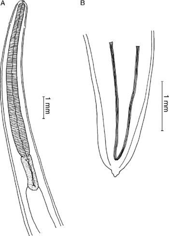
Description (fig. 1) and measurements (in millimetres) of the adult nematodes are given below.
Main morphological characteristics of Anisakis sp. A from Mesoplodon densirostris. (A) Anterior end; (B) posterior end of male (dorsal view).

Description. Body robust with transverse cuticular striae posterior to lips. Maximum width posterior but close to mid-body. Deirids papillate at level of the first sixth of the oesophagus. Dorsal and ventrolateral lips with low bi-lobed anterior projection bearing a dentigerous ridge on its inner surface. Dorsal lip with two sublateral double papillae at the base; each ventrolateral lip with one double papillae. Excretory pore between bases of ventrolateral lips. Triangular-shaped mouth opening. Oesophagus muscular. Thin-walled oblong ventriculus (sometimes sigmoid) connecting obliquely with the intestine.
Male (based on one male). Total length 95, maximum width 1.2; oesophagus 6.05 in length and 0.58 in width; ventriculus 1.62 in length and 0.41 in width; spicules subequal, tapering to rounded point; right spicule 2.42, left spicule 2.30; tail conical, 0.31 in length, distal extremity slightly pointed; caudal alae wide. The surface of the tail cuticle was slightly swollen, preventing correct visualization and description of the caudal papillae and plates.
Female (based on 22 females). Total length 74–165, maximum width 2–4; oesophagus 4.37–9.00 in length and 0.62–0.93 in width; ventriculus 1.12–2.37 in length and 0.25–0.60 in width; in most cases, vulva at the middle of the body.
Type host. Mesoplodon densirostris.
Habitat. Stomach.
Type locality. Atlantic coast of Galicia (NW Spain).
Deposition of type specimens. Holotype (one male; anterior and posterior ends): Natural History Museum, London, 2008.2.14.1. Paratypes (two females): Natural History Museum, London, 2008.2.14.2-3.
Discussion
PCR-RFLP and nuclear ribosomal DNA spacers and cox2 mitochondrial gene sequence analyses confirm that the nematodes found in the stomach of a Blainville's beaked whale stranded in the Galician coast; Anisakis sp. A, found as third-stage larvae in the fish species Aphanopus carbo and Scomber japonicus from Madeiran waters (Pontes et al., Reference Pontes, D'Amelio, Costa and Paggi2005); and Anisakis sp., detected as third-stage larvae in Aphanopus carbo from the Azores Islands, in Merluccius merluccius from the Iberian Atlantic coast and in Trachurus trachurus from New Zealand waters (Mattiucci & Nascetti, Reference Mattiucci and Nascetti2006), and as fourth-stage larvae in Mesoplodon mirus and M. grayi from South African and New Zealand coasts, respectively (Valentini et al., Reference Valentini, Mattiucci, Bondanelli, Webb, Mignucci-Giannone, Colom-Llavina and Nascetti2006), belong to the same species.
Unfortunately, the improper preservation of the male tail cuticle prevented a complete description of this species, since, as is well known, the number and arrangement of caudal papillae is one of the most important taxonomic characters in Ascaridoidea (Fagerholm, Reference Fagerholm1991). Therefore, until new male specimens are available for study, we propose to retain the name Anisakis sp. A for this new taxon.
The form and measurements of the ventriculus as well as the length and shape of the male spicules are used for identifying the morphospecies belonging to the genus Anisakis (Davey, Reference Davey1971; Paggi et al., Reference Paggi, Nascetti, Webb, Mattiucci, Cianchi and Bullini1998; Mattiucci et al., 2005). In spite of the bad condition of the tail cuticle of the holotype male, Anisakis sp. A can be easily distinguished from A. physeteris, A. brevispiculata and A. paggiae by the shape and length of the ventriculus (oblong, always longer than broad). In addition, it can also be distinguished from the three sibling species in the Anisakis simplex complex (i.e. A. simplex s.s., A. pegreffii and A. simplex C) and from A. typica for having subequal spicules (ratio 1 : 1.05). Finally, regardless of the morphological similarity between Anisakis sp. A and A. ziphidarum, the spicules of Anisakis sp. A (length 2.30–2.42 mm) are longer than those of Anisakis ziphidarum (1.55 mm or shorter; Paggi et al., Reference Paggi, Nascetti, Webb, Mattiucci, Cianchi and Bullini1998). In addition, Anisakis sp. A specimens are usually shorter than 140 mm (except for one female that was 165 mm in length) while A. ziphidarum specimens are 160 mm or longer.
Although Anisakis species were considered to be host generalist, the recognition of new species (both sibling and morphospecies) within the genus has allowed a more precise picture about significant host preference, especially of adult stages to definitive host species. In particular, A. physeteris, A. brevispiculata and A. paggiae display a strong association with sperm whales (Physeter and Kogia species) while Anisakis sp. A, described herein, and A. ziphidarum seem to be parasites of cetaceans belonging to the family Ziphiidae (i.e. Ziphius and Mesoplodon species). The molecular studies support this host specificity, since analysis of ITS sequences of Anisakis sp. A indicated a close relationship with A. ziphidarum (genetic distance: 0.0335), rather than with the other species (genetic distances ranging from 0.0494 to 0.0827; Pontes et al., Reference Pontes, D'Amelio, Costa and Paggi2005).
In any case, the finding of Anisakis sp. A in members of the genus Mesoplodon (this work; Valentini et al., Reference Valentini, Mattiucci, Bondanelli, Webb, Mignucci-Giannone, Colom-Llavina and Nascetti2006) and the close relationship with A. ziphidarum seem to support the hypothesis of Mattiucci & Nascetti (Reference Mattiucci and Nascetti2006) that the members of the genus Anisakis show a strict correlation between genetic similarities between parasite species and the sharing of the same or closely related host species, as a result of host–parasite co-evolution.
Acknowledgements
This work was supported in part by grants from the Consellería de Innovación, Industria e Comercio (Xunta de Galicia, Spain; PGIDIT04RAG310015PR) and from the University of Vigo.