Introduction
The mammalian nasal cavity has two primary functional divisions: olfactory and respiratory (Moore, Reference Moore1981; Hillenius, Reference Hillenius1992, Reference Hillenius1994; Novacek, Reference Novacek, Hanken and Hall1993; Rowe et al., Reference Rowe, Eiting, Macrini and Ketcham2005; Clarke and Pörtner, Reference Clarke and Pörtner2010; Laaß et al., Reference Laaß, Hampe, Schudack, Hoff, Kardjilov and Hilger2011; Crompton et al., Reference Crompton, Musinsky, Owerkowicz, Dial, Shubin and Brainerd2015, Reference Crompton, Owerkowicz, Bhullar and Musinsky2017). The respiratory region, related to air filtration and conservation of heat and water, includes the maxilloturbinal ossifications and is located mostly in the anteriormost part of this cavity. The olfactory region is located in the middle and posterior portions of the nasal cavity and contains the ethmoturbinals and nasoturbinals covered by olfactory epithelium, which is innervated by the olfactory nerve (cranial nerve I) that transmits the sense of smell to the brain. In crown mammals, an ossified cribriform plate formed by the ethmoid bone separates the nasal cavity from the olfactory fossa of the brain cavity (Reznik, Reference Reznik1990; Hillenius, Reference Hillenius1992, Reference Hillenius1994; Van Valkenburgh et al., Reference Van Valkenburgh, Theodor, Friscia, Pollack and Rowe2004; Crompton et al., Reference Crompton, Owerkowicz, Bhullar and Musinsky2017).
The evolutionary history of the complex mammalian nasal region, including its complex system of ossified turbinals and sensory and nonsensory soft tissues, can be traced in nonmammaliaform therapsids, and especially in nonmammaliaform cynodonts (Hillenius, Reference Hillenius1992, Reference Hillenius1994; Ruf et al., Reference Ruf, Maier, Rodrigues and Schultz2014; Crompton et al., Reference Crompton, Musinsky, Owerkowicz, Dial, Shubin and Brainerd2015, Reference Crompton, Owerkowicz, Bhullar and Musinsky2017; Rowe, Reference Rowe and Kass2017; Rowe et al., Reference Rowe, Macrini and Luo2011). Significant morphological changes have been documented in the nasal cavity of nonmammaliaform cynodonts associated with physiological modifications that occurred during the Triassic Period (ca. 251–201 Ma) (Hillenius, Reference Hillenius1992, Reference Hillenius1994; Crompton et al., Reference Crompton, Musinsky, Owerkowicz, Dial, Shubin and Brainerd2015; Rowe, Reference Rowe and Kass2017). In these forms, the turbinals and cribriform plate were cartilaginous (Crompton et al., Reference Crompton, Musinsky, Owerkowicz, Dial, Shubin and Brainerd2015, Reference Crompton, Owerkowicz, Bhullar and Musinsky2017), and the limit between the respiratory and olfactory regions is not clear in the fossils. Consequently, our knowledge rests on the interpretation of the internal anatomy of the rostral bones and the mold of the cavity.
Traversodontidae is a clade of Triassic cynodonts included within Cynognathia, which together with Probainognathia forms the Eucynodontia clade (Hopson and Kitching, Reference Hopson and Kitching2001; Abdala and Gaetano, Reference Abdala, Gaetano and Tanner2018; Abdala, Reference Abdala, Alderton and Elias2019). Cynognathia comprises the carnivorous cynognathids plus gomphodonts (diademodontids, trirachodontids, and traversodontids), characterized by their labiolingually expanded postcanine teeth related to an herbivorous/omnivorous diet (Chinsamy and Abdala, Reference Chinsamy and Abdala2008; Liu and Abdala, Reference Liu, Abdala, Kammerer, Angielczyk and Fröbisch2014; Abdala and Gaetano, Reference Abdala, Gaetano and Tanner2018; Hendrickx et al., Reference Hendrickx, Gaetano, Choiniere, Mocke and Abdala2020). Several studies have examined the morphology of the nasal cavity, rostral recesses, nasolacrimal duct, and maxillary canal of nonmammaliaform cynodonts (e.g., Watson, Reference Watson1913; Brink, Reference Brink1955; Kühne, Reference Kühne1956; Crompton, Reference Crompton1958; Bonaparte, Reference Bonaparte1966; Fourie, Reference Fourie1974; Kemp, Reference Kemp1979, Reference Kemp1980, Reference Kemp2006; Hillenius, Reference Hillenius1994, Reference Hillenius2000; Ruf et al., Reference Ruf, Maier, Rodrigues and Schultz2014; Crompton et al., Reference Crompton, Musinsky, Owerkowicz, Dial, Shubin and Brainerd2015, Reference Crompton, Owerkowicz, Bhullar and Musinsky2017; Benoit et al., Reference Benoit, Manger and Rubidge2016, Reference Benoit, Ruf, Miyamae, Fernandez, Rodrigues and Rubidge2019; Hendrickx et al., Reference Hendrickx, Gaetano, Choiniere, Mocke and Abdala2020; Kerber et al., Reference Kerber, Martinelli, Rodrigues, Ribeiro, Schultz and Soares2020), but although traversodontids are taxonomically diverse, the nasal cavity of these animals has been poorly studied (e.g., Bonaparte, Reference Bonaparte1966; Kemp, Reference Kemp1980; Hillenius, Reference Hillenius1994; Crompton et al., Reference Crompton, Owerkowicz, Bhullar and Musinsky2017; Hendrickx et al., Reference Hendrickx, Gaetano, Choiniere, Mocke and Abdala2020).
In southern Brazil, a rich fossil record of Late Triassic cynodonts has been documented (Abdala and Ribeiro, Reference Abdala and Ribeiro2010; Martinelli and Soares, Reference Martinelli, Soares., Agnolin, Lio, Egli, Chimento and Novas2016). Exaeretodon riograndensis Abdala, Barberena, and Dornelles, Reference Abdala, Barberena and Dornelles2002 and Siriusgnathus niemeyerorum Pavanatto et al., Reference Pavanatto, Pretto, Kerber, Müller, Da-Rosa and Dias-da-Silva2018 represent two abundant and well-known traversodontids. Both taxa are the largest traversodontids found in Brazil, with adult skulls reported up to 25 cm long. The external osteology of E. riograndensis was studied by Abdala et al. (Reference Abdala, Barberena and Dornelles2002), Oliveira et al. (Reference Oliveira, Schultz and Soares2007), and Liparini et al. (Reference Liparini, Oliveira, Pretto, Soares and Schultz2013), and that of S. niemeyerorum by Pavanatto et al. (Reference Pavanatto, Pretto, Kerber, Müller, Da-Rosa and Dias-da-Silva2018) and Miron et al. (Reference Miron, Pavanatto, Pretto, Müller, Dias-da-Silva and Kerber2020). Recently, the brain endocast morphology of both cynodonts was studied by Pavanatto et al. (Reference Pavanatto, Kerber and Dias-da-Silva2019). In the present study, we aim to describe the morphology of the nasal cavities of E. riograndensis and S. niemeyerorum using computed tomography (CT) scanning and explore their paleobiological implications.
Materials and methods
Analyzed specimens
Exaeretodon riograndensis (CAPPA/UFSM 0030 and 0227) and Siriusgnathus niemeyerorum (CAPPA/UFSM 0032) from the Upper Triassic of southern Brazil. The specimens are housed in the Paleontological Collection of the Centro de Apoio à Pesquisa Paleontológica da Quarta Colônia/Universidade Federal de Santa Maria (CAPPA/UFSM), São João do Polêsine, Rio Grande do Sul, Brazil.
Location and stratigraphic information
The fossils were collected from Upper Triassic strata in southern Brazil. The specimens CAPPA/UFSM 0030 and 0227 of E. riograndensis were excavated from the Janner site on the outskirts of Agudo, State of Rio Grande do Sul (29°39′10.89″S; 53°17′34.2″W), which is biostratigraphically positioned in the upper portion of the Hyperodapedon Assemblage Zone (i.e., Exaeretodon sub-AZ), Candelária Sequence, and is late Carnian in age (Oliveira et al., Reference Oliveira, Schultz and Soares2007; Pretto et al., Reference Pretto, Schultz and Langer2015; Müller and Garcia, Reference Müller and Garcia2019). The specimens come from the reddish mudstone within the basal two-thirds of the outcrop; the upper levels are composed of massive sandstones, with no records of fossil vertebrates so far (Müller et al., Reference Müller, de Araújo-Júnior, Aires, Roberto-da-Silva and Dias-da-Silva2015). In addition to E. riograndensis, the Janner site has yielded several other fossil vertebrates, such as the sauropodomorph dinosaurs Pampadromaeus barberenai Cabreira et al., Reference Cabreira, Schultz, Bittencourt, Soares, Fortier, Silva and Langer2011 and Bagualosaurus agudoensis Pretto et al., Reference Pretto, Langer and Schultz2019, the probainognathian Trucidocynodon riograndensis Oliveira et al., Reference Oliveira, Soares and Schultz2010, and the pseudosuchian ornithosuchid Dynamosuchus collisensis Müller et al., Reference Müller, von Baczko, Desojo and Nesbitt2020 (see Oliveira et al., Reference Oliveira, Soares and Schultz2010; Cabreira et al., Reference Cabreira, Schultz, Bittencourt, Soares, Fortier, Silva and Langer2011; Stefanello et al., Reference Stefanello, Müller, Kerber, Martínez and Dias-da-Silva2018; Pretto et al., Reference Pretto, Langer and Schultz2019; Müller et al., Reference Müller, von Baczko, Desojo and Nesbitt2020).
The specimen assigned to S. niemeyerorum (CAPPA/UFSM 0032) was collected from the Niemeyer site, also located in Agudo (29°40′25″S; 53°14′4.20″W). The stratigraphic column comprises a 23 m thick lithological sequence of reddish mudstones, sandstones, and intraformational conglomerates, with carbonatic concretions and hydromorphic levels (Pavanatto et al., Reference Pavanatto, Pretto, Kerber, Müller, Da-Rosa and Dias-da-Silva2018). Siriusgnathus niemeyerorum is the most abundant fossil found at this site, which also contains remains of probainognathians, dinosaurs, and indeterminate archosauromorphs (Pavanatto et al., Reference Pavanatto, Pretto, Kerber, Müller, Da-Rosa and Dias-da-Silva2018; Müller, Reference Müller2020; Stefanello et al., Reference Stefanello, Kerber, Martinelli and Dias-da-Silva2020). The strata exposed at this site are considered younger than those of the Janner site (for further details, see Martinelli et al., Reference Martinelli, Escobar, Francischini, Kerber, Müller, Rubert, Schultz and Da-Rosa2020; Miron et al., Reference Miron, Pavanatto, Pretto, Müller, Dias-da-Silva and Kerber2020).
Scanning procedures and three-dimensional modeling
The three specimens were scanned at the DIX medical clinic (Diagnóstico por Imagem do Hospital de Caridade) in Santa Maria, Rio Grande do Sul, using a Philips Brilliance 16-slice CT scanner and a Philips Brilliance 64-slice CT scanner (see Pavanatto et al., Reference Pavanatto, Kerber and Dias-da-Silva2019). The raw scanning data were exported from the scanner computer in DICOM format. The scanning parameters are available in Table 1.
Scanning parameters of the analyzed specimens.

The endocranial structures were observed through analysis of the tomographic slices and by using tridimensional reconstructions of the cavities. The slices were manually segmented using 3D Slicer 3.8 to generate three-dimensional (3D) models of the regions of interest. The same software was employed to measure the volumes of the nasal cavity and the dorsal recess of the nasal cavity. The dorsal recess of the nasal cavity was artificially extracted (the ventral limit of the structure is a straight plane, and its contour is delimited by the edges of the recess) from the rest of the nasal cavity endocast, and its volume was estimated and compared with the total volume of the cavity. Design Spark Mechanical 2.0 was employed to render the models.
Comparison
For comparison, we collected data from the literature (e.g., Watson, Reference Watson1913; Brink, Reference Brink1955; Fourie, Reference Fourie1974; Kemp, Reference Kemp1980; Hillenius, Reference Hillenius1994; Ruf et al., Reference Ruf, Maier, Rodrigues and Schultz2014; Crompton et al., Reference Crompton, Owerkowicz, Bhullar and Musinsky2017; Kerber et al., Reference Kerber, Martinelli, Rodrigues, Ribeiro, Schultz and Soares2020) and analyzed the specimen MACN-Pv 18114 of Exaeretodon argentinus Bonaparte, Reference Bonaparte1962 (Ischigualasto Formation, late Carnian, Argentina; Bonaparte, Reference Bonaparte1966) and CT scans of Diademodon cf. D. tetragonus Seeley, Reference Seeley1894 (UCMP 42446, Cynognathus Assemblage Zone, early Anisian, South Africa; Macrini, Reference Macrini2006) (http://digimorph.org/specimens/Diademodon_sp/); Massetognathus pascuali Romer, Reference Romer1967 (UFRGS-PV-0968-T, Chañares Formation, early Carnian, Argentina; Rodrigues, Reference Rodrigues2005); Massetognathus ochagaviae Barberena, Reference Barberena1981 (MCP 3871 PV, Pinheiros-Chiniquá Sequence, late Ladian–early Carnian, Brazil; Hoffmann et al., Reference Hoffmann, Rodrigues, Soares and Andrade2019); Ecteninion lunensis Martínez et al., Reference Martínez, May and Forster1996 (PVSJ 422, Ischigualasto Formation, late Carnian, Argentina; Martínez et al., Reference Martínez, May and Forster1996) (http://digimorph.org/specimens/Ecteninion_lunensis/); Trucidocynodon riograndensis (CAPPA/UFSM 0029, Candelária Sequence, Carnian, Brazil; Stefanello et al., Reference Stefanello, Müller, Kerber, Martínez and Dias-da-Silva2018); Pseudotherium argentinus Wallace et al., Reference Wallace, Martínez and Rowe2019 (PVSJ 882, Ischigualasto Formation, late Carnian, Argentina; Wallace et al., Reference Wallace, Martínez and Rowe2019) (http://digimorph.org/specimens/Pseudotherium_argentinus/); and Didelphis virginiana (Kerr, Reference Kerr1792) (TMM M-2517, Recent; Macrini et al., Reference Macrini, de Muizon, Cifelli and Rowe2007) (http://digimorph.org/specimens/Didelphis_virginiana/).
Except for T. riograndensis (CAPPA/UFSM 0029), M. pascuali (UFRGS-PV-0968-T), and M. ochagaviae (MCP 3871 PV), the CT-scan data sets for the specimens are available on digimorph.com. The CT scan of T. riograndensis (CAPPA/UFSM 0029) was studied by Stefanello et al. (Reference Stefanello, Müller, Kerber, Martínez and Dias-da-Silva2018); the brain endocast morphology of M. pascuali (UFRGS-PV-0968-T) was described by Rodrigues (Reference Rodrigues2005) and Rodrigues et al. (Reference Rodrigues, Ruf and Schultz2013) and that of M. ochagaviae (MCP 3871 PV) by Hoffmann et al. (Reference Hoffmann, Rodrigues, Soares and Andrade2019). Scan parameters for the comparative specimens are available at digimorph.com and in the respective studies mentioned, and for brevity, they are not repeated here.
Terminology
The anatomical nomenclature follows Crompton et al. (Reference Crompton, Owerkowicz, Bhullar and Musinsky2017) for the structures of the nasal cavity, Hillenius (Reference Hillenius2000) for the nasolacrimal duct, and Benoit et al. (Reference Benoit, Ruf, Miyamae, Fernandez, Rodrigues and Rubidge2019) for the maxillary canal.
Repositories and institutional abbreviations
CAPPA/UFSM, Centro de Apoio à Pesquisa Paleontológica da Quarta Colônia, Universidade Federal de Santa Maria, São João do Polêsine, Brazil; MACN-Pv, Museo Argentino de Ciencias Naturales “Bernardino Rivadavia,” Colección de Paleontología de Vertebrados, Buenos Aires, Argentina; MCP, Museu de Ciência e Tecnologia, Pontifícia Universidade Católica do Rio Grande do Sul, Porto Alegre, Brazil; PVSJ, Museu de Ciências Naturais, Universidade Nacional de San Juan, San Juan, Argentina; TMM, Texas Memorial Museum, Austin, Texas, USA; UCMP, University of California Museum of Paleontology, Berkeley, California, USA; UFRGS, Universidade Federal do Rio Grande do Sul, Porto Alegre, Brazil.
Results
Description of the nasal cavity
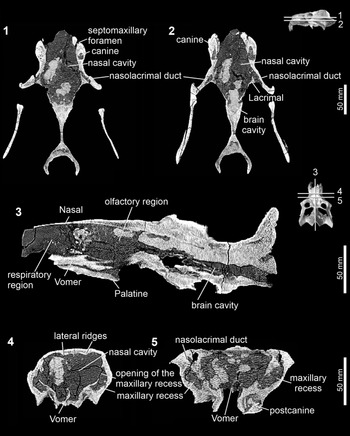
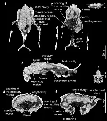
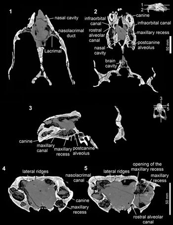
The nasal cavities of Exaeretodon riograndensis and Siriusgnathus niemeyerorum (Figs. 1–8) are confluent with the brain cavity (Figs. 1.3, 2.3, 3.3), without an ossified cribriform plate, following the pattern found in all other nonmammaliaform cynodonts (Crompton, Reference Crompton1958; Crompton et al., Reference Crompton, Owerkowicz, Bhullar and Musinsky2017; Pavanatto et al., Reference Pavanatto, Kerber and Dias-da-Silva2019). The estimated volumes of the nasal cavities of the analyzed specimens are 157.199 cm3 in CAPPA/UFSM 0030 (skull length: 22.4 cm), 103.304 cm3 in CAPPA/UFSM 0227 (skull length: 16.8 cm), and 137.710 cm3 in CAPPA/UFSM 0032 (skull length: 22.04 cm).
Computed tomography slices of the nasal cavity of Exaeretodon riograndensis (CAPPA/UFSM 0030). The 3D models of the cranium show the positions of the slices 1–5. (1, 2) Transverse slices; (3) sagittal slice; (4, 5) coronal slices.

Computed tomography slices of the nasal cavity of Exaeretodon riograndensis (CAPPA/UFSM 0227). The 3D models of the cranium show the positions of the slices 1–5. (1, 2) Transverse slices; (3) sagittal slice; (4, 5) coronal slices.

Computed tomography slices of the nasal cavity of Siriusgnathus niemeyerorum (CAPPA/UFSM 0032). The 3D models of the cranium show the positions of the slices 1–5. (1, 2) Transverse slices; (3) parasagittal slice; (4, 5) coronal slices.

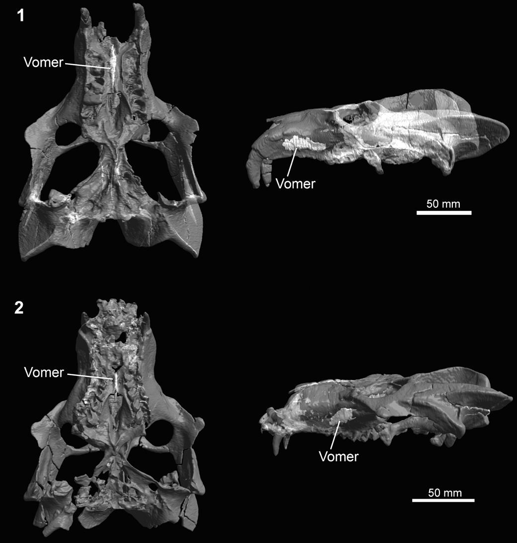
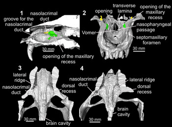
Translucent cranium of Exaeretodon riograndensis highlighting the vomer, in ventral and lateral views, respectively. (1) CAPPA/UFSM 0030; (2) CAPPA/UFSM 0227.

Sectioned crania of Traversodontidae evidencing the internal morphology of the nasal cavity. (1, 4) Siriusgnathus niemeyerorum (CAPPA/UFSM 0032); (2, 3) Exaeretodon riograndensis: (2) CAPPA/UFSM 0227; (3) CAPPA/UFSM 0030). (1) Cranium sagittally sectioned; (2) cranium transversally sectioned in anterior view; (3, 4) cranium transversally sectioned, evidencing the internal surface of the roof of the nasal cavity.

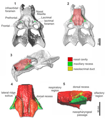
(1–3) Cranium and (4, 5) endocranial casts of the nasal cavity, maxillary recess, and nasolacrimal duct of Exaeretodon riograndensis (CAPPA/UFSM 0030). (1, 2, 4) Dorsal views; (3) lateral oblique view; (5) lateral view.

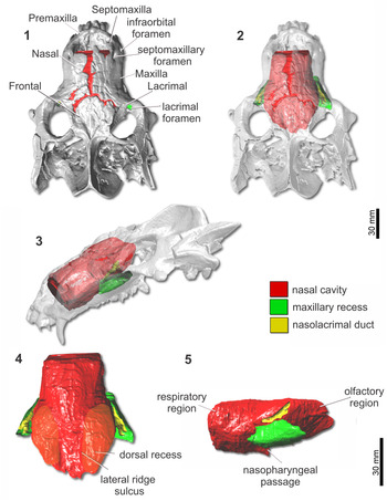
(1–3) Cranium and (4, 5) endocranial casts of the nasal cavity, maxillary recess, and nasolacrimal duct of Exaeretodon riograndensis (CAPPA/UFSM 0227). (1, 2, 4) Dorsal views; (3) lateral oblique view; (5) lateral view.

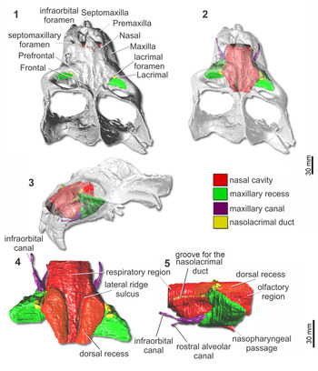
(1–3) Cranium and (4, 5) endocranial casts of the nasal cavity, maxillary recess, maxillary canal, and nasolacrimal duct of Siriusgnathus niemeyerorum (CAPPA/UFSM 0032). (1, 2, 4) Dorsal views; (3) lateral oblique view; (5) lateral view.

The external nasal opening of the nasal cavity is anteriorly delimited by the nasal and a portion of the premaxilla (dorsally) and by the premaxilla and septomaxilla (lateroventrally) (Figs. 1–3, 6.1, 7.1, 8.1). Anterodorsally, the nasal cavity is covered by the nasal and posterodorsally, by the prefrontal, frontal, and postorbital (Figs. 1–3, 6.1, 7.1, 8.1). The lateral wall of the nasal cavity is formed by the premaxilla (anteriorly), maxilla, and lacrimal (posteriorly). Ventrally, it is delimited by the premaxilla, maxilla, and palatine. The intranarial process is absent in E. riograndensis and S. niemeyerorum (Pavanatto et al., Reference Pavanatto, Pretto, Kerber, Müller, Da-Rosa and Dias-da-Silva2018) (Figs. 5.1, 7, 8). The vomer is preserved only in E. riograndensis (Figs. 1, 2, 4, 5.2) and is better preserved in CAPPA/UFSM 0030 than in CAPPA/UFSM 0227, which preserves only the posterior portion of this bone (Figs. 4.2, 5.2). It is a long and thin plate-like bone located in the middle region of the nasal cavity.
The transverse lamina is located in the posterior region of the nasal cavity. According to Crompton et al. (Reference Crompton, Owerkowicz, Bhullar and Musinsky2017), it is formed by the vomerine plate and palatine flange (Fig. 5.2). This structure consists of an obliquely (dorsoventrally) oriented lamina that converges medially with its counterpart, forming the floor of the olfactory chamber and the roof of the nasopharyngeal passage, as in other nonmammaliaform cynodonts (Crompton et al., Reference Crompton, Musinsky, Owerkowicz, Dial, Shubin and Brainerd2015, Reference Crompton, Owerkowicz, Bhullar and Musinsky2017). Lateral to the transverse lamina is a rounded opening (Fig. 5.2) that Crompton et al. (Reference Crompton, Owerkowicz, Bhullar and Musinsky2017; figs. 7B, 8A) identified as the entrance of branch V2 of the trigeminal nerve into the nasal cavity.
On the internal surface of the nasal and frontal, bones that form the roof of the nasal cavity in E. riograndensis and S. niemeyerorum, are two well-marked and convergent lateral ridges, anteroposteriorly oriented, which are almost as long as the entire extension of the cavity (Figs. 1–3, 5.3, 5.4). They are more evident in the posterior region of the nasal cavity. There is no median ridge on the internal surface of the nasal and frontal in both taxa (Figs. 1–3, 5.3, 5.4). The lateral ridges form sulci on the endocast of the nasal cavity (Figs. 6.4, 7.4, 8.4).
Lateral to the lateral ridge is a wide recess on the ventral surface of the bones of the roof of the nasal cavity (i.e., frontal, prefrontal, nasal, and maxilla) (Fig. 5.1). The recess forms a salient elevation on the dorsal aspect of the nasal cavity endocast, being wider in its middle portion and narrower posteriorly, similar to a scalene triangle. In one specimen of E. riograndensis (CAPPA/UFSM 0030) (length: 55 mm; width: 19 mm; volume: 9.99 cm3) (Fig. 6.4) and in S. niemeyerorum (CAPPA/UFSM 0032) (length: 49 mm; width: 21.6 mm; volume: 8.95 cm3) (Fig. 8.4), the recess is more marked than in the other specimen of E. riograndensis (CAPPA/UFSM 0227) (length: 40 mm; width: 20 mm; volume: 6.45 cm3) (Fig. 7.4), which is smaller in cranial size (and thus probably represents an ontogenetic difference).
In E. riograndensis, the width between the right and left recesses is the same along the entire extension of the structure (Figs. 5.3, 7.4) (24 mm in the anteriormost region and 25 mm in the posteriormost in CAPPA/UFSM 0030; 14 mm and 16 mm, respectively, in CAPPA/UFSM 0227). By contrast, the width between the two recesses becomes narrower posteriorly in S. niemeyerorum (Figs. 5.4, 8.4) (23 mm in the anteriormost region and 10 mm in the posteriormost).
On the internal surface of the maxilla is a groove for the nasolacrimal duct (sensu Hillenius, Reference Hillenius2000) extending anteroposteriorly from the point where the lacrimal duct enters the nasal cavity up to the septomaxillary foramen (Fig. 5.1). This groove is more clearly visible in CAPPA/UFSM 0032 (Fig. 5.1) and CAPPA/UFSM 0227 and is reflected on the surface of the endocast of CAPPA/UFSM 0032 (Fig. 8.5).
Located ventrally is the nasopharyngeal passage, which opens into the secondary choana. The 3D reconstruction of the nasopharyngeal passage reveals a posteroventrally oriented duct (Figs. 6.5, 7.5, 8.5).
Maxillary recess
All specimens have a well-developed maxillary recess. In E. riograndensis, the recess is similar in both CAPPA/UFSM 0030 and CAPPA/UFSM 0227 (Figs. 6, 7). It is less dorsoventrally extended than in S. niemeyerorum (Figs. 3.5, 8.5) but anteroposteriorly longer. In S. niemeyerorum, this recess is pyramidal, transversally thin, and dorsoventrally elongated (Fig. 8). In all specimens, the recess is connected to the nasal cavity by a wide opening (Figs. 1–3, 5.1).
Nasolacrimal duct and maxillary canal
In the studied taxa, the opening of the nasolacrimal duct, the lacrimal foramen, is located in the anterolateral region of the orbit, within the lacrimal bone (Figs. 1–3, 5.1). In E. riograndensis, the nasolacrimal duct is longer and less obliquely oriented than in S. niemeyerorum. It is longer in the largest E. riograndensis specimen CAPPA/UFSM 0030 than in CAPPA/UFSM 0227, probably reflecting an ontogenetic difference. In CAPPA/UFSM 0227, it is bifurcated posteriorly (Fig. 7.5).
In S. niemeyerorum, the nasolacrimal duct is posterodorsally oblique (Fig. 8.5). This canal is located dorsally to the maxillary recess and anterolaterally to the region of the dorsal recess of the nasal cavity (Fig. 8.5). The nasolacrimal duct is anteriorly continuous with a groove on the internal face of the maxilla, which is extended up to the septomaxillary foramen (Fig. 5.1).
The maxillary canal is the passage for the maxillary branch of the trigeminal nerve (V2) (Benoit et al., Reference Benoit, Ruf, Miyamae, Fernandez, Rodrigues and Rubidge2019). In our sample, it was possible to reconstruct the maxillary canal only in S. niemeyerorum. This canal leaves the maxillary recess and extends anteriorly in parallel and dorsolaterally to the postcanine dental series (Fig. 8.4, 8.5). At the level of the first postcanine, this canal splits into two separate canals, the rostral alveolar canal and the infraorbital canal (Figs. 3.2, 8.5) (Benoit et al., Reference Benoit, Ruf, Miyamae, Fernandez, Rodrigues and Rubidge2019). The rostral alveolar canal extends ventromedially, almost reaching the canine alveolus (posterior to this alveolus; Fig. 8.5), and the longer infraorbital canal is directed dorsolaterally up to one of the infraorbital foramina (there are at least two infraorbital foramina; see Pavanatto et al., Reference Pavanatto, Pretto, Kerber, Müller, Da-Rosa and Dias-da-Silva2018, fig. 5, where the authors identified these openings as “maxillary foramina”) located on the lateral surface of the maxilla. Other ramifications of the maxillary canal are possibly obliterated by the low resolution of the CT scans.
Discussion
Comparative morphology of the nasal cavity
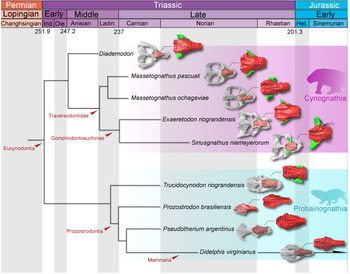
In comparison with other nonmammaliaform cynodonts (Fig. 9), the external morphology of the nasal cavities of the gomphodontosuchines Exaeretodon riograndensis and Siriusgnathus niemeyerorum is characterized by the absence of the internarial process, differing from probainognathians (e.g., Kühne, Reference Kühne1956; Hopson and Kitching, Reference Hopson and Kitching2001; Ruf et al., Reference Ruf, Maier, Rodrigues and Schultz2014; Crompton et al., Reference Crompton, Owerkowicz, Bhullar and Musinsky2017; Stefanello et al., Reference Stefanello, Müller, Kerber, Martínez and Dias-da-Silva2018) and nongomphodontosuchine traversodontids (e.g., Massetognathus ochagaviae, MCP 3871 PV). The absence of this process is a synapomorphy of gomphodontosuchines (Liu and Olsen, Reference Liu and Olsen2010; Liu and Abdala, Reference Liu, Abdala, Kammerer, Angielczyk and Fröbisch2014).
Phylogenetic relationships of selected cynodonts (based on Liu and Olsen, Reference Liu and Olsen2010; Pavanatto et al., Reference Pavanatto, Pretto, Kerber, Müller, Da-Rosa and Dias-da-Silva2018; Wallace et al., Reference Wallace, Martínez and Rowe2019). Cynognathia: Diademodon cf. D. tetragonus (UCMP 42446); Massetognathus pascuali (UFRGS-PV-0968-T); Massetognathus ochagaviae (MCP 3871 PV); Exaeretodon riograndensis (CAPPA/UFSM 0030); Siriusgnathus niemeyerorum (CAPPA/UFSM 0032). Probainognathia: Trucidocynodon riograndensis (CAPPA/UFSM 0029); Prozostrodon brasiliensis (UFRGS-PV-0543-T; after Kerber et al., Reference Kerber, Martinelli, Rodrigues, Ribeiro, Schultz and Soares2020); Pseudotherium argentinus (PVSJ 882); Didelphis virginiana (TMM M-2517). Not to scale.

There is no evidence of an ossified cribriform plate in E. riograndensis and S. niemeyerorum, as in other nonmammaliaform cynodonts in which the internal nasal cavity is known. The ossified condition of this structure is formed by the ethmoid bone in the crown Mammalia (Crompton et al., Reference Crompton, Owerkowicz, Bhullar and Musinsky2017). On the internal surface of the bones of the roof of the nasal cavity, in E. riograndensis, E. argentinus (Bonaparte, Reference Bonaparte1966), and S. niemeyerorum, are two lateral ridges that could have supported the cartilaginous turbinals (see Hillenius, Reference Hillenius1994; Crompton et al., Reference Crompton, Musinsky, Owerkowicz, Dial, Shubin and Brainerd2015, Reference Crompton, Owerkowicz, Bhullar and Musinsky2017). No median ridge was observed in the studied specimens (Fig. 5.3, 5.4). A median ridge, probably for the attachment of a cartilaginous nasal septum, is present in most cynodonts and is reflected on the endocast of the nasal cavity, as seen in early epicynodonts Galesaurus planiceps, Owen, Reference Owen1859 and Thrinaxodon liorhinus Seeley, Reference Seeley1894, the gomphodonts Diademodon tetragonus, Massetognathus pascuali, and Etjoia dentitransitus Hendrickx et al., Reference Hendrickx, Gaetano, Choiniere, Mocke and Abdala2020 (Watson, Reference Watson1913; Fourie, Reference Fourie1974; Crompton et al., Reference Crompton, Musinsky, Owerkowicz, Dial, Shubin and Brainerd2015; Pusch et al., Reference Pusch, Kammerer and Fröbisch2019a; Hendrickx et al., Reference Hendrickx, Gaetano, Choiniere, Mocke and Abdala2020), and the probainognathians Oligokyphus major Kühne, Reference Kühne1956, Probainognathus jenseni Romer, Reference Romer1970, Elliotherium kersteni Sidor and Hancox, Reference Sidor and Hancox2006, Prozostrodon brasiliensis (Barberena et al., Reference Barberena, Bonaparte and Teixeira1987), and Brasilodon quadrangularis Bonaparte et al., Reference Bonaparte, Martinelli, Schultz and Rubert2003 (Kühne, Reference Kühne1956; Ruf et al., Reference Ruf, Maier, Rodrigues and Schultz2014; Crompton et al., Reference Crompton, Owerkowicz, Bhullar and Musinsky2017; Kerber et al., Reference Kerber, Martinelli, Rodrigues, Ribeiro, Schultz and Soares2020). In the specimen CAPPA/UFSM 0029 assigned to Trucidocynodon riograndensis, the median ridge is not visible, and neither is its impression on the endocast surface. However, this condition could be related to the poor preservation of the internal cavities of the specimen. The nasal cavity of Brasilodon quadrangularis, the sister group of Mammaliaformes (Bonaparte et al., Reference Bonaparte, Martinelli, Schultz and Rubert2003, Reference Bonaparte, Martinelli and Schultz2005), was studied by Ruf et al. (Reference Ruf, Maier, Rodrigues and Schultz2014), and among other features, the authors inferred the presence of partially ossified turbinals identified as the remnants of the nasoturbinal and first ethmoturbinal, on the basis of thin bone fragments, as well as a ridge on the ventral surface of the frontal identified as the mesethmoid. Crompton et al. (Reference Crompton, Owerkowicz, Bhullar and Musinsky2017), however, argued that without having a more complete documentation of these bones, it is not possible to confirm whether the interpreted elements are indeed ossified turbinals. They also argued that the ridge that Ruf et al. (Reference Ruf, Maier, Rodrigues and Schultz2014) described as the mesethmoid is quite similar to the median ridge found in other nonmammaliaform cynodonts and may not represent an independent ossification. The earliest mammaliaform Morganucodon has ridges on the internal surface of the nasal cavity, which would support turbinals (Kermack et al., Reference Kermack, Mussett and Rigney1981; Kielan-Jaworowska et al., Reference Kielan-Jaworowska, Cifelli and Luo2004), but no unequivocal evidence of ossification of these structures exists (Crompton et al., Reference Crompton, Owerkowicz, Bhullar and Musinsky2017). The oldest fossil with signs of ossification of the internal structures of the nasal cavity is Haldanodon exspectatus Kühne and Krusat, Reference Kühne and Krusat1972 from the Upper Jurassic of Portugal (Lillegraven and Krusat, Reference Lillegraven and Krusat1991). However, as noted by Crompton et al. (Reference Crompton, Owerkowicz, Bhullar and Musinsky2017), these structures were not figured in detail.
The vomer of E. riograndensis is thin and tall, similar to that of M. pascuali (MCZ 3798; Crompton et al., Reference Crompton, Owerkowicz, Bhullar and Musinsky2017). In some probainognathians, the vomer is lower and robust (Y-shaped), showing a well-marked dorsal sulcus, probably for the insertion of a cartilaginous septum, as seen in P. brasiliensis, P. argentinus, and B. quadrangularis (Ruf et al., Reference Ruf, Maier, Rodrigues and Schultz2014; Wallace et al., Reference Wallace, Martínez and Rowe2019; Kerber et al., Reference Kerber, Martinelli, Rodrigues, Ribeiro, Schultz and Soares2020).
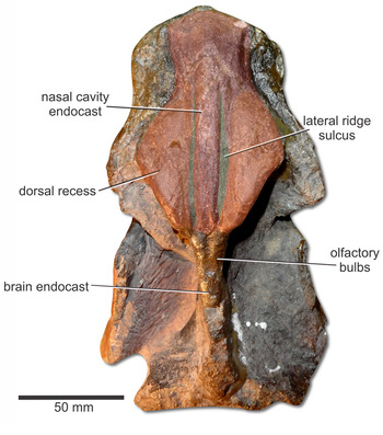
In the description of the nasal cavity of E. argentinus, Bonaparte (Reference Bonaparte1966) pointed out the presence of globular longitudinal elevations on the dorsal region of a natural endocast, close to the orbital edges (Fig. 10), which he interpreted as cranial sinuses. The anterior region of the elevation was interpreted as the cast of a “maxillary sinus” (not the same cavity present in the lateral of the nasal cavity, i.e., maxillary recess) and the posterior region as the “frontal sinus” although there is no indication of bone separation between these regions. This cavity, which is reflected on the endocast of the nasal cavity as a globular longitudinal elevation, is also present in E. riograndensis and S. niemeyerorum. Notwithstanding, sinuses are cavities resulting from bone remodeling during ontogeny between two layers of bones, whereas recesses are cavities that do not necessarily form between bony layers or relate to the removal of bone tissue (Farke, Reference Farke2010a; Sharp, Reference Sharp2016; Billet et al., Reference Billet, Hautier, De Thoisy and Delsuc2017). In addition, sinuses and recesses are named according to the bones they excavate although they may invade adjacent elements (Novacek, Reference Novacek, Hanken and Hall1993). However, as this cavity excavates more than a single bone, and the homology with other structures is not clear, we opted to identify it generically as a dorsal recess of the nasal cavity, at least until further studies are conducted.
Specimen MACN-Pv 18114 of a small-sized specimen of Exaeretodon argentinus (Ischigualasto Formation, Ischigualasto-Villa Unión Basin, Argentina) with cranial bones detached, showing the natural endocasts of the brain and nasal cavity. It was originally described by Bonaparte (Reference Bonaparte1966).

Except for Massetognathus, which also shows a recess on the dorsal region of the nasal cavity, but less marked than in Exaeretodon spp. and S. niemeyerorum (see Crompton et al., Reference Crompton, Musinsky, Owerkowicz, Dial, Shubin and Brainerd2015, fig. 11.4, identified as part of the olfactory chamber), other cynodonts in which the nasal cavity has been studied (Watson, Reference Watson1913; Brink, Reference Brink1955; Fourie, Reference Fourie1974; Kemp, Reference Kemp1979, Reference Kemp1980; Ruf et al., Reference Ruf, Maier, Rodrigues and Schultz2014; Kerber et al., Reference Kerber, Martinelli, Rodrigues, Ribeiro, Schultz and Soares2020) do not show this structure (Fig. 9), at least not as developed as in traversodontids, especially the gomphodontosuchines here studied. By comparing specimens of different cranial sizes, and probably ontogenetic stages, of E. riograndensis (CAPPA/UFSM 0030 and CAPPA/UFSM 0227) (as well as in the small-sized specimen MACN-Pv 18114 of E. argentinus), it is possible to observe that the dorsal recess of the nasal cavity increases in size during ontogenetic maturation. In this region, it is also possible to observe interspecific variation between the two traversodontids. The space between the right and left dorsal recesses of the nasal cavity in E. riograndensis is the same along the entire length of the structure, whereas it becomes narrower posteriorly in S. niemeyerorum.
Both analyzed species show well-developed maxillary recesses in the lateral region of the nasal cavity within the maxilla. Such structures are present in most nonmammaliaform cynodonts (Crompton et al., Reference Crompton, Owerkowicz, Bhullar and Musinsky2017). This structure has been identified as a sinus (Fourie, Reference Fourie1974; Kemp, Reference Kemp1979, Reference Kemp1980), atrium (Crompton et al., Reference Crompton, Musinsky, Owerkowicz, Dial, Shubin and Brainerd2015), or antrum (Benoit et al., Reference Benoit, Manger and Rubidge2016), but according to Crompton et al. (Reference Crompton, Owerkowicz, Bhullar and Musinsky2017), it is better defined as a recess (see preceding discussion).
The nasolacrimal duct of both E. riograndensis and S. niemeyerorum is long and inclined dorsoposteriorly. In S. niemeyerorum, the duct is more inclined than in E. riograndensis, similar to Massetognathus pascuali. As observed in the taxa here studied, the nasolacrimal duct is connected by a groove to the septomaxillary foramen, which is close to the place where the vomeronasal organ was located (Hillenius, Reference Hillenius2000). In this sense, the nasolacrimal duct supplied orbital fluids to the vomeronasal organ, increasing its function (i.e., chemosensory system) (Hillenius, Reference Hillenius2000).
The maxillary canal found in S. niemeyerorum displays the plesiomorphic pattern of cynodonts, consisting of a long canal enclosed within the facial process of the maxilla, with two to three external openings (Benoit et al., Reference Benoit, Ruf, Miyamae, Fernandez, Rodrigues and Rubidge2019). By contrast, mammaliaforms have a shorter canal, and the innervations of the trigeminal nerve (V2) are not distributed within the bone. This 3D reconstruction does not show a high degree of ramification inside the maxilla like other therapsids and nonprobainognathian cynodonts (Benoit et al., Reference Benoit, Manger and Rubidge2016, Reference Benoit, Ruf, Miyamae, Fernandez, Rodrigues and Rubidge2019; Pusch et al., Reference Pusch, Kammerer and Fröbisch2019a, Reference Pusch, Ponstein, Kammerer and Fröbischb). However, the existence of small branches cannot be excluded, their absence being perhaps a result of the resolution of the tomography.
Functional inferences
The delimitation of the space for the respiratory and olfactory chambers in the nasal cavity of nonmammaliaform cynodonts is not clear, and only some speculations can be made about the limits of both areas (see Crompton et al., Reference Crompton, Musinsky, Owerkowicz, Dial, Shubin and Brainerd2015, fig. 11.4). The transverse lamina is considered the ventral limit of the olfactory chamber, and the space anterior to this lamina is occupied by the respiratory chamber, which was probably filled by cartilaginous turbinals attached on the ventral surface of the nasal and frontal bones (Hillenius, Reference Hillenius1994; Crompton et al., Reference Crompton, Musinsky, Owerkowicz, Dial, Shubin and Brainerd2015, Reference Crompton, Owerkowicz, Bhullar and Musinsky2017).
Due to the similarity of the nasal cavities of E. riograndensis, S. niemeyerorum, and Massetognathus (with the gross anatomy fairly similar among eucynodonts), it is possible that breathing in these animals was similar to that inferred for Massetognathus by Crompton et al. (Reference Crompton, Owerkowicz, Bhullar and Musinsky2017; see also Hillenius, Reference Hillenius1992, Reference Hillenius1994). According to Crompton et al. (Reference Crompton, Owerkowicz, Bhullar and Musinsky2017), the route taken by the airflow during the respiratory process could have been either directly into the nasopharyngeal passage or through the respiratory chamber, flowing into the maxillary recess before exiting the chamber. The path that most air takes to reach the lungs depends on the respiratory state. When in a state of low activity (resting phase), the air is inhaled through the nostrils, travels through the nasal cavity (passing through the recesses), and is exhaled through the nostrils. During periods of high activity (e.g., running phase), most of the air enters through the nostrils but is exhaled through the mouth, resulting in wheezing, which also has the purpose of reducing body temperature.
As discussed in the preceding, gomphodontosuchines show a recess in the dorsal region of the nasal cavity and a well-developed maxillary recess. The presence of sinuses/recesses around the nasal cavity (i.e., paranasal sinuses) has been found in other vertebrate groups, especially mammals (Witmer, Reference Witmer, Koppe, Nagai and Alt1999; Rossie, Reference Rossie2006; Farke, Reference Farke2010a; Curtis and Van Valkenburgh, Reference Curtis and Van Valkenburgh2014; Ranslow et al., Reference Ranslow, Richter, Neuberger, Van Valkenburgh, Rumple, Quigley, Pang, Krane and Craven2014; Mori et al., Reference Mori, Hanida, Kumahata, Miyabe-Nishiwaki, Suzuki, Matsuzawa and Nishimura2015; Ruf et al., Reference Ruf, Janßen and Zeller2015; Sharp, Reference Sharp2016; Kerber et al., Reference Kerber, Ferreira and Negri2019), but sinuses/recesses are rare in nonmammaliaform cynodonts (with the exception of the maxillary recess present in most forms). These structures could originate, opportunistically, through the pneumatization of areas where the bone is not mechanically necessary during ontogenetic development (epithelial hypothesis; Witmer, Reference Witmer1997). In parallel, possible particular functions of these structures could be the reduction of the mass of the cranium; an increase in the volume of the nasal cavity, increasing olfaction and immune defenses in the nasal cavity; the dissipation of stress across the skull during mastication and shock absorption from some behaviors such as head-butting; a heat-regulating effect and thermal insulation; an increase in the surface area for the attachment of muscles; and as vocal resonators, among others (Moore, Reference Moore1981; Novacek, Reference Novacek, Hanken and Hall1993; Witmer, Reference Witmer, Koppe, Nagai and Alt1999; Farke, Reference Farke2008, Reference Farke2010a, Reference Farkeb; Márquez, Reference Márquez2008; Keir, Reference Keir2009; Fernicola et al., Reference Fernicola, Toledo, Bargo and Vizcaíno2012; Curtis et al., Reference Curtis, Lai, Wei and Van Valkenburgh2015; Sharp, Reference Sharp2016; Crompton et al., Reference Crompton, Owerkowicz, Bhullar and Musinsky2017). Hence, it is plausible that potential functions for such structures are particular to each taxonomic group.
Analogous to the distribution of the paranasal sinuses in other vertebrates, which are more common in large animals, in nonmammaliaform cynodonts, the recess in the dorsal region of the nasal cavity is present in large-sized traversodontids and the maxillary recess is not present or is quite reduced in small forms, such as prozostrodonts (Ruf et al., Reference Ruf, Maier, Rodrigues and Schultz2014; Benoit et al., Reference Benoit, Manger and Rubidge2016, Reference Benoit, Ruf, Miyamae, Fernandez, Rodrigues and Rubidge2019). Although these endocranial cavities in traversodontids are quite simple when compared with those of large mammals, for example, they can be useful in reducing the weight of the head. However, the extreme size variation of these forms is not the only explanation for the presence of these structures, since Diademodon is also a large cynodont but does not have an well-developed dorsal recess of the nasal cavity. Nevertheless, it is possible that it evolved subsequently in the lineage, appearing in traversodontids (Fig. 9).
Although it is not possible to know precisely the function of the dorsal recess of the nasal cavity, or whether it had functions, it is here inferred that it probably expanded the volume of the nasal cavity, especially the olfactory chamber, as the recess is located closer to this region. Comparing the estimated volume of the left and right recesses and the total volume of the nasal cavity (see values in the description section), it is suggested that these structures correspond to ~12.5% in E. riograndensis (12.7% in CAPPA/UFSM 0030 and 12.4% in CAPPA/UFSM 0227) and ~13% in S. niemeyerorum of the total volume of the nasal cavity. In Massetognathus ochagaviae (total volume of the nasal cavity: 29.595 cm3; recess volume: 1.97 cm3), both recesses also represent ~13% of the total volume of the nasal cavity. In living tetrapods, the olfactory chamber is covered by olfactory epithelium (Moore, Reference Moore1981; Hillenius, Reference Hillenius1992; Novacek, Reference Novacek, Hanken and Hall1993; Rowe et al., Reference Rowe, Eiting, Macrini and Ketcham2005), and a posterior expansion of the nasal cavity has been interpreted as indicative of a possible increase in the sense of smell in vertebrates (Witmer and Ridgeley, Reference Witmer and Ridgely2009; Fonseca et al., Reference Fonseca, Martinelli, Marinho, Ribeiro, Schultz and Soares2020). Concerning the biological role of the maxillary recess, Crompton et al. (Reference Crompton, Owerkowicz, Bhullar and Musinsky2017) suggested it was involved in the respiration of cynodonts, increasing the area of the respiratory chamber.
Final remarks
As in all known nonmammaliaform cynodonts, the internal structures of the nasal cavities of Exaeretodon riograndensis and Siriusgnathus niemeyerorum were probably cartilaginous, without ossified turbinals and a cribriform plate separating them from the olfactory fossa of the brain cavity. These traversodontids lack a median ridge on the ventral surface of the roof of the nasal cavity. Interspecific differences in the nasal cavities of E. riograndensis and S. niemeyerorum are observed, such as the space between the right and left recesses, which is uniform in E. riograndensis, and the nasolacrimal canal, which is less inclined anteroposteriorly in this taxon.
The traversodontids E. riograndensis, E. argentinus, and S. niemeyerorum (as well as Massetognathus) show a dorsal recess located on the dorsal region of the nasal cavity. This recess could have increased the volume of the nasal cavity, and as it is located in the posterior portion where the olfactory chamber was probably located; it is suggested that it could have improved the olfactory systems of these omnivorous animals. Furthermore, the plesiomorphic condition of the maxillary canals of S. niemeyerorum indicates lower facial sensibility compared with probainognathians.
Acknowledgments
We thank the Niemeyer family for kindly allowing the paleontological prospection and field work at their property; CAPPA/UFSM team for the support in the field work; Digimorph.org, UTCT, T. Rowe, J. Rodgers for access to the CT scans of comparative specimens; A.E.B. Pavanatto and L.R. Miron, who prepared the specimens; Coordenação de Aperfeiçoamento de Pessoal de Nível Superior (CAPES) scholarship for A.S.F.; Fundação de Amparo à Pesquisa do Estado do Rio Grande do Sul (FAPERGS 17/2551-0000816-2) and Conselho Nacional de Desenvolvimento Científico e Tecnológico (CNPq 422568/2018-0; research grant 309414/2019-9) to L.K. Finally, we thank F. Abdala and J. Schultz for their useful comments that greatly improved the manuscript.