The fate of patients with transposition of the great arteries has changed completely after the introduction and wide adaptation of the arterial switch operation. Over time, experience with the technique and many surgical modifications have led to a point where most potential risk factors from the early era of arterial switches have lost their significance. Even coronary anomalies and intramural patterns have been recently reported to have no significant impact on survival after an arterial switch operation.Reference Fricke, Bulstra and Naimo1 In most centres, after an arterial switch operation, patients with transposition are treated as almost completely healthy without any restrictions related to their daily activities.Reference Villafañe, Lantin-Hermoso and Bhatt2 A recently published meta-analysis reported that the incidence of sudden cardiac death in this population is so low that routine coronary imaging is not justified.Reference van Wijk, van der Stelt and Ter Heide3 Based on current guidelines, we can even qualify these patients for all competitive sport activities when clinical, electrocardiography and echocardiography examinations, and laboratory and exercise tests show no abnormalities.Reference Villafañe, Lantin-Hermoso and Bhatt2,Reference Van Hare, Ackerman and Evangelista4
In our centre, we initially had a programme for routine catheterisation in every patient at approximately the age of 6 years after an arterial switch operation, followed by repeated perfusion scintigraphy. With increasing experience, this routine protocol was waived and no coronary imaging was performed. After near-fatal incidents in previously asymptomatic patients, in 2015, we returned to the routine imaging of coronary arteries with a modified protocol.Reference Szymczyk, Moll and Sobczak-Budlewska5
The aim of this study was to describe the frequency of coronary abnormalities found by routinely performed coronary CT angiography and to discover whether any potential red flags can be observed by basic studies, that is, electrocardiography, echocardiography, perfusion scintigraphy examinations, and laboratory and exercise tests.
Patients and methods
Since the arterial switch operation was first applied in our Cardiac Surgery Department in 1991 till the end of 2018, we have performed 803 arterial switch operations in patients with transposition of the great arteries. All survivors are followed in our Cardiology Department according to the dedicated post-operative management protocol. This protocol is continuously modified according to data analysis, the results of performed studies, and new modalities that can replace previously used techniques. In 2015, we reintroduced post-operative coronarography in patients after arterial switch operation at the age of 6 years, but only for those patients with complex coronary anomalies; thus, patients showing usual patterns and configurations with the circumflex coronary artery arising from the right coronary artery were excluded from routine catheterisation. We also standardised the age at which patients undergo routine perfusion scintigraphy (rest and stress), which is currently performed in patients at approximately 11 years of age. Additionally, a new routine assessment of the coronary arteries by coronary CT angiography was introduced for patients older than 16 years. This study is performed together with MRI to evaluate ventricular volume and function, arterial valve function, and flow at the anastomosis level.Reference Szymczyk, Moll and Sobczak-Budlewska5 The introduction of routine coronary CT angiography as a standard routine assessment was mainly based on our own experience with asymptomatic patients who had serious coronary problems detected after near-fatal incidents, and one case ended with massive infarct of the left ventricle and eventually heart transplant. Additionally, numerous similar case reports where complex anatomic features remained undiscovered until the serious event occurred and results were presented by Ou et al,Reference Ou, Khraiche and Celermajer6,Reference Ou, Mousseaux and Azarine7 with relatively high frequencies of coronary abnormalities, allow us to introduce routine coronarography in complex cases and tomographic scans in all adolescents after arterial switch. CT is reliable for the detection of most frequent coronary pathologies in this group of patients – especially as they are related mostly to the proximal part of coronary arteries and to other surrounding structures. Current equipment and dose reduction policies make this modality relatively safe and effective.Reference Watson, Mah, Joseph Schoepf, King, Huda and Hlavacek8 Our studies were performed using a Philips Brilliance iCT 256 scanner (Brilliance iCT; Philips Healthcare, Cleveland, Ohio, United States of America). After initial calcium score assessment, the range of coronary CT angiography scanning was selected. Retrospective gating was used in patients with heart rates more than 65 beats per minute; in patients with heart rates below 65, prospective triggering was used. For further radiation dose reduction, acquisition parameters were selected according to the patient’s weight, including the use of post-processing iterative protocols (IDOS).
An initial cohort of 50 consecutive patients who underwent routine coronary CT angiography long-term after an arterial switch operation was included in this study. The frequency of abnormal results and potentially dangerous features related to the coronary anatomy was evaluated. In all cases, the potential correlations of these findings with the medical interview data, surgical report details, and significant abnormalities detected by clinical, electrocardiography, echocardiography, Holter electrocardiography, and perfusion scintigraphy examinations and laboratory, exercise stress, and cardiopulmonary exercise tests were assessed.
Surgical technique
Our surgical technique was presented before, Reference Szymczyk, Moll and Sobczak-Budlewska5,Reference Moll, Michalak and Młudzik9 and here we focus on important aspects for coronary transfer. All of our arterial switch operations were performed by one cardiac surgery team lead by JJM. For the coronary transfer, a punch technique was initially used, but after 1996, the trap door technique was introduced and performed in all of the cases, including all coronary anomalies. In our institution, the direct anastomosis of the pulmonary trunk is a method of choice and to make it possible, the aorta is transected higher above the valve and pulmonary trunk just above the commissures. The aortic arch shortens and shifts back and provides enough tissue for wide and tensionless anastomosis of the pulmonary trunk, which is crucial to avoid the pressure on the translocated coronary arteries. A patch from fresh autologous pericardium was used for pulmonary reconstruction only in selected cases when the risk of coronary compression was high or when direct reconstruction was unsuccessful.
Data analysis
Coronary CT angiography data were reviewed to describe the frequency of potentially dangerous coronary abnormalities, the selection of which was made on the basis of existing literature regarding patients with transposition after arterial switch and patients with isolated coronary anomalies:Reference Ou, Khraiche and Celermajer6,Reference Ou, Mousseaux and Azarine7,Reference Lee, Uppu and Lytrivi10–Reference Monroy-Gonzalez, Alexanderson-Rosas and Prakken13
stenosis was related mostly to the ostial acute angle or the proximal interarterial course of the coronary artery, including all patients with reduction of the coronary lumen of over 30%Reference Ou, Mousseaux and Azarine7;
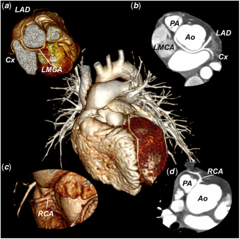
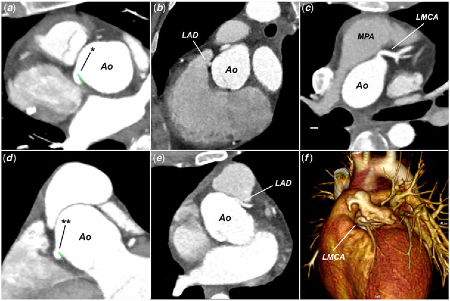
acute proximal angulation of at least one coronary artery was described when the proximal branching angle of the coronary artery was <30 degrees (Figs 1 and 2);
the interarterial course was described when any part of the coronary artery had a pattern between the pulmonary trunk and aorta;
coronary compression with high ellipticity index was described for arteries with a height-to-width ratio over 2 (ratio between the larger and the smaller diameter of the coronary artery);
any other abnormalities detected in CT scans with potential impact on myocardial perfusion were also analysed like muscular bridge, proximal kinking pattern without stenosis, and coronary fistula.
Volume-rendered 3D reconstructions of CT image. Central picture: patient with transposition after arterial switch operation associated with complex coronary anatomy – posterior looping of LMCA and anterior looping of the RCA. Acute branching angle of LMCA and retrograde pattern between aorta and left atrium are shown on 3D (a) and 2D (b) reconstructions. Complex pattern of the right coronary artery is presented on 3D (c) and 2D (d) reconstructions. Ao = ascending aorta; Cx = circumflex coronary artery; LAD = Left anterior descending artery; LMCA = left main coronary artery; PA = pulmonary artery; RCA = right coronary artery.

Examples of 2D and 3D CT angiograms with selected coronary features: (a) acute angulation of the LMCA in transverse plane of aortic sinus (* LMCA ostium with acute branching angle); (b) high ellipticity index of left anterior descending artery located between pulmonary artery and aorta; (c) proximal kinking of the LMCA; (d) acute angulation of the LMCA in coronal plane of aortic sinus (** LMCA ostium with acute branching angle); (e) interarterial course of LAD; and (f) 3D reconstruction – proximal kinking of the LMCA. Ao = ascending aorta; LAD = left anterior descending artery; LMCA = left main coronary artery; MPA = main pulmonary artery.

In all cases, the clinical data and basic screening test results were evaluated to identify potential “red flags,” that is, abnormalities that could facilitate the selection of high-risk patients without direct coronary imaging. Clinical data included the following:
Medical history of chest pain while at rest or during exertion or any symptoms related to the cardiovascular system, such as collapse, palpitation, and dyspnea.
Surgical technique – initially, we used the punch technique for coronary transfer until 1996, when the trap door technique was introduced, which has since been used with our own operator’s modifications in all switch operations. All cases with deviations from the usual coronary transfer technique were classified as complex coronary transfers and included transplantation of the two vessels to the one coronary sinus, transplantation very close to the commissure, or transplantation high in the coronary sinus. In our department, direct pulmonary anastomosis without patch is the method of choice; only in selected, high-risk patients is a fresh pericardial patch used for pulmonary reconstruction.Reference Moll, Michalak and Młudzik9,Reference Moll, Michalak and Sobczak-Budlewska14
Additional defects associated with transposition – the analysed defects include ventricular septal defects, aortic arch anomalies, coronary anomalies, bicuspid native pulmonary valve (neoaortic), and non-facing commissures.
Aortic root diameters obtained by CT and normalised to the body surface area by z scores calculated for tomographic normal values.Reference Hegde, Lensing and Greenberg15
Spatial position of the great arteries calculated as the angle between the line connecting the middle of the aortic and pulmonary valves and the line connecting the middle of the sternum and spine.Reference Szymczyk, Moll and Sobczak-Budlewska5
The results of the basic examinations were evaluated to establish the presence of any potential indicators that could be used as substitutes for direct imaging studies. Non-invasive, standard screening tests were evaluated in the context of potential relations to coronary problems. Any detected abnormality that could indicate coronary problems meant that the results were classified as abnormal:
electrocardiography – abnormal Q wave, ST segment, or T wave;
echocardiography – any regional myocardial dysfunction outside regions related to the patch closure of a ventricular septal defect, a reduction in the global ejection fraction, left ventricle dilatation, and significant mitral regurgitation;
exercise test – chest pain or discomfort, ischemic electrocardiography changes, ventricular arrhythmia induced by exercise, or Duke Treadmill Score below 5;
perfusion scintigraphy – any ischemic regions present while at rest or during exertion constituting more than 5% of the left ventricular myocardium.
Statistical analysis
Statistical analyses were performed using Statistica 13 software (StatSoft, Inc., Tulsa, Oklahoma, United States of America). Quantitative data are presented as the mean with 95% confidence interval and standard deviation or the median and interquartile range (25th–75th percentile) depending on the normality of the distribution. Qualitative data are presented as percentages. The normality of the distribution was tested using the Shapiro–Wilk and Lilliefors tests. Associations between quantitative datasets were tested using Spearman’s correlation test. Differences between the groups were assessed using the Mann–Whitney U-test. To evaluate the statistical relation between qualitative datasets, the Chi square test or Fisher exact test was used. A p value <0.05 was considered statistically significant.
Results
The median age at the time of coronary CT angiography was 19.8 years (interquartile range: 17.9–23 years, range: 14–25 years). Among this group, four patients had CT scans earlier than was planned in our post-operative protocol because of additional clinical reasons. Abnormalities were frequently discovered on coronary CT angiography: acute branching of at least one coronary artery was found in 25 patients (50%); ostial stenosis was present in 3 patients (6%); muscular bridge was present in 8 patients (16%); interarterial course was observed in 4 patients (8%); high ellipticity index was present in 5 patients (10%); and proximal kinking was observed in 12 patients (24%). Additionally, one case of coronary artery fistula was detected. In summary, 30 out of 50 patients (60% of our study group) showed some kind of coronary abnormality.
None of the above-mentioned coronary features were significantly related to defects associated with transposition of the great arteries, the presence of coronary anomalies, non-facing commissures, or patch reconstruction of the pulmonary artery. In all cases of interarterial course, present in the coronary artery arising from the left coronary sinus, the patients had undergone surgery using the punch technique before the trap door technique had been introduced. This correlation was statistically significant (0.046, Fisher exact test). The majority of patients with a high ellipticity index had also undergone surgery with the punch technique (four out of five), but this relation was not statistically significant (p = 0.15). Proximal coronary stenosis was significantly correlated with a history of complex coronary transfer during the arterial switch operation (p = 0.048, Fisher exact test).
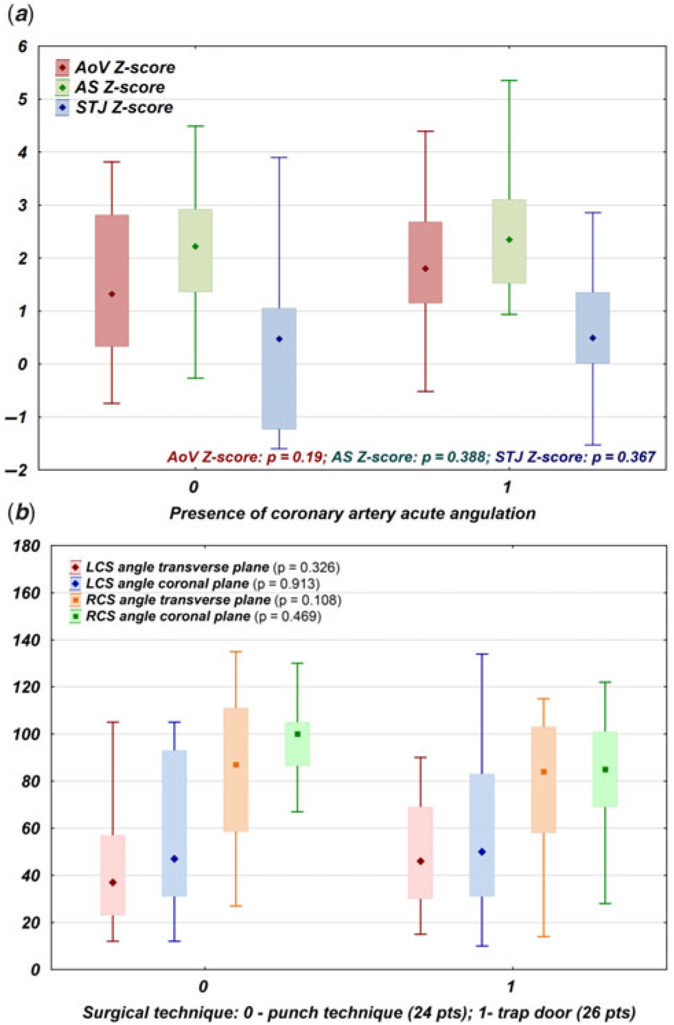
The presence of unfavourable coronary features was not significantly correlated with the spatial configuration of the great arteries or the aortic root diameter. The root diameter normalised using tomographic z scores was not significantly different between patients with and without acute proximal coronary angulation (Fig 3a). The branching angle of coronary arteries was similar regardless of the surgical technique (Fig 3b), but coronary arteries arising from the left coronary sinus had significantly lower branching angles than those arising from the right coronary sinus (p < 0.001, Kruskal–Wallis test).
( a ) Comparison of the neoaortic root z scores calculated for the AoV, AS, and STJ in relation to the presence of the acute proximal angulation of at least one coronary artery. Middle point: mean, box: interquartile range, whiskers: range. ( b ) Comparison of the coronary artery branching angle separately for the left and right coronary sinuses and transverse and coronal planes in relation to the surgical technique used for coronary transplantation. Middle point: mean, box: interquartile range, whiskers: range. AoV = neoaortic valve; AS = aortic sinus; STJ = sinotubular junction.

All patients in the study group were asymptomatic during the examinations, and in only three cases were the symptoms identified in the medical interview classified as abnormal. Those patients had non-specific chest pain sometimes associated with exertion (two patients) or radiation to the left arm (one patient). No significant correlations were detected between these symptoms and the coronary CT angiography. All patients had troponin T levels within the normal range. In six patients, the creatinine kinase (myocardial band) level was slightly increased (below two times the normal value in four patients, below three times the normal value in two patients). This elevation was not related to clinical symptoms, basic test results, or coronary CT angiography results. In all patients, the ejection fraction was within the normal range and only three patients showed regional hypokinesis; in two of these patients, the muscular band was detected on coronary CT angiography, but this relationship was not statistically significant (p = 0.06, Fisher’s exact test). None of our patients had electrocardiography criteria for myocardial ischemia, and non-specific criteria were present in 13 cases: non-specific T wave abnormalities, such as flattened or inverted T waves or a non-specific ST elevation (1 mm or below); these findings were not correlated significantly with the coronary CT angiography findings. The exercise test (performed in 46 patients from the study group) did not reveal clinical symptoms or electrocardiography changes characteristic of coronary problems. In two patients, exercise induced premature ventricular beats, but their presence was not significantly correlated with any abnormalities detected by coronary CT angiography. Single-photon emission CT at rest and during exercise was performed in 21 patients, mostly in patients with abnormal coronary CT angiography results, including all patients exhibiting stenosis, interarterial course, fistula, and myocardial bridge. A non-specific perfusion decrease (below 5%) was commonly detected; significant lesions were detected in three patients, but these results were not significantly correlated with any specific abnormalities detected on coronary CT angiography. All three patients with coronary stenosis detected on coronary CT angiography had normal single-photon emission CT results.
Discussion
Currently, the first wave of patients with transposition of the great arteries treated with a neonatal arterial switch operation is reaching the age of 30 years. They are still young and mostly asymptomatic, and because they underwent surgery at the beginning of the arterial switch operation era, they were strictly followed after the operation, most likely with more control visits and examinations than they truly required. Because the incidence of adverse events was low, most centres decided to significantly reduce the post-operative protocol for asymptomatic patients, starting with invasive studies and associated with radiation, such as coronarography and CT. Reference Sarris, Balmer and Bonou16,Reference Cohen, Eidem and Cetta17 Current guidelines for these patients do not support the need for routine post-operative coronary imaging in asymptomatic patients, and they can be qualified for competitive sports based on medical interviews, electrocardiography and echocardiography examinations, and exercise tests. Reference Van Hare, Ackerman and Evangelista4,Reference Sarris, Balmer and Bonou16 Both doctors and patients are beginning to consider that planned, routine follow-up visits and examinations are not required and would not be beneficial for future decision-making. Therefore, many patients who have undergone an arterial switch operation have been or will soon be lost to follow-up. Only recent guidelines provided by the EACVI group support the idea of direct visualisation of the coronary arteries reimplanted during arterial switch operation. Reference Di Salvo, Miller and Babu Narayan18 With many surgical modifications, increased experience and improvements in prenatal diagnosis, neonatal, and post-operative care, the number of patients with transposition of the great arteries after an arterial switch operation has increased and the survival rate of most complex cases has improved. Reference Villafañe, Lantin-Hermoso and Bhatt2 Coronary anomalies are common in this patient population, Reference Moll, Michalak and Sobczak-Budlewska14 and a surgically created pattern after coronary transplantation can promote some unfavourable features, such as acute proximal angulation, interarterial course, high ellipticity index, coronary compression, or kinking stenosis, which may change during child growth and development. Reference Ou, Khraiche and Celermajer6 The direction of such changes is still unpredictable, but now we know that a significant proportion of these patients are reaching adulthood with some potentially dangerous features, which may be clinically significant and definitely require careful follow-up studies accompanied by lifestyle modifications and competitive sports restrictions. Reference Szymczyk, Moll and Sobczak-Budlewska5,Reference Ou, Khraiche and Celermajer6 Our data show that these patients with potentially dangerous coronary features with higher risks for adverse coronary events cannot be identified indirectly based on routinely performed examinations, including medical histories, laboratory and stress tests, or electrocardiography or echocardiography examinations; even single-photon emission CT cannot exclude complex coronary anatomy. A review of the surgical reports is helpful because the potential problems may be associated more frequently with a complex coronary transfer, the surgical technique, or a complex coronary anatomy, but the sensitivity of such data is still limited, and a large proportion of the patients had absolutely no “red flags” in the interview or screening tests; nevertheless, the coronary CT angiography results were abnormal.
The selection of potentially dangerous features for our study was made based on an existing literature regarding patients with transposition after arterial switch and a healthy population with isolated coronary anomalies, Reference Ou, Khraiche and Celermajer6,Reference Ou, Mousseaux and Azarine7,Reference Lee, Uppu and Lytrivi10–Reference Monroy-Gonzalez, Alexanderson-Rosas and Prakken13 where these abnormalities usually show their significance during excessive exertion. Reference Eckart, Scoville and Campbell11 The acute branching angle (below 30 degrees), stenosis over 50%, and interarterial course were shown to be associated with the risk of adverse cardiac events. Factors such as high ellipticity index, intramural bridge, and coronary fistula may be potentially dangerous, but their significance, especially in the pediatric population, is not yet established. The extrapolation of such calculated risk to patients with transposition of the great arteries after an arterial switch operation is impossible. These patients have a more complex anatomy, with different relationships between the aorta and pulmonary artery, a complex neoaortic root anatomy, and unique proximal coronary patterns. Reference Villafañe, Lantin-Hermoso and Bhatt2 Additionally, in approximately 5% of cases, the exact coronary anatomy is unknown because that is the frequency of significant disagreement between the post-operative coronary anatomy detected during coronary CT angiography and what has been described in surgical reports. Reference Szymczyk, Moll and Sobczak-Budlewska5 In our series, acute angulation of the proximal coronary artery was not significantly correlated with the root diameter or surgical technique used, which may indicate that the initial pattern and anatomy influence the post-operative coronary pattern more significantly. The left-sided coronary ostium is usually closer to the pulmonary trunk than is the right-sided coronary ostium. Thus, the interarterial course, high ellipticity index, ostial stenosis, and acute angulation are significantly more frequently related to the left coronary artery or a solitary left anterior descending artery.
Arterial switch operations have dramatically changed the fates of patients with transposition of the great arteries. Short- and mid-term survival rates are currently excellent, and most patients have no limitations on daily activities. These patients are completely asymptomatic and participate in a variety of sports activities. However, the coronary anatomy sometimes obscures dangerous features related to coronary transplantation during surgery and unique spatial relations of the great arteries post-operatively. Reference Szymczyk, Moll and Sobczak-Budlewska5 We know that the frequency of such features is significant, Reference Szymczyk, Moll and Sobczak-Budlewska5,Reference Ou, Khraiche and Celermajer6,Reference Ou, Mousseaux and Azarine7,Reference Ashrafpoor, Danchin, Houyel, Ramadan, Belli and Paul19 and our study shows that they cannot be reliably predicted using basic screening tests. A similar conclusion was published by Tsuda et al. Reference Tsuda, Bhat, Robinson, Baffa and Radtke20 For responsible patient management, for example, in terms of sports qualification, further follow-up visits and establishing the real impact of the coronary anatomy during the post-operative follow-up are needed to determine the exact coronary pattern in every single patient after an arterial switch operation.
Conclusions
Abnormal coronary CT angiography results are very frequent in patients with transposition of the great arteries after an arterial switch operation. Potentially dangerous features, such as proximal kinking stenosis, interarterial course, coronary compression with high ellipticity index, and acute proximal angulation, are frequently asymptomatic according to routinely performed screening tests (medical interviews, laboratory tests, electrocardiography and echocardiography examinations, and stress tests). For accurate sports and lifestyle advising, scheduled visits, and additional diagnostic tests, routine coronary imaging is necessary.
Acknowledgements
None.
Financial Support
This work was supported by an internal institutional grant (Polish Mother’s Memorial Hospital, Research Institute, grant no. Nr 2017/III/30-GW).
Conflicts of Interest
None.
Ethical Standards
This retrospective study was approved by the institutional ethics board (52/2018), which waived the need for informed consent.