Background
Bone is composed of inorganic (calcium phosphate crystals) and organic (collagen, albumin, osteocalcin) compounds and is a dynamic organ due to the continuous remodelling process. Although bone growth in humans largely ceases at the end of adolescence, bone is continually renewed through adulthood, enabling the skeleton to adapt to the changing stresses of life.Reference Poulsen, Moughan and Kruger1
The cells involved in bone turnover/remodelling are osteoclasts, osteoblasts and osteocytes. Osteoclasts are the cells responsible for bone resorption. Active osteoclasts reside in the mineralized surface of the bone where they produce and release lysosomal enzymes and free radicals into the space in contact with the bone. These products dissolve the minerals and degrade the bone matrix.Reference Blair, Schlesinger, Ross and Teitelbaum2 Bone remodelling involves the sequential and coupled resorption of a small area of bone tissue by specialized macrophage-like cells known as osteoclasts, followed by replacement of this tissue with new bone synthesized by osteoblasts.Reference Pead, Skerry and Lanyon3–Reference Manolagas5
In humans, an estimated 10% of bone is remodelled each year.Reference Theill, Boyle and Penninger6 Bone remodelling is regulated by controlling osteoblast and osteoclast cell number and activity. Osteoclastogenesis is largely regulated by a triad of proteins consisting of a ligand (i.e. receptor-activated nuclear-kappa B ligand, RANK-L), its receptor (i.e. RANK) and a decoy receptor (i.e. osteoprotegerin, OPG). RANK is a membrane-bound protein expressed on osteoclast precursors and mature osteoclasts.Reference Theoleyre, Wittrant, Tat, Fortun, Redini and Heymann7 RANK-L exists in both a membrane-bound and soluble form and is produced by a range of cells including osteoblasts and activated T-cells.Reference Schoppet, Preissner and Hofbauer8 OPG is a soluble protein secreted by a range of different cells, including osteoblasts.Reference Theoleyre, Wittrant, Tat, Fortun, Redini and Heymann7
Binding of RANK-L to RANK promotes osteoclastogenesisReference Walsh, Kim, Kadono, Rho, Lee, Lorenzo and Choi9, Reference Asagiri and Takayanagi10 and inhibits osteoclast apoptosis.Reference Asagiri and Takayanagi10 Binding of RANK-L to OPG prevents RANK-L/RANK-induced osteoclastogenesis. Increased OPG protein levels lead to a rapid reduction in osteoclast number.Reference Theoleyre, Wittrant, Tat, Fortun, Redini and Heymann7 Hence the balance between RANK, RANK-L and OPG is a major factor controlling osteoclast number. Various signalling pathways converge to regulate osteoblastogenesis. These include the canonical WntReference Bonewald12 and bone morphogenic protein (BMP) signalling pathways.Reference Kanaan and Kanaan13
Osteoporosis
The WHO Working Group defined osteoporosis as a disease characterized by low bone mass and microarchitectural deterioration of bone tissue leading to enhanced bone fragility and a consequent increase in fracture incidence. Osteoporosis can be identified according to measurements of bone mineral density (BMD), using dual-energy X-ray absorptiometry (DEXA). According to WHO criteria, osteoporosis is defined as a bone density T score at or below 2.5 standard deviations (T score) below normal peak values for young adults. Of note, the standard BMD was that measured in young adult women who were at the age of peak bone mass.
Primary or idiopathic osteoporosis has been classified as type I or type II. Type I osteoporosis occurs in women between 51 and 75 years of age and is related to oestrogen deficiency seen in the menopausal transition. Type II or senile osteoporosis occurs in people older than 60 years of age and affects both men and women. Age-related bone loss results from changes at cellular level, such as decreased osteoblastogenesis, shortened osteoblast lifespan, increased adipogenesis, as well as from hormonal changes, including decreased levels of sex steroid hormones and vitamin D, and increased levels and activity of parathyroid hormone (PTH).
Secondary osteoporosis is the result of an underlying disease (kidney and liver disease, coeliac diseases, inflammatory bowel diseases, diabetes, nutrition deficiency, sarcopenia, endocrine abnormalities, cancer, alcoholism) and/or medications (glucocorticoids, long-term heparin treatment, oral anticoagulants, aromatase inhibitors for breast cancer, anti-androgen therapy for prostate cancer, high doses of thyroxine, thiazolinediones, antiretroviral drugs, selective inhibitors of serotonin reuptake, anticonvulsants, proton pump inhibitors) that adversely affect bone.
Low bone mass is the main factor underlying osteoporosis. Any factor that influences the development of peak bone mass or the loss of bone in middle age will affect later fracture risk. These factors can be divided into unmodifiable factors, such as gender, age, genetics and ethnicity, and modifiable factors including mechanical and local factors, hormonal status, nutrition and lifestyle factors (physical activity, smoking, alcohol consumption and diet).
Mechanical factors
The apposition of bone and endochondral growth depends on the mechanical forces generated by muscles. Osteocytes are specialized ‘mechano-sensing’ cells that reside within bone. They detect mechanical strain and initiate signalling pathways, leading to both osteoclastogenesis and osteoblastogenesis.Reference Cherian, Siller-Jackson, Gu, Wang, Bonewald, Sprague and Jiang14 There is some evidence that, after mechanical load, microfractures may occur in bone with subsequent activation of interleukins and growth factors that stimulate bone turnover.
Lipid mediators play a critical role in the mechano-signalling pathway. Within seconds of mechanical loading of bone, the lipid mediator prostaglandin E2 (PGE2) is released by osteocytes and mature osteoblasts.Reference Quinn and Gillespie11 Phospholipase-mediated membrane release of fatty acids, notably arachidonic acid (AA, 20:4n-6), the substrate for PGE2 synthesis, and expression of the inducible form of cyclo-oxygenase (COX), COX-2, which oxidizes AA to PGE2, are up-regulated as an early response to strain.Reference Smith and Clark15 PGE2 promotes osteoclastogenesis by stimulating expression of both RANK-L and RANK and inhibiting expression of OPG. PGE2 also activates the Wnt signalling pathwayReference Quinn and Gillespie11 and promotes cbfa-1 Reference Yoshida, Oida, Kobayashi, Maruyama, Tanaka, Katayama, Yamaguchi, Segi, Tsuboyama, Matsushita, Ito, Ito, Sugimoto, Ushikubi, Ohuchida, Kondo, Nakamura and Narumiya16, Reference Watkins, Li, Lippman and Feng17 and insulin-like growth factor 1 (IGF-1)Reference Celil and Campbell18 expression, thereby stimulating osteoblastogenesis. Although the importance of AA and PGE2 in regulating bone remodelling is well established, the involvement of long chain polyunsaturated fatty acids (LCPUFAs) in the control of bone metabolism may be much more extensive than is currently recognized.
Local factors
These are produced by bone cells and bone marrow cells. Some, such as macrophage colony-stimulating factor (M-CSF) or granulocyte colony-stimulating factor (G-CSF), exert their effect by regulating cell differentiation. IGF-1, IGF-2, interleukin-1 (IL-1), interleukin-6 (IL-6) and tumour necrosis factor alpha (TNF-α) are mediators and regulators of cell–cell interaction.
Systemic hormones
Vitamin D, PTH, calcitonin and sex-steroid hormones (mainly oestrogens) affect bone metabolism. Oestrogens induce osteoclast apoptosis and regulate the action of IL-1, IL-6 and TNF-α.
Nutrition factors
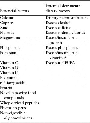
Nutrients and nutrient intake may influence bone quality by various mechanisms. Nutrient intake affects the bone structure, the rate of bone metabolism, modulates the endocrine and/or paracrine system and influences the homeostasis of other bone-active elements. Many of the nutrients and food components we consume as part of a Westernized diet could have a positive or negative impact on bone health (Table 1).Reference Cashman19
Potential nutritional determinants of bone health: modified from Cashman (Reference Cashman2007).Reference Cashman19 Some nutrients could be categorized as being both beneficial and detrimental depending on dietary exposure level: insufficient or in excess

Nutrition and osteoporosis
Diet may influence bone by various mechanisms, including alteration of bone structure, the rate of bone metabolism, the endocrine and/or paracrine system and homeostasis of calcium and possibly of other bone-active mineral elements.Reference Cashman, Remacle and Reusens20
These dietary factors range from inorganic minerals (e.g. calcium, magnesium, phosphorus, sodium, potassium and various trace elements) and vitamins (vitamins A, D, E, K, C and certain B vitamins) to macronutrients, such as protein and fatty acids. In addition, the relative proportions of these dietary factors derived from different types of diets (vegetarian vs. omnivorous) may also affect bone health and thus osteoporosis risk. Furthermore, in recent years a number of bioactive food components have been proposed as being beneficial for bone health. A review of all of these dietary factors is beyond the scope of the present article.
Three important reports (the European Commission's Report on Osteoporosis in the European Community: Action for Prevention;21 the US Surgeon General's Report on Bone Health and Osteoporosis;Reference Cashman, Remacle and Reusens20 and the World Health Organization's report on Diet, Nutrition and the Prevention of Chronic Diseases 22) have stressed the importance of calcium and vitamin D in promoting bone health. Also, selected nutrients/food components play important roles in bone health (such as vitamin K and dietary phytoestrogens). However, at present, the evidence-based indications for these latter nutritional factors is less well developed.21, Reference Prentice23, 24
Nutrients associated with fruit and vegetable intake (potassium and magnesium) have been associated with greater bone mineral density (BMD) in late premenopausal women and older subjects. It is well known that calcium and vitamin D attenuate postmenopausal bone loss.Reference Weaver25, Reference Weaver26
An important role in the pathogenesis of osteoporosis may be exerted by a decrease in plasma antioxidants. Vitamin C, an antioxidant vitamin, is an essential co-factor in the formation of stable collagen.Reference Oeffinger27, Reference Peterkofsky28 In preosteoblastic and osteoblast-like cell lines, vitamin C promotes collagen genesis, vitamin D-stimulated expression of alkaline phosphatase and mineralization.Reference Sugimoto, Nakada, Fukase, Imai, Kinoshita and Fujita29, Reference Franceschi and Young30 Several epidemiological studies have found an association between dietary vitamin C intake and bone mineral density in postmenopausal women.Reference Hall and Greendale31, Reference Wang, Luz Villa, Marcus and Kelsey32 In addition, it has been recently shown that lower dietary intake of vitamin C and vitamin E may increase the risk of hip fracture.Reference Melhus, Michaëlsson, Holmberg, Wolk and Ljunghall33 A recent study shows that older osteoporotic women have lower levels of all natural antioxidants tested than controls. This difference is significant across the panel of exogenous (dietary vitamin A, vitamin C, vitamin E) and endogenous molecules (uric acid, superoxide dismutase and glutathione peroxidase) with scavenging activity. Taken together, these data seem to point to a negative role for antioxidant deficit in age-related bone-loss.Reference Maggio, Barabani, Pierandrei, Polidori, Catani, Mecocci, Senin, Pacifici and Cherubini34
There is increasing evidence that lack of certain fatty acids in the diet contributes to bone loss.Reference Kruger and Horrobin35–Reference Das38 Moreover, osteoclastogenesis is regulated by a number of cytokines, growth factors and eicosanoids. Eicosanoids (prostaglandins, leukotrienes and related compounds) are formed from long chain polyunsaturated fatty acids released from membrane phospholipids. The amount and type of produced eicosanoids reflect the type of fatty acids available. The membrane fatty acid composition is in turn influenced by the dietary source of fatty acids.Reference Lewis, Austen and Soberman39–Reference Fernandes, Lawrence and Sun41
Polyunsaturated fatty acids (PUFAs)
The most common source of n-6 fatty acids is linoleic acid (LA), which is found in high concentrations in various vegetable oils. Arachidonic acid (AA), the 20-carbon n-6 fatty acid, is obtained largely by synthesis from LA in the body. The n-3 fatty acids eicosapentaenoic acid (EPA) and docosahexaenoic acid (DHA) are found instead in fish and fish oils.
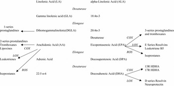
PUFAs are precursors of a wide range of metabolites that are oxidation products formed by the activity of cyclo-oxygenases (COX) and lipoxygenases (LOX). There are two main kinds of PUFA metabolites: eicosanoids (prostaglandins, leukotrienes, lipoxins and resolvins) and docosanoid (Figure 1).
Metabolism of long-chain polyunsaturated fatty acids: modified diagram from Kruger.Reference Kruger, Coetzee, Haag and Weiler111 LOX, lipoxygenases; COX, cyclo-oxygenases; ROS, reactive oxgen series; 13R-HDHA, 13R hydroxylated DHA; 17R-HDHA, 17R- hydroxylated DHA.

COX converts AA in thromboxane A 2 (TXA2) and catalyses the conversion of AA and EPA into prostaglandins of the 1-, 2- and 3- series, respectively. COX has greater specificity for AA than EPA and consequently synthesizes 2-series rather than 3-series prostaglandins.
LCPUFAs and lipid mediators derived from changes in dietary PUFAs can affect composition of different tissues including bone cells and consequently can modulate osteoblast and osteoclast function.
Hypothetical mechanisms underlying the relationship between omega-3 and osteoporosis
There are at least six mechanisms by which omega-3 fatty acids may affect bone remodelling. Omega-3 may modulate calcium absorption, inflammatory cytokines, peroxisome proliferators-activated receptor γ (PPARγ), NO synthesis, production of growth factors and prostaglandins.
Calcium absorption
Dietary intake of fats affects calcium absorption and urinary calcium.Reference Schlemmer, Coetzer, Claassen and Kruger42, Reference Claassen, Coetzer, Steinmann and Kruger43 Studies suggest that LCPUFAs may promote intestinal calcium absorption, thereby increasing the overall calcium balance by Ca2+-ATPase, the enzyme responsible for active calcium absorption in the intestine. The activity of Ca2+-ATPase in basolateral membranes from duodenal enterocytes treated with DHA is increased compared with non-treated and EPA-treated membranes.Reference Haag, Magada, Claassen, Bohmer and Kruger44 The stimulatory effect of DHA is only evident in membranes deprived of calmodulin, suggesting that DHA may only have a physiologically relevant effect on active calcium transport when dietary calcium intake is low. However, dietary supplementation with either fish oil or evening primrose oil in rats receiving a calcium-adequate diet results in increased calcium transport across the basolateral membraneReference Coetzer, Claassen, Papendorp and Kruger45 and decreased faecal calcium excretion.Reference Claassen, Potgieter, Seppa, Vermaak, Coetzer, Vanpapendorp and Kruger46, Reference Kruger, Coetzer, de Winter and Claassen47 It is possible that physiological changes brought about by increased membrane content of LCPUFAs led to increased passive, as well as active, calcium transport. The n-6 LCPUFA may be less effective than n-3 LCPUFA in promoting calcium absorption because an increase in overall calcium balance has only been observed with fish oil supplementation.Reference Claassen, Potgieter, Seppa, Vermaak, Coetzer, Vanpapendorp and Kruger46, Reference Kruger, Coetzer, de Winter and Claassen47
There is some evidence that one of the mechanisms by which 1,25-dihydroxyvitamin D promotes intestinal calcium absorption is by increasing the concentration of highly unsaturated fatty acids in membrane phospholipids.Reference Kruger, Coetzer, de Winter and Claassen47 Membrane LCPUFAs content is known to affect the structure, fluidity and polarity of membranes, as well as the relative proportion of membrane-bound proteins.Reference Stillwell and Wassall48 Structurally, membranes are composed of ‘liquid disordered’ phospholipids interspersed with tightly packed, more orderly ‘lipid rafts’ that consist of sphingolipids and cholesterol. Cholesterol is essential for the formation of lipid rafts.Reference Simons and Ehehalt49 Highly unsaturated long chain fats, such as DHA, have a strong aversion to cholesterol. Incorporation of DHA into a membrane region results in complete expulsion of cholesterol, hence reducing the proportion of lipid rafts in the membrane.Reference Stillwell and Wassall48 Altering the dispersion of lipid rafts within membranes modulates the activity of membrane proteins. Examples of lipid raft-associated proteins include Ca2+-ATPaseReference Sepulveda, Berrocal-Carrillo, Gasset and Mata50 and components of the NF-B kinase complex.Reference Tu, Huang, Bae, Slaughter, Whitelegge, Crother, Bickel and Nel51
Incorporation of unsaturated fats into cellular membranes increases membrane fluidity.Reference Valentine and Valentine52 The greatest increase occurs with the addition of two and three double bonds, with little change in fluidity occurring with more than three double bonds.Reference Stillwell and Wassall48 Membrane permeability and the speed of membrane flip-flop (movement of membrane constituents between layers in the membrane bilayer) are increased as the number of double bonds in the fatty acyl chains increases.Reference Armstrong, Brzustowicz, Wassall, Jenski and Stillwell53 Increased membrane unsaturation may expedite cellular uptake of nutrients and other molecules, particularly by passive transport.
Inflammatory cytokines
Inflammatory cytokines play an active role in the pathogenesis of osteoporosis by stimulating bone resorption and inhibiting bone formation. Previous studies have shown that various proteins play a role in the interaction between osteoblasts and osteoclasts. There is evidence that, in bone cells, omega-6 PUFA and AA increase pro-inflammatory cytokine production (TNFα, IL-1) while omega-3 fatty acids can reverse this effect.Reference Priante, Bordin, Musacchio, Clari and Baggio54 Bordin suggested that a protein kinase C-dependent mechanism could be responsible for the effects of AA on cytokine production.Reference Bordin, Priante, Musacchio, Giunco, Tibaldi and Clari55
Reactive oxygen species (ROS) and oxidative stress are key factors of bone metabolism. Both n-3 and n-6 fatty acids are incorporated into cellular phospholipid membranes, where they are precursors of eicosanoids such as prostaglandins, leukotrienes and thromboxanes. Eicosanoid metabolites of n-3 fatty acids are less atherogenic, pro-inflammatory and vasocostrictive than eicosanoids derived from n-6 fatty acids.
The dietary intake of long chain n-3 fatty acids (EPA, DHA) partially replaces the n-6 fatty acids, particularly AA, in the cell membranes. The increased omega-3/omega-6 fatty acid ratio results in change of membrane function with subsequent decrease in IL-1, IL-6 and TNF-α.Reference Chandrasekar and Fernandes56, Reference Ferrucci, Cherubini, Bandinelli, Bartali, Corsi, Lauretani, Martin, Andres-Lacueva, Senin and Guralnik57
Supporting the beneficial role of omega-3, Georghiade reported that 133 patients with non-ischaemic heart failure on EPA+DHA treatment compared with placebo had a significant reduction of the inflammatory markers TNFα, IL-1 and IL-6.Reference Gheorghiade58
Animal and human studies suggest that ω-3 supplements may protect from arthritis, lupus and IgA nephropathy, and may prevent bone loss probably through anti- inflammatory activity.Reference Kehn and Fernandes59
Peroxisome proliferators-activated receptor γ
An additional mechanism can link free fatty acids and osteoporosis. During ageing an increased marrow adiposity usually accompanies osteoporosis. In addition, osteoblasts derive from pluripotent mesenchymal stem cells that can differentiate into mature osteoblasts or adipocytes.
A regulatory mechanism controlling the differentiation of stem cells into osteoblasts is the PPARγ nuclear transcription factor. PPARγ is expressed in adipose tissue and bone marrow. Heterozygous PPARγ-deficient mice exhibit enhanced osteoblastogenesis, increased bone volume and decreased adipogenesis.Reference Akune, Ohba, Kamekura, Yamaguchi, Chung, Kubota, Terauchi, Harada, Azuma, Nakamura, Kadowaki and Kawaguchi60, Reference Ali, Weinstein, Stewart, Parfitt, Manolagas and Jilka61 Elderly women with type 2 diabetes under PPARγ agonist treatment had greater bone loss than those who are not on this medication.Reference Schwartz, Sellmeyer, Vittinghoff, Palermo, Lecka-Czernik, Feingold, Strotmeyer, Resnick, Carbone, Beamer, Park, Lane, Harris and Cummings62
AA, LA and other mediators derived from n-6 fatty acids from enzymatic conversion are known activators of PPARγ; in contrast, EPA and DHA are not known as activators of PPARγ. Lowering the dietary ratio of n-6 to n-3 fatty acids may diminish PPARγ activation and promote osteoblastogenesis instead of bone marrow adiposity.Reference Vanek and Connor63
Prostaglandin secretions
PGE2 is derived from AA and is a modulator of bone remodelling interacting both in bone resorption and formation.Reference Collins and Chambers64, Reference Jee and Ma65 Increased synthesis of PGE2 impacts adversely on bone remodelling.Reference Corwin66 Several studies have shown an increase in osteoblastic markers when PGE2 production was decreased or inhibited.Reference Evans, Bunning and Russell67–Reference Kajii, Suzuki and Yoshikawa71
Because conversion to prostaglandin from n-3 PUFAs is less effective than from n-6 PUFAs,Reference Raisz, Alander and Simmons72, Reference Laneuville, Breuer, Xu, Huang, Gage and Watson73 lowering the n-6/n-3 PUFA ratio by increasing n-3 intake can also reduce PGE2 conversion.Reference Kruger and Horrobin35, Reference Watkins, Li, Allen, Hoffman and Seifert74, Reference Li, Seifert, Ney, Grahn, Grant and Allen75 Accordingly, animal studies demonstrated a reduction of PGE2 production and pyridinoline levels in rats fed with n-3 PUFA.Reference Shen, Peterson, Tatum and Dunn76 Li observed an increased PGE2 production in rats on diet enriched with n-6 fatty acids compared with those on n-3 PUFA treatment.Reference Li, Seifert, Ney, Grahn, Grant and Allen75 Other in vivo animal studies demonstrated that in rats the continuous infusion of PGE2 led to an increased bone loss and bone resorption.Reference Tian, Zhang, Setterberg, Zeng, Iturria and Ma77
Different hypotheses have been made to explain the mechanism by which PGE2 is involved in bone remodelling. Some authors reported a stimulatory effect of PGE2 on osteoclastogenesis, increasing osteoclast size and inhibiting osteoclast apoptosis.Reference Ono, Kaneko, Choudhary, Pilbeam, Lorenzo and Akatsu78, Reference Tsutsumi, Xie, Wei, Zhang, Zhang and Flick79 PGE2 has also been shown to stimulate IL-6 production.Reference Calder80 PGE2 could promote osteoclastogenesis by stimulating expression of both RANK-L and RANK by fibroblasts and inhibiting expression of OPG.Reference Quinn and Gillespie11, Reference Liu, Kirschenbaum, Yao and Levine81 Other authors demonstrated that COX-1 and COX-2 have greater specificity for AA than EPA and synthesize 2-series rather than 3-series prostaglandins.Reference Watkins, Lippman, Le Boutellier, Li and Seifert82 n-6 fatty acids up-regulate COX-2 expression and promote 2-series prostaglandin formation. n-3 LCPUFA inhibit COX-2 expression,Reference Hamid, Singh, Reddy and Cohen83 possibly modulating toll-like receptor signalling pathways.Reference Shen and Tai84
PUFAs and growth factors
Growth factor β1 (TGF β1) and insulin-like growth factor 1 (IGF-1) are anabolic factors involved in bone turnover. TGFβ1 induces proliferation and has anabolic properties in osteoblasts.
Ramirez-Yanez et al. reported in vitro a dose-dependent effect of PGE2 on TGF β1 receptor synthesis: low concentration of PGE2 increased expression of TGF β1 receptor in osteoblasts,Reference Ramirez-Yanez, Hamlet, Jonarta, Seymour and Symons85 which means increased binding of TGF β1 to this receptor and consequently an increased osteblastic activity, but higher concentrations of PGE2 led to opposite effects.Reference Martinez-Ramirez, Palma, Martinez-Gonzalez, Delgado-Martinez, De la Fuente and Delgado-Rodriques86 Also IGF-1 promotes bone matrix formation, stimulates osteoblasts activityReference Watkins and Seifert87 and seems to be a key factor in maintaining a balance between osteoblast and osteoclast activity during physiological bone remodelling through the regulation of RANK–RANK-L expression.Reference Wang, Nishida, Elalieh, Long, Halloran and Bikle88
The role of PUFAs in regulation of IGF-1 and IGF-1 binding protein (IGFBP) production is still unclear. Probably modulation of local PGE2 could down-regulate or up-regulate the secretion of this anabolic hormone.Reference Li, Seifert, Ney, Grahn, Grant and Allen75, Reference Weiler and Fitzpatrick-Wong89–Reference Delany, Pash and Canalis90 In rats, n-3 PUFA enhanced IGF-1 production, which in turn stimulates osteoblast differentiation.Reference Shen, Peterson, Tatum and Dunn76
PUFAs, NO secretion and NO synthase
An increased production of constitutive NO is associated with decreased number and diminished activity of osteoclastic cells and decreased bone resorption.Reference Das91 Activation of the inducible nitric oxide synthase (iNOS) by inflammation has been related to enhanced osteoclastic activity.Reference Armour, van't Hof and Grabowski92 Impact of n-3 PUFA on NO may have a positive effect on bone metabolism; n-3 fatty acids are able to elevate the production of constitutive NO, while suppressing the production of the inducible form probably inhibits the action of inflammatory cytokines.Reference Watkins, Li, Lippman and Feng17 It has been shown that NO decreases the RANK-L/OPG equilibrium, suggesting an inhibitory effect on osteoclastogenesis in murine stromal cells.Reference Fan, Roy, Zhu, Murphy, Ackert-Bicknell and Hart93
Das hypothesized that the effects of NO may depend on its local concentration: at low concentrations NO promotes the proliferation of osteoblasts, whereas high concentrations (associated with up-regulation of iNOS) may enhance bone resorption.Reference Das91 It has been reported that AA significantly up-regulated iNOS mRNA expression in human osteoblast-like cells, stimulating osteoclastic activity.Reference Priante, Musacchio, Pagnin, Calo and Baggio94
Omega-3, omega-6 and osteoporosis
Animal studies
There is a wealth of evidence from animal studies showing a positive effect of dietary supplementation with fatty acids on bone loss.
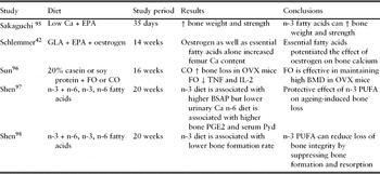
Dietary supplementation with fish oil rich in omega-3 PUFAs has been shown to enhance bone density as well as duodenal calcium uptake in rats. The latter process is supported by membrane ATPases.Reference Haag, Magada, Claassen, Bohmer and Kruger44 In ovariectomized (OVX) rats on low calcium diet, adding EPA to the diet prevents the loss of bone weight and increases bone strength.Reference Sakaguchi, Morita and Murota95 In addition, OVX rats supplemented with GLA and EPA show a significant increase in bone calcium content.Reference Schlemmer, Coetzer, Claassen and Kruger42
Fish oil (n-3 fatty acids), in contrast to corn oil (n-6 fatty acids), is also effective in maintaining high BMD in OVX mice. In the same study, the analysis of RANK-L expression and cytokine production in activated T cells support the notion that osteoclast generation and activation may be one mode of action of n-3 fatty acids in decreasing bone loss.Reference Sun, Krishnan, Zaman, Lawrence, Bhattacharya and Fernandes96
Shen also demonstrated a protective effect of long chain n-3 PUFAs on age-related bone loss due to modulation of local factors and hormones such as IGF-1, PTH and vitamin D.Reference Shen, Yeh, Rasty, Li and Watkins97 The same authors found that a diet rich in long chain n-3 PUFA lessens loss of bone integrity in male rats through reducing the bone turnover rate by suppressing both bone formation and resorption.Reference Shen, Yeh, Rasty, Chyu, Dunn, Li and Watkins98 Excess LCPUFAs can also have a detrimental effect on bone mass (Table 2).
Effects of n-3 fatty acids on osteoporosis in animal studies

EPA, eicosapentaneic acid; GLA, gamma-linolenic acid; FO, fish oil; CO, corn oil; OVX, ovariectomized; BSAP, bone-specific alkaline phosphatase activity; Pyd, pyridinoline; EPO, evening primrose oil; EPFO, evening primrose and fish oil; OO, olive oil.
In conclusion, numerous animal studies notwithstanding, the protective ratio of dietary n-6/n-3 PUFA is not well established.
Human studies
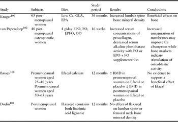
In spite of increasing evidence of the positive effects of dietary fats on bone metabolism from animal and in vitro studies, few studies have been conducted in humans. Most human data derive from observational studies conducted in postmenopausal women (Table 3). These studies show that the total amount of fat consumed is positively associated with fracture risk and negatively associated with bone density.Reference Michaëlsson, Holmberg, Mallmin, Wolk, Bergström and Ljunghall99
Human studies testing the effects of n-3 fatty acids on osteoporosis in the elderly: modified from Salari et al. Reference Salari, Rezaie, Larijani and Abdollahi2008Reference Salari, Rezaie, Larijani and Abdollahi110 and Poulsen et al. Reference Poulsen, Moughan and Kruger2007Reference Poulsen, Moughan and Kruger1

Kruger conducted a study in which older osteoporotic or osteopenic women (mean age 79.5 years) with habitually low dietary calcium intakes were supplemented with 6 g of LCPUFA-rich oil (3.6 g LA, 480 mg GLA, 240 mg EPA and 180 mg DHA) in conjunction with 600 mg of calcium carbonate per day for 18 months. Controls who received 600 mg of calcium carbonate and 6 g of coconut oil per day exhibited a 3.2% decrease in lumbar spine BMD over the 18-month period; however, BMD was maintained in the LCPUFA-supplemented group.Reference Kruger, Coetzer, de Winter, Gericke and van Papendorp100
In another study, 12 months of EPA supplementation increased bone density in postmenopausal women.Reference Terano101 In old (mean age 80 years) osteoporotic women, daily supplementation with 4 g of fish oil containing 16% EPA and 11% DHA or a mixture of fish and evening primrose oils containing 60% linoleic acid, 8% GLA, 4% EPA and 3% DHA for 16 weeks resulted in decreased serum alkaline phosphatase activity and increased serum concentration of procollagen.Reference Van Papendorp, Coetzer and Kruger102
A longitudinal study was conducted on 891 women during menopausal transition using a Food Frequency Questionnaire (FFQ) to evaluate food intake and DEXA to assess BMD. The authors found a negative relationship between increased intake of fatty acids and femoral BMD loss.Reference Macdonald, New, Golden, Campbell and Reid103 Data from the Rancho Bernardo study showed that a higher ratio of n-6 to n-3 fatty acids is associated with lower BMD at the hip in both sexes.Reference Weiss, Barrett-Connor and von Mühlen104
A recent observational study in older adults showed a positive association between self-reported intake of omega-3 fatty acids and total femoral BMD.Reference Griel, Kris-Etherton, Hilpert, Zhao, West and Corwin105
A recent case-control study conducted on 165 patients aged over 65 with low-energy fracture reported a significant association between intake of omega-6 fatty acids and risk of fracture. However, in the same study the authors failed to find a significant relationship between osteoporotic fracture and intake of omega-3 fatty acids or with omega-3/omega-6 ratio.Reference Martinez-Ramirez, Palma, Martinez-Gonzalez, Delgado-Martinez, De la Fuente and Delgado-Rodriques86
Griel observed a significant reduction in markers of bone resorption (N-telopeptides and bone-specific alkaline phosphatase) when men reduce dietary SFA and increase PUFA consumption.Reference Griel, Kris-Etherton, Hilpert, Zhao, West and Corwin105 Using data from NHANES III, Corwin found that saturated fat intake is negatively associated with BMD at several hip sites and the greatest effects are seen among men less than 50 years old.Reference Corwin, Hartman, Maczuga and Graubard106 In a recent observational study assessing fatty acid concentration in 95 healthy young men, Hogstrom found a positive association between serum n-3 fatty acid levels and BMD.Reference Högström, Nordström and Nordström107 In contrast, two more recent studies showed no effect of LCPUFA supplementation on bone during menopause. In one study, pre- and postmenopausal women (age range 25–40 years and 50–65 years, respectively) during a 12-month follow-up period were supplemented with Efacal, a Scotia Pharmaceuticals product containing a combination of evening primrose oil (4.0 g/day providing approximately 430 mg GLA/day), fish oil (440 mg/day providing approximately 70 mg EPA/day) and calcium (1.0 g/day). No additional benefit of LCPUFA supplementation on total body BMD over calcium supplementation alone was observed.Reference Bassey, Littlewood, Rothwell and Pye108
Consistent with these findings, 12-month treatment of 40 g flaxseed oil supplement per day (a source of ALA but also of other bioactive components, such as lignans) in menopausal women (age range 45–65 years) did not show any significant difference in BMD compared with women supplemented with a wheat germ placebo. However, the composition of LCPUFAs in this supplement (namely 18-carbon n-3 LCPUFA rather than the longer chain 20- and 22-carbon LCPUFAs) differed considerably from the supplements used in the previous studies.Reference Dodin, Lemay, Jacques, Legare, Forest and Masse109 Importantly, not only the total amount of LCPUFA but also the composition of dietary LCPUFAs is important for optimizing bone formation.
Many studies have focused on determining the optimal ratio of n-6:n-3 fats in the diet for maximizing bone mass. Watkins et al. reported that a ratio of 1.2:1 n-6:n-3 LCPUFAs resulted in a higher rate of bone formation during growth compared with ratios of 23.8:1, 9.8:1 and 2.6:1.Reference Kajii, Suzuki and Yoshikawa71
Conclusion
In spite of increasing evidence of the positive effects of omega-3 fatty acids on bone metabolism from animal and in vitro studies, the few studies conducted in humans do not allow any definitive conclusions on the therapeutic use of these compounds in clinical practice at this time. The dietary ratio of n-6/n-3 PUFA deserves more attention in future intervention studies. Several authors hypothesize that lowering the n-6/n-3 ratio might be a reliable therapeutic target to induce positive effects on bone metabolism. However, there are at least two different strategies to reach this goal. Based on the positive effects of n-6 on bone health, there is evidence to increase n-3 rather than reduce n-6 intake. Given the difficulty of an accurate n-3 and n-6 assessment, future studies in the field of bone health should also include monounsaturated (MUFA) and saturated (SFA) fatty acids.
Conflicts of interest
The authors declare that they have no conflict of interest to disclose concerning this manuscript.