INTRODUCTION
The genus Leishmania contains parasite species causing a spectrum of clinical phenotypes in humans. Understanding the factors that underlie this pleomorphism is important for the design of rational diagnosis and surveillance strategies, as well as for the development of new drugs and vaccines. Obviously, parasite and host factors interact in producing a clinical phenotype (de Almeida et al. Reference de Almeida, Vilhena, Barral and Barral-Netto2003) and their elucidation requires, among others, good natural and experimental models.
The 2 neotropical species Leishmania (Viannia) braziliensis and L. (V.) peruviana fit perfectly in this context. The former is the most aggressive species of subgenus Viannia, causing severe cutaneous and mucosal lesions, in patients throughout the Amazonian basin (Guerra, Reference Guerra, Walton, Wijeyaratne and Modabber1988). In contrast, L. (V.) peruviana causes benign cutaneous lesions and it was never found to be associated with the mucosal phenotype (Lucas et al. Reference Lucas, Franke, Cachay, Tejada, Cruz, Kreutzer, Barker, McCann and Watts1998). To date, these species have only been encountered in Peru, being (essentially) endemic in the Andean highlands (between 800 and 3000 metres above sea level). Despite these major clinical differences, the 2 parasites are genetically very similar: a few markers only allow their discrimination (Dujardin et al. Reference Dujardin, Bañuls, Victoir, De Doncker, Arevalo, Llanos-Cuentas, Tibayrenc and Le Ray1995a; Victoir et al. Reference Victoir, Dujardin, De Doncker, Barker, Arevalo, Hamers and Le Ray1995; Bañuls et al. Reference Bañuls, Dujardin, Guerrini, De Doncker, Jaquet, Arevalo, Noël, Le Ray and Tibayrenc2000; Garcia et al. Reference Garcia, Kindt, Quispe-Tintaya, Bermudez, Llanos-Cuentas, Arevalo, Bañuls, De Doncker, Le Ray and Dujardin2005; Zhang et al. Reference Zhang, Miranda-Verastegui, Arevalo, Ndao, Ward, Llanos-Cuentas and Matlashewski2006). On the basis of karyotype data, it has been hypothesized that L. (V.) peruviana would descend from L. (V.) braziliensis and would have acquired its ‘peruviana’ character during the southward colonization of the transmission cycle in the Peruvian Andes (Dujardin et al. Reference Dujardin, Llanos-Cuentas, Cáceres, Arana, Dujardin, Guerrini, Gomez, Arroyo, De Doncker, Jacquet, Hamers, Guerra, Le Ray and Arevalo1993, Reference Dujardin, Bañuls, Victoir, De Doncker, Arevalo, Llanos-Cuentas, Tibayrenc and Le Ray1995a, Reference Dujardin, Dujardin, Tibayrenc, Timperman, De Doncker, Jacquet, Arevalo, Llanos-Cuentas, Guerra, Bermudez, Hamers and Le Rayb, Reference Dujardin, Bañuls, Arevalo, Tibayrenc and Le Ray1998, Reference Dujardin, Henriksson, Victoir, Brisse, Gamboa, Arevalo and Le Ray2000, Reference Dujardin, Victoir, De Doncker, Guerbouj, Arevalo and Le Ray2002).
Molecular studies showed that some of the genetic characters distinguishing both species concerned genes encoding putative virulence factors (virulence being defined as the degree of pathogenicity of a microorganism genetically endowed with that capacity, as manifested against a host with an intact immune system under normal conditions; Chang et al. Reference Chang, Reed, McGwire and Soong2003): the metalloprotease gp63 (Victoir et al. Reference Victoir, Dujardin, De Doncker, Barker, Arevalo, Hamers and Le Ray1995) and the cysteine proteinase b (Garcia et al. Reference Garcia, Kindt, Quispe-Tintaya, Bermudez, Llanos-Cuentas, Arevalo, Bañuls, De Doncker, Le Ray and Dujardin2005). This might support the contribution of parasite factors to the difference in pathogenicity observed in humans. However, these molecular markers might be confounding factors and further documentation of the role of the parasite and host should come from experimental studies in vitro and in vivo but these were mainly done for Leishmania strains from subgenus Leishmania (mostly L. (L.) major) (Garin et al. Reference Garin, Sulahian, Pratlong, Meneceur, Gangneux, Prina, Dedet and Derouin2001; Achour et al. Reference Achour, Chenik, Louzir and Dellagi2002; Baldwin et al. Reference Baldwin, Elso, Curtis, Buckingham and Handman2003; Vladimirov et al. Reference Vladimirov, Badalová, Svobodová, Havelková, Hart, Blazková, Demant and Lipoldová2003; Sádlová et al. Reference Sádlová, Volf, Victoir, Dujardin and Votỳpka2006).
In the present study, we developed in vitro and in vivo models for experimental infections and compared the phenotypic differences between L. (V.) braziliensis and L. (V.) peruviana strains. To that end, we selected 4 clinical isolates from Peru: (i) 2 representative strains of L. (V.) braziliensis isolated from patients with cutaneous and mucosal lesions, respectively and (ii) 2 representative strains of L. (V.) peruviana originating from the North and the South of the Andean endemic area. A key issue for this type of experimental study is to work with populations that are as homogenous as possible. Therefore, a procedure was adapted to enrich infective-stage parasites (metacyclics) in culture. These purified parasites (homogeneous population) were used to infect macrophage cell lines as well as golden hamsters. We showed in vitro as well as in vivo that under our experimental conditions the L. (V.) braziliensis isolates used here were more infective than the L. (V.) peruviana ones.
MATERIALS AND METHODS
Leishmania strains
The 4 representative strains used in the present study were isolated from Peruvian patients with cutaneous or mucosal lesions. The L. (V.) braziliensis were sympatric isolates originating from the Amazonian basin: MHOM/PE/91/LC2043 (mucosal origin), and MHOM/PE/91/LC2177 (cutaneous origin); the L. (V.) peruviana isolates were both of cutaneous origin: MHOM/PE/90/HB86 (Andes from the North) and MHOM/PE/90/LCA08 (Andes from the South). In order to ensure the homogeneity of the parasite population, the strains were cloned by the micro-drop method (Van Meirvenne et al. Reference Van Meirvenne, Janssens and Magnus1975) and characterized by multi-locus enzyme electrophoresis (13 enzymes, Bañuls, Reference Bañuls1998). Identity of the strains was confirmed by Pulsed Field Gradient Electrophoresis before the beginning of the experiments, as described elsewhere (Dujardin et al. Reference Dujardin, Gajendran, Hamers, Mathijsen, Urjel, Recacoechea, Villaroel, Bermudez, Desjeux, De Doncker, Le Ray and Hart1987). For each strain, cryostabilates made from parasites with a minimum number of subinoculations were thawed and first cultivated at 26°C in a biphasic agar medium supplemented with 15% defibrinated rabbit blood and 0·85% saline solution. They were then adapted to grow at 26°C in an enriched medium, M199 (Sigma), supplemented with 20% (v/v) heat-inactivated fetal calf serum (FCS) and adjusted to pH 7·4. In order to have enough parasites for the experiments, promastigotes at early stationary phase (1×10⁶ parasites/ml) were subcultivated into 2 bottles (50 ml each) with medium M199, one at pH7·4 and the other adjusted to pH 5·5 (modified method by Bates and Tetley, Reference Bates and Tetley1993; Zakai et al. Reference Zakai, Chance and Bates1998; Almeida et al. Reference Almeida, Cuba, de Sa, Pharoah, Howard and Miles1993), and both were incubated at 26°C. The growth curve was monitored by daily counting of parasites using a Neubauer brightline haemocytometer.
Ficoll centrifugation gradient
In order to obtain purified metacyclic parasites from the pH 5·5 culture, we adapted the method described by Späth and Beverley (Reference Späth and Beverley2001). Ficoll Type 400 (Sigma) was used to prepare a 20% stock solution. The gradient was formed using Ficoll 20% at the bottom and Ficoll 10% on the top of a 15 ml or 50 ml tube, according to the volume of parasite culture to be purified. The parasite culture at early stationary phase (4th day of culture, according to the growth curve) was resuspended in M199 (2–6 ml according to the parasite concentration) and poured slowly on the top of the Ficoll 10% and centrifugated at 2500 rpm for 15 min. After centrifugation, the fraction above or inside the Ficoll 10% was recovered, and an aliquot was observed under the microscope to count and verify the morphology and viability of the parasites. The remaining fraction of the purified parasites was used to evaluate their resistance to complement, and their infectivity in vitro and in vivo. From now on, these purified parasites will be called ‘Met-pH5·5’. The metacyclic population has become relatively more homogeneous in comparison with the stationary promastigotes where mixed populations coexist. The homogenization process achieved by the Ficoll gradient centrifugation experiment facilitates separation of the parasites based on the physical properties: body and flagellum size, body shape (characteristics of the metacyclic forms) (Bates and Tetley, Reference Bates and Tetley1993).
Complement lysis test
Resistance to complement lysis of the Met-pH5·5 parasites and promastigotes obtained from stationary phase culture at pH 7·4 (4th day according to the growth curve, and from now on called ‘Pro-pH7·4’) was tested. The parasites (3×10⁶) were exposed to serial dilutions of human serum from a healthy individual (from 1:1 to 1:128 in phosphate buffer solution). In order to estimate the percentage of surviving parasites, we counted in duplicate the parasites showing flagellar movement using a Neubauer brightline haemocytometer. With the program Probit SPSS 3.2, the concentration of serum that produces 50% parasite lysis (LD50) was estimated. This experiment was carried out in duplicate to ensure the reproducibility of the counting.
In vitro infections
For the in vitro infections 3 independent experiments were performed using the macrophage cell line RAW 264 (murine origin), which were infected with the Met-pH5·5 or Pro-pH7·4 parasites in a ratio of 30:1 (parasites:macrophage). After 2 h incubation at 34°C and 5% CO2, free parasites were removed by repeated washings and the cultures were incubated for an additional 3 days at 34°C and 5% CO2. The infection was monitored (24 h, 48 h and 72 h post-infection) using parallel culture plates which were fixed with 0·25% glutaraldehyde and stained with Giemsa and May-Grünwald (dilution 1:4) in order to visualize the intracellular amastigotes. Up to 200 cells were counted in order to determinate the percentage of infected macrophages and the average number of amastigotes by infected macrophages. The product of the 2 previous parameters was used to define an infection index.
In vivo infections
In this case, for each species, we used the strain with the higher rate of in vitro infectivity. For each strain, 15 male golden hamsters (Mesocricetus auratus) of 4 weeks age, obtained from the Peruvian National Institute of Health, were randomly distributed in 3 groups, 5 animals in each, (i) one group inoculated at the right footpad with 1×10⁶ promastigotes at stationary phase (Pro-pH7·4) resuspended in 100 μl of saline solution, (ii) the second group inoculated with 1×10⁶ purified metacyclic promastigotes (Met-pH5·5) resuspended in 100 μl of saline solution and (iii) the third group inoculated at the left footpad with 100 μl of saline solution as a control. The diameter of the lesions was measured with a micrometer (0–25 mm), with a precision of 0·01 mm, every 2 weeks at the beginning of the experiment, and every week later on.
Statistical analysis
The data were reported as the average±s.d. and using the PROBIT program to calculate the LD50 for the complement test. The in vitro and in vivo data did not have a normal distribution, so a non-parametric test i.e. the Two-sample Wilcoxon Rank-sum (Mann-Whitney) was used for comparing both in vitro and in vivo data. The differences were considered as statistically significant at P<0·05.
RESULTS
Enrichment and purification of metacyclics
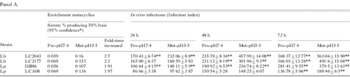
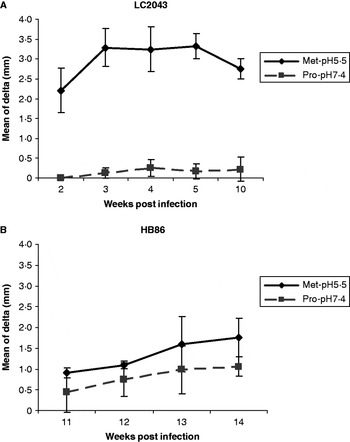
For the 4 representative strains used here, enrichment and purification of metacyclics parasite forms (by parasite culture at pH 5·5 and using the Ficoll centrifugation gradient, respectively) were verified by comparing 3 parameters between Pro-pH7·4 and Met-pH5·5 parasites (Table 1). (1) The complement lysis test showed that twice as many Met-pH5·5 parasites survived serum concentrations compared to Pro-pH7·4 parasites. (2) Macrophage in vitro infection showed infection indices that were mostly higher for Met-pH5·5 parasites, and that the effect was more pronounced for the 48 h and 72 h infection groups, with differences that were statistically significant for all strains except for LCA08 at 48 h post-infection (95% confidence interval). (3) In vivo infection of hamsters revealed that lesions induced by Met-pH5·5 parasites were larger than those induced by Pro-pH7·4 parasites. For LC2043, differences were statistically significant (ANOVA, P<0·05) from the 2nd week post-infection until the end of the experiment (6 weeks follow up). Metacyclics of HB86 also showed significant differences at the 14th week post-infection (ANOVA, P<0·05). The maximum difference between Met-pH5·5 and Pro-pH7·4 size lesion was 3·16 mm (5th week post-infection) and 0·70 mm (14th week post-infection) for LC2043 and HB86 respectively. The difference between lesion pattern produced by Pro-pH7·4 and Met-pH5·5 parasites was larger in L. (V.) braziliensis than in L. (V.) peruviana, which could be explained by a higher proportion of metacyclics in Pro-pH7·4 parasites of HB86 used for the infection than the corresponding parasites of LC2043.
Results for in vitro (panel A) and in vivo (panel B) infections using stationary non-purified promastigotes (Pro-pH7·4) and purified metacyclics (Met-pH5·5) from Leishmania (Viannia) braziliensis (Lb) and L. (Viannia) peruviana (Lp)
(*Program Probit SPSS 3.2 used to calculate the LD50 for the complement test. ** Differences in the in vitro infections using 95% confidence interval. *** Differences statistically significant in the in vivo infections (ANOVA; P<0·05, n=5).)
Panel A.

Panel B.

Infection differences between L. (V.) braziliensis and L. (V.) peruviana representative strains
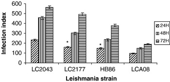
To compare virulence between L. (V.) braziliensis and L. (V.) peruviana, we compared in vitro and in vivo results obtained with Met-pH5·5 parasites from the representative strains assayed here. These representative strains were chosen because of their genomic and genetic characteristics very well described elsewhere (Dujardin et al. Reference Dujardin, Dujardin, Tibayrenc, Timperman, De Doncker, Jacquet, Arevalo, Llanos-Cuentas, Guerra, Bermudez, Hamers and Le Ray1995b; Bañuls et al. Reference Bañuls, Dujardin, Guerrini, De Doncker, Jaquet, Arevalo, Noël, Le Ray and Tibayrenc2000). (i) The in vitro infectivity was expressed as the infection index rate at 24 h, 48 h, and 72 h post-infection of each representative strain. In all 3 independent experiments for each strain, the 4 strains ranked as following (from high to low): LC2043, LC2177, HB86 and LCA08 (Table 1 and Fig. 1). Noteworthy was that, despite the fact that the experiments were carried out with different culture batches of parasites and macrophages, the in vitro infection parameters were quite reproducible. Thus, for example, the average number of amastigotes per macrophage followed the same trend, at 72 h the values were 6·83±0·73 (LC2043), 6·3±0·26 (LC2177), 5·3±0·26 (HB86) and 3·2±0·26 (LCA08). A similar trend was observed with the percentage of infected macrophages (data not shown). Statistically, infection indices differed significantly between the 4 strains. This was observed at different times except for LC2177 and HB86 at 24 h post-infection (95% confidence interval). (ii) The in vivo infection experimental model was carried out with the more infective representative strain from each species observed after the in vitro experiments. In the case of the LC2043-infected animals, the development of the lesion was faster (2 weeks post-infection) than in the animals infected with HB86 (14 weeks post-infection). In addition, LC2043 lesions were larger in size than those produced by HB86 (maximum size lesion: 3·32 mm and 1·76 mm respectively) (Fig. 2A and B respectively). To assure that the lesion was due to the presence of the parasite, an aspirate from the lesion was taken after 1 month of infection and placed on blood agar medium, cultures were positives after 1 week of incubation (data not shown).
In vitro infection of macrophages with purified metacyclic promastigotes: infection indices at different times post-infection for the L. (V.) braziliensis (LC2043 and LC2177) and L. (V.) peruviana (HB86 and LCA08) strains. For a given time-point, differences between strains were statistically different (P=0·000, Mann-Whitney test) except for LC2177 and HB86 at 24 h (*).

In vivo infections of hamsters using purified metacyclic promastigotes from LC2043 (A) and HB86 (B). Differences were statistically significant from 2 weeks post-infection until the end of the experiment for LC2043 and only at week 14 post-infection for HB86 (ANOVA; P<0·05, n=5).

DISCUSSION
In the presented work, an experimental model was adapted and evaluated for analysing the in vitro and in vivo infectivity of L. (V.) braziliensis and L. (V.) peruviana representative strains. These two parasite species cause dramatically different pathologies in humans, despite their great genetic similarity.
The use of purified metacyclic promastigotes is very important for infectivity studies, not only because they indeed represent the infective forms inoculated by the vector, but also because they represent a well-defined and relatively homogeneous population. Such a population is essential to compare different Leishmania strains at a corresponding developmental stage. So far, most studies on metacyclogenesis have focused on parasites of subgenus Leishmania: L. (L.) major, L. (L.) donovani, L. (L.) tropica, L. (L.) infantum and L. (L.) mexicana (Bates and Tetley, Reference Bates and Tetley1993; Zakai et al. Reference Zakai, Chance and Bates1998). In comparison there are fewer reports on parasites of the subgenus Viannia (Almeida et al. Reference Almeida, Cuba, de Sa, Pharoah, Howard and Miles1993; Pinto-da-Silva et al. Reference Pinto-da-Silva, Camurate, Costa, Oliveira, da Cunha-e-Silva and Saraiva2002), despite the fact that the latter comprise some species highly pathogenic for humans. Our experimental procedure was adapted from that evaluated in subgenus Leishmania species and combined the acidic induction of metacyclogenesis (Bates and Tetley, Reference Bates and Tetley1993; Zakai et al. Reference Zakai, Chance and Bates1998) with the purification of metacyclics by a Ficoll gradient centrifugation method (Späth and Beverley, Reference Späth and Beverley2001). Production of L. (V.) braziliensis and L. (V.) peruviana metacyclics was verified by measuring 3 parameters (and comparison with non-purified stationary promastigotes): (i) increased resistance to complement lysis, (ii) higher infection indices in macrophages and (iii) larger lesions in hamsters. All parameters indicated an homogeneous metacyclic population in the strains tested here. Previous work done by our group identified a molecular marker (the infective-insect stage protein also known as the Meta 1 protein) using metacyclic-enriched preparations (without purification) of L. (V.) braziliensis LC2043 (also produced by acid induction, Gamboa et al. Reference Gamboa, Van Eys, Victoir, Torres, Adaui, Arevalo and Dujardin2007), one of the strains here studied. This marker, and maybe others identified in the future, might be studied by measuring their expression profile using purified preparations of metacyclic parasites from this and other Leishmania species from subgenus Viannia using the methodology described here (manuscript in preparation).
Using purified metacyclics as sources of infection, comparison of in vitro and in vivo patterns could be made between L. (V.) braziliensis and L. (V.) peruviana representative strains, giving us the real image of the infection capacity of these strains, that normally hide when mixed parasite populations are used. In vitro (tested for 4 representative strains) as well as in vivo (2 representative strains), it was clear that for the representative strains tested here, L. (V.) braziliensis was more infective than L. (V.) peruviana, showing higher infection indices and faster appearance and larger size of lesions respectively. This correlates with the clinical outcome observed for L. (V.) braziliensis, associated with severe cutaneous and mucosal lesions in the Amazonian basin (Guerra, Reference Guerra, Walton, Wijeyaratne and Modabber1988). Meanwhile, L. (V.) peruviana produces benign cutaneous lesions, never associated with the mucosal phenotype (Lucas et al. Reference Lucas, Franke, Cachay, Tejada, Cruz, Kreutzer, Barker, McCann and Watts1998). Thus our model was sensitive enough to detect phenotypic differences among the representative strains. Our results should be confirmed with a larger sample of the 2 species (from different geographical origin and clinical outcome); but at this preliminary stage, they already converged with different pathogenicities in humans: L. (V.) braziliensis, the most pathogenic in humans, being also more infective in our experimental models. Considering the higher average number of amastigotes observed by in vitro-infected macrophages in L. (V.) braziliensis, it would be interesting to measure if this is related to an increased resistance to nitric oxide. This feature could indeed confer a survival benefit for the parasites inside the macrophage and was shown to correlate with disease severity in American tegumentary leishmaniasis (Giudice et al. Reference Giudice, Camada, Leopoldo, Pereira, Riley, Wilson, Ho, de Jesus, Carvalho and Almeida2007).
A more extensive analysis of the in vitro data revealed another interesting result. Indeed, the 4 strains ranked from high to low infectivity as follows: LC2043, LC2177, HB86 and LCA08. It is noteworthy that this corresponded to the gradient of karyotype dissimilarity previously observed among the same strains, which correlated itself with the distance between their geographical origins (Dujardin et al. Reference Dujardin, Llanos-Cuentas, Cáceres, Arana, Dujardin, Guerrini, Gomez, Arroyo, De Doncker, Jacquet, Hamers, Guerra, Le Ray and Arevalo1993, Reference Dujardin, Dujardin, Tibayrenc, Timperman, De Doncker, Jacquet, Arevalo, Llanos-Cuentas, Guerra, Bermudez, Hamers and Le Ray1995b, Reference Dujardin, Dujardin, Tibayrenc, Timperman, De Doncker, Jacquet, Arevalo, Llanos-Cuentas, Guerra, Bermudez, Hamers and Le Ray1998). We hypothesized that this dynamic picture would reflect a recent speciation of L. (V.) peruviana from L. (V.) braziliensis (Dujardin et al. Reference Dujardin, Llanos-Cuentas, Cáceres, Arana, Dujardin, Guerrini, Gomez, Arroyo, De Doncker, Jacquet, Hamers, Guerra, Le Ray and Arevalo1993, Reference Dujardin, Bañuls, Victoir, De Doncker, Arevalo, Llanos-Cuentas, Tibayrenc and Le Ray1995a, Reference Dujardin, Dujardin, Tibayrenc, Timperman, De Doncker, Jacquet, Arevalo, Llanos-Cuentas, Guerra, Bermudez, Hamers and Le Rayb, Reference Dujardin, Bañuls, Arevalo, Tibayrenc and Le Ray1998, Reference Dujardin, Henriksson, Victoir, Brisse, Gamboa, Arevalo and Le Ray2000, Reference Dujardin, Victoir, De Doncker, Guerbouj, Arevalo and Le Ray2002). According to our hypothesis, a L. (V.) braziliensis-like parasite would have colonized the Pacific slopes of the Andes, in the North of Peru (close to the lowest pass across the Andes along the whole Peruvian territory). Then, through a North-South migration and isolation, it would have increased its genomic differentiation. Further work on a larger set of strains is needed to verify this possible correlation between infectivity and geographical location of parasite populations. This may open new perspectives to understand the process of speciation in Leishmania and its implications in terms of pathogenicity.
In conclusion, we thus have a model able to demonstrate, in a reproducible way, differences in infectivity among representative strains from Leishmania of subgenus Viannia. Our results support the role of the parasite in the differences in human pathogenicity observed between L. (V.) braziliensis and L. (V.) peruviana. However, further extrapolation should be made with great caution. There is still a huge gap between experimental models and the real situation in humans. In vitro models lack the immunological context. On the other hand immunological response may be different between animals and humans and, last but not least, there is a major difference between an infection caused by a needle or by a sandfly bite (immuno-modulatory effect of saliva) (Almeida et al. Reference Almeida, Cuba, de Sa, Pharoah, Howard and Miles2003). Further work is thus needed to upgrade in vitro as well as in vivo models and gather information to extrapolate the data to human infection.
This study was funded by WHO-TDR, Research Training Grants (M8/181/4/G.261, ID 990815), UNESCO-L'OREAL fellowship for Women in Science 2003, EC (INCO-CT2005-015407), and by the Directorate-General for Development Cooperation (DGDC) of the Belgian Government (framework agreement 02, project 95501).