Introduction
Dementia is a progressive neurodegenerative disease, and is set to become the developed world's largest socio-economic healthcare burden over coming decades. According to different estimates, between 2 and 10% of all cases of dementia start before the age of 65 and the prevalence doubles with every 5-year increment in age after 65. The number of people globally who are living with dementia is estimated to be 35.6 million, and epidemiological studies indicate that this number is expected to grow at an alarming rate. It is estimated that numbers will nearly double every 20 years, to 65.7 million in 2030 and 115.4 million in 2050 (Alzheimer's Disease International, World Alzheimer Report 2010). Currently, in the UK, there are 800,000 people living with dementia and over 17,000 younger people (65 years of age or below) have dementia. As the size of the population is growing and people are living longer, by 2021 there will be over 1 million people living with dementia in the UK. This is costing the NHS, local authorities and families £23 billion a year and this will grow to £27 billion by 2018 (Dementia 2012: A national challenge).
The term ‘dementia’ is used to describe a syndrome that may be caused by a number of illnesses in which there is progressive decline in multiple areas of function, including decline in memory, reasoning, communication skills and the ability to carry out activities of daily living. Alongside this decline, individuals may develop behavioural and psychological symptoms such as depression, psychosis, aggression and wandering, which cause problems in themselves, which complicate care, and which can occur at any stage of the illness. Alzheimer's disease is the most common form of dementia, accounting for 62% of dementia cases. Other forms of dementia include vascular dementia (17%), mixed (AD and vascular) dementia (10%), dementia with Lewy bodies (4%), fronto-temporal dementia (2%) and Parkinson's dementia (2%) (Dementia 2012: A national challenge).
AD is a complex neurodegenerative disorder of which the exact cause is still unclear. There are currently no effective disease-modifying therapies and the only available treatments are symptomatic. The incurable nature of AD presents a great challenge for healthcare systems and considerable resources have focused on developing therapeutic agents to directly delay the pathophysiological processes. As we persist in the search for a cure, there has been significant progress in the understanding of the pathophysiological events that cause the disease with a focus on amyloid formation. The underlying pathology of Alzheimer's disease is believed to precede the onset of clinical symptoms by many years.Reference Thal, Rub, Orantes and Braak1 The clinical diagnosis of AD is defined by NINDS-ADRDAReference McKhann, Drachman, Folstein, Katzman, Price and Stadlan2 and DSM-IV criteria. However, these criteria have been revised to include the biomarkers, and revised criteria have been developed by the joint National Institute of Ageing and Alzheimer's Association (NIA-AA criteria) for the purpose of research.Reference McKhann, Knopman, Chertkow, Hyman, Jack, Kawas, Klunk, Koroshetz, Manly, Mayeux, Mohs, Morris, Rossor, Scheltens, Carrillo, Thies, Weintraub and Phelps3
Mild cognitive impairment (MCI) refers to a transitional state between the cognition of normal ageing and mild dementia. Amnestic mild cognitive impairment (aMCI) refers to individuals who have isolated impairment of short-term recall but otherwise intact higher function and do not meet DSM-IV criteria for dementia. The initial criteria were set out by Petersen et al.,Reference Petersen, Smith, Waring, Ivnik, Tangalos and Kokmen4 and include: (1) memory complaint, preferably corroborated by an informant; (2) impaired memory function for age and education; (3) preserved general cognitive function; and (4) intact activities of daily living. Patients with aMCI are at a higher risk of developing Alzheimer's disease, and 10–15% convert to AD per year, compared with healthy control subjects who convert at a rate of 1–2% per year.Reference Petersen, Smith, Waring, Ivnik, Tangalos and Kokmen4–Reference Petersen, Doody, Kurz, Mohs, Morris, Rabins, Ritchie, Rossor, Thal and Winblad6 NIA-AA criteria divide MCI into early and late MCI depending on the appearance of imaging and CSF biomarkers.
Amyloid cascade hypothesis
The most widely accepted hypothesis to describe the aetiology of AD remains the amyloid cascade hypothesis. Within this hypothesis it is assumed that the amyloid β peptide (Aβ) is the prime initiator of AD pathology, where altered amyloid precursor protein (APP) processing leads to generation of toxic Aβ species. Aβ is considered as the primary influence that triggers significant biochemical and histological alterations driving tau hyperphosphorylation and other downstream events including inflammation, ultimately resulting in neuronal dysfunction and death. These changes manifest clinically in the form of dementia, which is irreversibly progressive.Reference Hardy and Selkoe7 In line with the amyloid cascade hypothesis, anti-amyloid therapies have been tried in AD, without great success. This could be because amyloid deposition putatively causes severe damage to neurons many years ahead of the clinical onset of dementia, hence a therapeutic window of opportunity may have already been lost, where the neuronal damage has already set in. Hence anti-amyloid therapies are now focusing on MCI stage. Interestingly, pathological studies have demonstrated that amyloid deposition can also occur in normal, healthy ageing. However, pathological studies that are almost always cross-sectional suffer from a limitation in that no longitudinal outcome data are attainable. This implies that it is virtually impossible to determine the sequence of events occurring early on in the disease process, and one cannot study the temporal relationship between key events. In quest of the pathophysiological conspirators in the disease process, the onus has now shifted towards identifying the disease early on in its pathogenesis, and to accurately monitor its progression. A major advancement in this arcane field of medicine has been the timely advent of amyloid imaging, which allows us to critically evaluate the complex relationship between Aβ aggregation and AD in vivo. Most importantly, amyloid imaging has the potential to detect Aβ in mildly affected as well as asymptomatic individuals, when the therapeutic window of opportunity might still be open to pharmacological intervention. In this review we focus on amyloid imaging in different diseases and the advances made in this field.
Search strategy and selection criteria
PubMed was searched for papers that were published before June 2012. The following key words were used in the search: ‘positron emission tomography’, ‘amyloid imaging’, ‘[11C]PIB’, ‘dementia’, ‘Alzheimer's disease’, ‘mild cognitive impairment’, ‘Lewy body disease’, ‘frontotemporal dementia’ and ‘non-demented’. The selected articles were examined for additional references not revealed by automated searches. Only articles published in English were considered.
Amyloid imaging
Amyloid imaging is the principal method for the detection of Aβ pathology; however, it is well known that no in vivo assessment of pathology is as sensitive as post-mortem histological and biochemical assessment.Reference Klunk8 During the past 8 years amyloid imaging has established itself alongside magnetic resonance imaging (MRI) and fluorodeoxyglucose positron emission tomography (FDG-PET) as an important neuroimaging tool for the investigation of brain ageing and dementia.Reference Herholz, Carter and Jones9
Amyloid imaging agents will detect beta-sheet rich fibrillar deposits of Aβ protein in plaques in AD and cerebrovascular amyloid (CAA).Reference Klunk8 Positron emission tomography (PET) is a well-established imaging modality. PET employs biologically active molecules in micromolar or nanomolar concentrations that have been labelled with short-lived positron-emitting isotopes such as Reference Koole, Lewis, Buckley, Nelissen, Vandenbulcke, Brooks, Vandenberghe and Van Laere15O (half-life 2 min), Reference Klunk, Engler, Nordberg, Wang, Blomqvist, Holt, Bergström, Savitcheva, Huang, Estrada, Ausén, Debnath, Barletta, Price, Sandell, Lopresti, Wall, Koivisto, Antoni, Mathis and Långström11C (20 min) and 18F (110 min). The very short half-lives of O-15 and C-11 restrict their use to fully equipped PET centres with a cyclotron and radiopharmaceutical laboratory. F-18-labelled tracers can be produced in specialized centres and distributed regionally to hospitals running a PET scanner only. The physical characteristics of these isotopes and the molecular specificity of labelled molecules, combined with the high detection efficacy of modern PET scanners, provide sensitivity for human in vivo measurement of indicator concentrations that is several orders of magnitude higher than with the other imaging techniques.Reference Herholz, Carter and Jones9
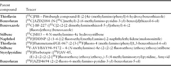
Tracers that have so far progressed into humans include [18F]FDDNP,Reference Shoghi-Jadid, Small, Agdeppa, Kepe, Ercoli, Siddarth, Read, Satyamurthy, Petric, Huang and Barrio10 [11C]PIB,Reference Klunk, Engler, Nordberg, Wang, Blomqvist, Holt, Bergström, Savitcheva, Huang, Estrada, Ausén, Debnath, Barletta, Price, Sandell, Lopresti, Wall, Koivisto, Antoni, Mathis and Långström11 [11C]AZD2184,Reference Nyberg, Jönhagen, Cselényi, Halldin, Julin, Olsson, Freund-Levi, Andersson, Varnäs, Svensson and Farde12 [11C]BF-227,Reference Kudo, Okamura, Furumoto, Tashiro, Furukawa, Maruyama, Itoh, Iwata, Yanai and Arai13 [11C]SB13,Reference Verhoeff, Wilson, Takeshita, Trop, Hussey, Singh, Kung, Kung and Houle14 [18F]Flutemetamol,Reference Koole, Lewis, Buckley, Nelissen, Vandenbulcke, Brooks, Vandenberghe and Van Laere15 [18F]AV1 ([18F]Florbetaben),Reference Rowe, Ackerman, Browne, Mulligan, Pike, O'Keefe, Tochon-Danguy, Chan, Berlangieri, Jones, Dickinson-Rowe, Kung, Zhang, Kung, Skovronsky, Dyrks, Holl, Krause, Friebe, Lehman, Lindemann, Dinkelborg, Masters and Villemagne16 [18F]FlorbetapirReference Zhang, Oya, Kung, Hou, Maier and Kung17 and [18F]AZD4694Reference Juréus, Swahn, Sandell, Jeppsson, Johnson, Johnström, Neelissen, Sunnemark, Farde and Svensson18, Reference Cselényi, Jönhagen, Forsberg, Halldin, Julin, Schou, Johnström, Varnäs, Svensson and Farde19 (Table 1). Of these tracers, only PIB has received an extensive biochemical profiling of its binding to a range of in vitro amyloid filamentsReference Klunk, Wang, Huang, Debnath, Holt, Shao, Hamilton, Ikonomovic, DeKosky and Mathis20–Reference Fodero-Tavoletti, Smith, McLean, Adlard, Barnham, Foster, Leone, Perez, Cortés, Culvenor, Li, Laughton, Rowe, Masters, Cappai and Villemagne22 and neuropathological lesions.Reference Fodero-Tavoletti, Smith, McLean, Adlard, Barnham, Foster, Leone, Perez, Cortés, Culvenor, Li, Laughton, Rowe, Masters, Cappai and Villemagne22, Reference Ye, Velasco, Fraser, Beach, Sue, Osredkar, Libri, Spillantini, Goedert and Lockhart23
Amyloid PET tracers

PIB
Currently the most widely studied PET tracer and an analogue of the amyloid-binding dye thioflavin-T is Pittsburgh Compound B (PIB). Modification of the polar amyloid binding histological dye, thioflavin-T, led to the finding that neutral benzothiazoles also bind to amyloid with high affinity and additionally cross the blood–brain barrier. It is shown benzothiazole amyloid binding agent 2-(4’-methyl amino phenyl) benzothiazole (called BTA-1) and related compounds bind to amyloid with low nanomolar affinity, enter the brain at levels sufficient for yielding a PET signal and clear rapidly from normal brain tissue. At the low nanomolar concentrations typically administered during PET studies, BTA-1 binds to extracellular amyloid plaques in post-mortem brain slices but not to intracellular neurofibrillary tangles or Lewy bodies. In vitro studies suggest that, while BTA-1 binds to fibrillar Aβ deposits found in the cortex and striatum, it does not bind to amorphous Aβ deposits such as those found in cerebellum.Reference Klunk, Wang, Huang, Debnath, Holt, Shao, Hamilton, Ikonomovic, DeKosky and Mathis20 The first direct in vivo visualization of brain amyloid was carried out in mild AD patients, where uptake patterns were in keeping with amyloid plaque deposition described in post-mortem studies of AD brains.Reference Klunk, Engler, Nordberg, Wang, Blomqvist, Holt, Bergström, Savitcheva, Huang, Estrada, Ausén, Debnath, Barletta, Price, Sandell, Lopresti, Wall, Koivisto, Antoni, Mathis and Långström11 [11C]PIB binds to fibrillar amyloid with no detectable binding to soluble Aβ forms or neurofibrillary tangles at PET tracer concentrations.Reference Mathis, Lopresti and Klunk24
The aim of the present review is to discuss the current state of amyloid imaging, focusing on [11C]PIB in AD, MCI and healthy control subjects.
Amyloid imaging in Alzheimer's disease
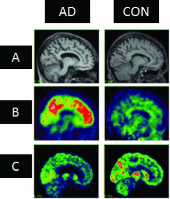
Within the past decade, several [11C]PIB-PET studies performed on AD subjects have demonstrated 2-fold increases or more in [11C]PIB retention in predominantly cortical areas compared with healthy control subjects (Figure 1), as judged either by visual assessment or quantitative analysis of tracer uptake.Reference Klunk, Engler, Nordberg, Wang, Blomqvist, Holt, Bergström, Savitcheva, Huang, Estrada, Ausén, Debnath, Barletta, Price, Sandell, Lopresti, Wall, Koivisto, Antoni, Mathis and Långström11, Reference Jack, Lowe, Senjem, Weigand, Kemp, Shiung, Knopman, Boeve, Klunk, Mathis and Petersen25–Reference Rowe, Ng, Ackermann, Gong, Pike, Savage, Cowie, Dickinson, Maruff, Darby, Smith, Woodward, Merory, Tochon-Danguy, O'Keefe, Klunk, Mathis, Price, Masters and Villemagne27 More specifically, using the region of interest and/or voxel-wise analysis, the highest tracer uptake is consistently observed in prefrontal cortex, precuneus and posterior cingulate cortex, followed intimately by lateral parietal and temporal cortex along with striatum in AD subjects compared with healthy controls.Reference Klunk, Engler, Nordberg, Wang, Blomqvist, Holt, Bergström, Savitcheva, Huang, Estrada, Ausén, Debnath, Barletta, Price, Sandell, Lopresti, Wall, Koivisto, Antoni, Mathis and Långström11, Reference Jack, Lowe, Senjem, Weigand, Kemp, Shiung, Knopman, Boeve, Klunk, Mathis and Petersen25–Reference Kemppainen, Aalto, Wilson, Någren, Helin, Brück, Oikonen, Kailajärvi, Scheinin, Viitanen, Parkkola and Rinne28 This is in keeping with the neuropathological observations of Aβ fibrillar plaques.Reference Thal, Rub, Orantes and Braak1, Reference Braak and Braak29 A relatively lower tracer uptake is found in the occipital cortex, globus pallidus and thalamus, whilst primary sensorimotor and visual cortices in addition to medial temporal lobe regions are relatively spared.Reference Rabinovici and Jagust30 Cerebellar grey matter exhibits very little if any [11C]PIB binding, allowing the cerebellum to be used frequently as a reliable reference region. Although graphical Logan analysis which uses arterial plasma input function has been suggested to be the gold standard for [11C]PIB-PET analysis, the use of target to cerebellum uptake ratios generates comparable results to Logan plot derived distribution volume ratios.Reference Lopresti, Klunk, Mathis, Hoge, Ziolko, Lu, Meltzer, Schimmel, Tsopelas, DeKosky and Price31, Reference Price, Klunk, Lopresti, Lu, Hoge, Ziolko, Holt, Meltzer, DeKosky and Mathis32 It is a very effective method, which is simpler to generate and shortens the scan time considerably. Interestingly, pathological studies examining cases with Prion disease have demonstrated significant amyloid deposits in the cerebellum.Reference Watanabe and Duchen33, Reference Mead, Poulter, Beck, Webb, Campbell, Linehan, Desbruslais, Joiner, Wadsworth, King, Lantos and Collinge34 Moreover, a recent [11C]PIB study reported significant amyloid deposition in the cerebellum of familial AD subjects.Reference Klunk, Price, Mathis, Tsopelas, Lopresti, Ziolko, Bi, Hoge, Cohen, Ikonomovic, Saxton, Snitz, Pollen, Moonis, Lippa, Swearer, Johnson, Rentz, Fischman, Aizenstein and DeKosky35 The cerebellum especially in these cases, is not always a suitable non-specific reference for quantification of [11C]PIB-PET, hence the pons should be considered.Reference Edison, Hinz, Ramlackhansingh, Thomas, Gelosa, Archer, Turkheimer and Brooks36, Reference Weiner, Veitch, Aisen, Beckett, Cairns, Green, Harvey, Jack, Jagust, Liu, Morris, Petersen, Saykin, Schmidt, Shaw, Siuciak, Soares, Toga and Trojanowski37
Representative images from Alzheimer's disease (AD) subjects and healthy control (CON) subjects showing median sagittal MRI (A) and PET with [11C]PIB (B) and [18F]FDG (C). Increased [11C]PIB retention is observed in AD subjects, with the highest tracer retention observed in frontal, temporal and posterior cingulate cortices compared with healthy control subjects (B). Glucose hypometabolism is most consistently observed in the temporoparietal regions in AD subjects compared with healthy controls (C).

Several longitudinal studies using [11C]PIB have been conducted to monitor the disease progression in AD. Engler et al. re-scanned 16 patients with mild AD who were included in their original study after 2.0 ± 0.5 years.Reference Engler, Forsberg, Almkvist, Blomquist, Larsson, Savitcheva, Wall, Ringheim, Långström and Nordberg38 [11C]PIB retention remained stable between the baseline and the 2-year follow-up period, suggesting the notion that amyloid deposition reaches a plateau by the early clinical stages of the disease, in line with the amyloid cascade hypothesis. There was a clear dissociation between the amyloid load, which remained relatively stable with minor variations, and clinical status, which continued to decline in a number of subjects. Furthermore, the cognitive decline was more accurately associated and correlated with the progressive deterioration in glucose metabolism measured by [18F]FDG.Reference Engler, Forsberg, Almkvist, Blomquist, Larsson, Savitcheva, Wall, Ringheim, Långström and Nordberg38 Jack and colleagues reported similar results when examining alterations in [11C]PIB uptake and brain atrophy over a year in a study comprising 21 healthy controls, 32 MCI and 8 AD subjects. They found small, insignificant increases in [11C]PIB retention in AD; however, this value was identical to the other diagnostic groups across the same brain regions. In contrast, the rates of brain atrophy as measured by changes in ventricular volume differed significantly between the groups and perhaps not surprisingly were highest in AD followed sequentially by MCI and controls. Moreover, the clinical decline revealed by longitudinal analysis strongly correlated with brain atrophy rates but not with changes in global [11C]PIB, and no association between brain atrophy and global [11C]PIB rates was detected.Reference Jack, Lowe, Weigand, Wiste, Senjem, Knopman, Shiung, Gunter, Boeve, Kemp, Weiner and Petersen39 The above observations are in agreement with other follow-up [11C]PIB studies, which found no elevations in [11C]PIB uptake after 2 yearsReference Scheinin, Aalto, Koikkalainen, Lötjönen, Karrasch, Kemppainen, Viitanen, Någren, Helin, Scheinin and Rinne40 and 5 years.Reference Kadir, Almkvist, Forsberg, Wall, Engler, Långström and Nordberg41 In accordance with previous studies,Reference Engler, Forsberg, Almkvist, Blomquist, Larsson, Savitcheva, Wall, Ringheim, Långström and Nordberg38 Kadir et al.Reference Kadir, Almkvist, Forsberg, Wall, Engler, Långström and Nordberg41 found considerable variability between subjects, with some demonstrating small increases in [11C]PIB and others exhibiting reduced [11C]PIB uptake. The latter finding is interesting and warrants further mention. Although the exact proportionality of fibrillar and oligomeric species of Aβ in AD remains ambiguous, the existence of a continuous dynamic equilibrium between amyloid plaques and oligomers cannot be ruled out.Reference Benilova, Karran and De Strooper42 Since an exchange between fibrillar and oligomeric Aβ surrounding plaques has been reported in transgenic APP mice,Reference Koffie, Meyer-Luehmann, Hashimoto, Adams, Mielke, Garcia-Alloza, Micheva, Smith, Kim, Lee, Hyman and Spires-Jones43 this suggests that a similar mechanism might be operant in AD and might be the reason for the observed decrease in levels of fibrillar Aβ forms.Reference Yan, Bero, Cirrito, Xiao, Hu, Wang, Gonzales, Holtzman and Lee44 More interestingly, recent data do indicate that benign insoluble aggregates may slowly leach to form toxic oligomers.Reference Meyer-Luehmann, Spires-Jones, Prada, Garcia-Alloza, de Calignon, Rozkalne, Koenigsknecht-Talboo, Holtzman, Bacskai and Hyman45 One of the caveats of the Kadir et al. study is that no correction for partial volume effects was performed and it cannot be ruled out that a proportion of the lower PIB binding observed may be due to the atrophy in later stages of the disease.Reference Kadir, Almkvist, Forsberg, Wall, Engler, Långström and Nordberg41
Amyloid imaging in mild cognitive impairment
It is believed that amyloid deposition occurs decades before any cognitive changes at a clinical level are established and begins to accelerate in the pre-clinical stage of AD, before reaching a relative plateau at the onset of symptoms. Presumably, MCI individuals with underlying AD pathology are at an increased risk of progressing to dementia. Amyloid imaging can potentially identify early AD pathology in MCI and can help predict the rate of progression from MCI to dementia of AD type or other dementias.
Several studies in MCI have demonstrated that [11C]PIB uptake is intermediate between AD and controls.Reference Rowe, Ng, Ackermann, Gong, Pike, Savage, Cowie, Dickinson, Maruff, Darby, Smith, Woodward, Merory, Tochon-Danguy, O'Keefe, Klunk, Mathis, Price, Masters and Villemagne27, Reference Okello, Edison, Archer, Turkheimer, Kennedy, Bullock, Walker, Kennedy, Fox, Rossor and Brooks46–Reference Mormino, Kluth, Madison, Rabinovici, Baker, Miller, Koeppe, Mathis, Weiner and Jagust48 In the majority of studies, [11C]PIB binding in MCI subjects is bi-modally distributed, with one subset of subjects demonstrating AD-like uptake levels, and the other showing low, control-like [11C]PIB values.Reference Jack, Lowe, Senjem, Weigand, Kemp, Shiung, Knopman, Boeve, Klunk, Mathis and Petersen25, Reference Rowe, Ng, Ackermann, Gong, Pike, Savage, Cowie, Dickinson, Maruff, Darby, Smith, Woodward, Merory, Tochon-Danguy, O'Keefe, Klunk, Mathis, Price, Masters and Villemagne27, Reference Lopresti, Klunk, Mathis, Hoge, Ziolko, Lu, Meltzer, Schimmel, Tsopelas, DeKosky and Price31, Reference Kemppainen, Aalto, Wilson, Någren, Helin, Brück, Oikonen, Kailajärvi, Scheinin, Viitanen, Parkkola and Rinne47–Reference Pike, Savage, Villemagne, Ng, Moss, Maruff, Mathis, Klunk, Masters and Rowe51 Depending on the criteria used to diagnose MCI and the threshold used to define [11C]PIB positivity, around 52–87% of MCI subjects demonstrate elevated [11C]PIB binding.Reference Jack, Lowe, Senjem, Weigand, Kemp, Shiung, Knopman, Boeve, Klunk, Mathis and Petersen25, Reference Rowe, Ng, Ackermann, Gong, Pike, Savage, Cowie, Dickinson, Maruff, Darby, Smith, Woodward, Merory, Tochon-Danguy, O'Keefe, Klunk, Mathis, Price, Masters and Villemagne27, Reference Lopresti, Klunk, Mathis, Hoge, Ziolko, Lu, Meltzer, Schimmel, Tsopelas, DeKosky and Price31, Reference Kemppainen, Aalto, Wilson, Någren, Helin, Brück, Oikonen, Kailajärvi, Scheinin, Viitanen, Parkkola and Rinne47–Reference Pike, Savage, Villemagne, Ng, Moss, Maruff, Mathis, Klunk, Masters and Rowe51 It has been established that individuals meeting the criteria for amnestic MCI are more likely to be [11C]PIB positive than patients diagnosed with non-amnestic MCI.Reference Forsberg, Engler, Almkvist, Blomquist, Hagman, Wall, Ringheim, Långström and Nordberg50, Reference Pike, Savage, Villemagne, Ng, Moss, Maruff, Mathis, Klunk, Masters and Rowe51 Moreover, ApoE4 carriers have a greater propensity to accumulate amyloid deposits and hence are more likely to show [11C]PIB positivity compared with non-ApoE4 carriers.Reference Pike, Savage, Villemagne, Ng, Moss, Maruff, Mathis, Klunk, Masters and Rowe51, Reference Okello, Koivunen, Edison, Archer, Turkheimer, Någren, Bullock, Walker, Kennedy, Fox, Rossor, Rinne and Brooks52 Interestingly, cross-sectional analysis of [11C]PIB-positive compared with [11C]PIB-negative MCI subjects reveal lower performance on episodic memory tasks in a few studiesReference Pike, Savage, Villemagne, Ng, Moss, Maruff, Mathis, Klunk, Masters and Rowe51 but not others.Reference Jack, Lowe, Senjem, Weigand, Kemp, Shiung, Knopman, Boeve, Klunk, Mathis and Petersen25, Reference Rowe, Ng, Ackermann, Gong, Pike, Savage, Cowie, Dickinson, Maruff, Darby, Smith, Woodward, Merory, Tochon-Danguy, O'Keefe, Klunk, Mathis, Price, Masters and Villemagne27
To date, a consensus regarding the cut-off values used to determine [11C]PIB positivity or negativity has not been reached due to paucity of available data on older cognitively normal individuals. This is because classification of subjects as either being [11C]PIB-positive or -negative relies on the distribution of [11C]PIB values present in the healthy control group under investigation. This may be the reason for the generation of different cut-off values and the lack of consensus reported amongst groups. Some groups treat amyloid deposition as a continuous variable,Reference Mormino, Kluth, Madison, Rabinovici, Baker, Miller, Koeppe, Mathis, Weiner and Jagust48, Reference Pike, Savage, Villemagne, Ng, Moss, Maruff, Mathis, Klunk, Masters and Rowe51 whereas other groups have dichotomized subjects into [11C]PIB-positive or -negative groups albeit with variability in their categorzation approaches.Reference Jack, Lowe, Senjem, Weigand, Kemp, Shiung, Knopman, Boeve, Klunk, Mathis and Petersen25, Reference Rowe, Ng, Ackermann, Gong, Pike, Savage, Cowie, Dickinson, Maruff, Darby, Smith, Woodward, Merory, Tochon-Danguy, O'Keefe, Klunk, Mathis, Price, Masters and Villemagne27, Reference Aizenstein, Nebes, Saxton, Price, Mathis, Tsopelas, Ziolko, James, Snitz, Houck, Bi, Cohen, Lopresti, DeKosky, Halligan and Klunk53 Although different research groups have different [11C]PIB cut-off values, most are within a few percentage points of each other. Recent analysis in conjunction with pathological studies suggest a biological relevance of slight [11C]PIB accumulations in ageing.Reference Mormino, Brandel, Madison, Rabinovici, Marks, Baker and Jagust54
Longitudinal studies investigating [11C]PIB uptake in MCI subjects have yielded mixed results. A recent 2-year longitudinal study involving 29 amnestic MCI subjects did not demonstrate any significant elevations in [11C]PIB uptake despite significant progression in hippocampal atrophy in the 17/29 (59%) subjects who progressed to develop AD,Reference Koivunen, Scheinin, Virta, Aalto, Vahlberg, Någren, Helin, Parkkola, Viitanen and Rinne55 consistent with other studies.Reference Jagust, Bandy, Chen, Foster, Landau, Mathis, Price, Reiman, Skovronsky and Koeppe56 Based on these observations, it may be suggested that amyloid deposition approaches a plateau by early stages of AD despite an ongoing decline in cognition. Contrary to this, Villemagne and colleagues have reported significant increases in overall amyloid burden in AD and MCI subjects at 2-year follow-up. AD subjects showed increases in [11C]PIB uptake of 91% over 20 months ranging from 1 to 18% with greater [11C]PIB retention in mild AD than more severe dementia. Meanwhile, the MCI group demonstrated an overall, significant increase of 2.1% (P < 0.02) in [11C]PIB uptake over a period of 20 months. Additionally, 12/65 MCI patients were followed up for 3 years and they demonstrated an increase of 5.5% in amyloid load.Reference Villemagne, Pike, Chételat, Ellis, Mulligan, Bourgeat, Ackermann, Jones, Szoeke, Salvado, Martins, O'Keefe, Mathis, Klunk, Ames, Masters and Rowe57 Jack et al.Reference Jack, Lowe, Weigand, Wiste, Senjem, Knopman, Shiung, Gunter, Boeve, Kemp, Weiner and Petersen39 reported an annual increase of 1.7% in 32 MCI subjects studied, and emerging studiesReference Ossenkoppele, van Berckel and Prins58 continue to challenge the theory that amyloid plaque load reaches a plateau in the prodromal stage of AD. The amyloid deposition begins in the pre-dementia phase but continues in subjects with mild to moderate AD and only halts in the latter stages of the disease.Reference Villemagne, Pike, Chételat, Ellis, Mulligan, Bourgeat, Ackermann, Jones, Szoeke, Salvado, Martins, O'Keefe, Mathis, Klunk, Ames, Masters and Rowe57
Early detection of AD pathology by way of amyloid imaging is of paramount importance as it could help identify MCI subjects who are more likely to progress over a specified time to AD. Forsberg et al. showed that 33% (7/21) of MCI subjects with increased [11C]PIB uptake converted to AD after a mean of 8.1 ± 6.0 months of clinical follow-up, whilst individuals with normal [11C]PIB uptake at baseline retained a diagnosis of MCI.Reference Forsberg, Engler, Almkvist, Blomquist, Hagman, Wall, Ringheim, Långström and Nordberg50 Similarly, a study showed that 38% (5/13) of the [11C]PIB-positive MCI subjects later converted to AD.Reference Wolk and Klunk59 Okello et al.Reference Okello, Edison, Archer, Turkheimer, Kennedy, Bullock, Walker, Kennedy, Fox, Rossor and Brooks46 analysed [11C]PIB scans of 31 amnestic MCI subjects over a period of 3 years and found that 14 out of the 17 (82%) [11C]PIB-positive subjects progressed to AD whilst only one of the 14 [11C]PIB-negative subjects converted to AD. Furthermore, the fastest converters demonstrated the highest [11C]PIB baseline uptake in the anterior cingulate and frontal cortex.Reference Okello, Koivunen, Edison, Archer, Turkheimer, Någren, Bullock, Walker, Kennedy, Fox, Rossor, Rinne and Brooks52 In a seminal study, it was found that 67% of the [11C]PIB-positive MCI subjects converted to AD over 2 years compared with only 5% of [11C]PIB-negative MCI subjects.Reference Villemagne, Pike, Chételat, Ellis, Mulligan, Bourgeat, Ackermann, Jones, Szoeke, Salvado, Martins, O'Keefe, Mathis, Klunk, Ames, Masters and Rowe57 Interestingly, of the five [11C]PIB-negative MCI subjects, only one developed AD, whilst the rest converted to other types of dementia including vascular dementia (one subject), frontotemporal lobar degeneration (FTLD, one subject) and dementia with Lewy bodies (two subjects). Villemagne et al. suggested that MCI subjects with increased Aβ deposition are 13 times more likely to develop AD compared with those with low Aβ deposition.Reference Villemagne, Pike, Chételat, Ellis, Mulligan, Bourgeat, Ackermann, Jones, Szoeke, Salvado, Martins, O'Keefe, Mathis, Klunk, Ames, Masters and Rowe57 More importantly, the fact that a few [11C]PIB-negative subjects can also progress to AD may suggest that amyloid plaques may not directly be responsible for the neuronal dysfunction of this disorder. An alternative explanation is that oligomeric Aβ species or non-fibrillar diffuse plaques that evade detection by [11C]PIB might be predominant in the [11C]PIB-negative MCI subjects, who might be in their earlier stages of the disease. Another plausible explanation is that the cause of the impairment may be due to a non-AD process, such as cerebrovascular pathology, tauopathy, etc. In summary, an elevated [11C]PIB retention implies that the baseline appears to be highly suggestive of future progression from MCI to AD. On the other hand, the few MCI subjects with negative [11C]PIB scans tend to progress to dementia of non-AD types.
Amyloid imaging in cognitively normal controls
Neuropathological studies carried out on post-mortem brain tissues have reported an increased prevalence of AD pathology in elderly non-demented individuals. For instance, post-mortem data has demonstrated that around 25–45% of the otherwise cognitively normal individuals above the age of 75 exhibit pathological changes indistinguishable from that observed in AD.Reference Price and Morris60 Consistent with this, depending on the age of the cohort and the threshold used to define [11C]PIB positivity, elevated [11C]PIB binding has been reported in 10–30% of the healthy control cases.Reference Klunk, Engler, Nordberg, Wang, Blomqvist, Holt, Bergström, Savitcheva, Huang, Estrada, Ausén, Debnath, Barletta, Price, Sandell, Lopresti, Wall, Koivisto, Antoni, Mathis and Långström11, Reference Jack, Lowe, Senjem, Weigand, Kemp, Shiung, Knopman, Boeve, Klunk, Mathis and Petersen25, Reference Mormino, Kluth, Madison, Rabinovici, Baker, Miller, Koeppe, Mathis, Weiner and Jagust48, Reference Pike, Savage, Villemagne, Ng, Moss, Maruff, Mathis, Klunk, Masters and Rowe51, Reference Aizenstein, Nebes, Saxton, Price, Mathis, Tsopelas, Ziolko, James, Snitz, Houck, Bi, Cohen, Lopresti, DeKosky, Halligan and Klunk53, Reference Villemagne, Pike, Chételat, Ellis, Mulligan, Bourgeat, Ackermann, Jones, Szoeke, Salvado, Martins, O'Keefe, Mathis, Klunk, Ames, Masters and Rowe57, Reference Mintun, Larossa, Sheline, Dence, Lee, Mach, Klunk, Mathis, DeKosky and Morris61–Reference Rodrigue, Kennedy, Devous, Rieck, Hebrank, Diaz-Arrastia, Mathews and Park64 On the contrary, no elevation in [11C]PIB binding has been reported in young normal controls.Reference Klunk, Engler, Nordberg, Wang, Blomqvist, Holt, Bergström, Savitcheva, Huang, Estrada, Ausén, Debnath, Barletta, Price, Sandell, Lopresti, Wall, Koivisto, Antoni, Mathis and Långström11, Reference Rodrigue, Kennedy, Devous, Rieck, Hebrank, Diaz-Arrastia, Mathews and Park64 [11C]PIB uptake in older controls falls along a continuum between the low binding observed in young controls and the high levels observed in AD. In [11C]PIB-positive subjects, the distribution of uptake is similar to AD and involves neocortical regions including prefrontal cortex, parietal regions, lateral temporal cortex and striatum.Reference Sojkova, Zhou, An, Kraut, Ferrucci, Wong and Resnick65, Reference Bennett, Schneider, Arvanitakis, Kelly, Aggarwal, Shah and Wilson66 However, focal binding in control subjects has been observed in occipital cortex but this pattern is suggestive of vascular amyloid deposition.Reference Mintun, Larossa, Sheline, Dence, Lee, Mach, Klunk, Mathis, DeKosky and Morris61
PET provides an excellent opportunity to analyse the regional distribution of amyloid plaques in vivo as a means to predict subjects who are at risk of acquiring AD through longitudinal observations. In addition, amyloid imaging allows for more rigorous quantification of distribution and burden of amyloid, and permits direct comparison of these measures to simultaneously derived cognitive metrics. This is in contrast to the post-mortem studies where significant delay between behavioural and histopathological assessment is observed.Reference Rabinovici and Jagust30
To the best of our knowledge, the largest study to date has utilized 408 cognitively normal non-demented older subjects to study the association between Aβ load and cognitive function and the effect of ApoE4 status on the aforementioned association.Reference Kantarci, Lowe, Przybelski, Weigand, Senjem, Ivnik, Preboske, Roberts, Geda, Boeve, Knopman, Petersen and Jack67 According to the appropriate cut-off values used to describe [11C]PIB positivity, 34% of the healthy subjects showed elevated uptake. In this seminal study, greater [11C]PIB uptake was associated with worse cognitive function and the decline in cognition appeared to be the steepest in ApoE4 carriers.Reference Kantarci, Lowe, Przybelski, Weigand, Senjem, Ivnik, Preboske, Roberts, Geda, Boeve, Knopman, Petersen and Jack67 ApoE4 is considered to be the most important genetic risk factor for AD as consistently confirmed by a wide range of [11C]PIB-PET studiesReference Rodrigue, Kennedy, Devous, Rieck, Hebrank, Diaz-Arrastia, Mathews and Park64, Reference Vlassenko, Mintun, Xiong, Sheline, Goate, Benzinger and Morris68 as well as genome wide association studies.Reference Bertram69 It may increase the risk of getting the disease partly by influencing Aβ clearance and fibrillogenesis. Furthermore, longitudinal studies have demonstrated that ApoE4 carriers have a faster rate of progression from MCI to ADReference Koivunen, Scheinin, Virta, Aalto, Vahlberg, Någren, Helin, Parkkola, Viitanen and Rinne55 and that MCI converters compared with non-MCI converters have a higher prevalence of ApoE4 allele.Reference Okello, Koivunen, Edison, Archer, Turkheimer, Någren, Bullock, Walker, Kennedy, Fox, Rossor, Rinne and Brooks52
A retrospective study carried out by Villemagne et al. in which they examined cognitive test scores over 6–10 years in 34 subjects, who underwent [11C]PIB-PET scans, showed that [11C]PIB positivity was found in 70% of subjects with declining cognitive scores compared with the 17% of those with stable scores.Reference Villemagne, Pike, Darby, Maruff, Savage, Ng, Ackermann, Cowie, Currie, Chan, Jones, Tochon-Danguy, O'Keefe, Masters and Rowe63 A recent longitudinal study by the same group demonstrated [11C]PIB positivity in 33 of the 106 (31%) cognitively healthy participants. During a follow-up period of 3 years, eight of the subjects with high amyloid load progressed to either MCI or AD. These subjects had significantly lower memory scores in the presence of higher baseline [11C]PIB retention and higher uptake at follow-up compared with those subjects who remained cognitively stable. Interestingly, one of the 73 [11C]PIB-negative subjects showed cognitive decline during the 3 years.Reference Villemagne, Pike, Chételat, Ellis, Mulligan, Bourgeat, Ackermann, Jones, Szoeke, Salvado, Martins, O'Keefe, Mathis, Klunk, Ames, Masters and Rowe57 Although the prediction is that an asymptomatic elderly individual with low [11C]PIB binding is less likely to convert to AD than a [11C]PIB-positive individual, the predictive value of [11C]PIB remains to be determined. This suggests that other insults besides Aβ load may play a role in the cognitive deterioration in these individuals. Moreover, there is no description of an amyloid plaque-only dementia, whilst tau-only pathology can cause neuronal loss in tauopathies such as FTLD.Reference Benilova, Karran and De Strooper42 Since post-mortem analysis is required to ascertain the diagnosis of AD, a [11C]PIB-negative scan might represent a misdiagnosed case, i.e. the cause of dementia might be due to non-AD-related processes. The other highly speculative explanations include lack of Aβ plaques or reduced uptake of [11C]PIB, reduced affinity for [11C]PIB for the Aβ plaques in a given individual, or a substance (e.g. medication) interfering with the binding. At present, there is no evidence to favour any of the explanations.Reference Mintun, Larossa, Sheline, Dence, Lee, Mach, Klunk, Mathis, DeKosky and Morris61 It can be suggested that Aβ oligomers might be mediating the toxicity at some distance from the established plaques. Moreover, they could subtly damage and predispose sensitive neurons to the development of intracellular tau aggregates.Reference Benilova, Karran and De Strooper42 The dilemma regarding oligomers is that its definition is misleading. In recent years, due to the lack of correlation between amyloid plaques and cognitive measures, the culprit is thought to be the amyloid oligomers; however, this assumption needs to be ascertained. To date, there is no mechanism to image amyloid oligomers, and the role of these oligomers in the disease pathogenesis needs to be elucidated.
Of particular note is the fact that although most [11C]PIB-positive elderly subjects have subtle cognitive deterioration and are likely to develop AD, nevertheless few [11C]PIB-positive individuals remain cognitively intact.Reference Rodrigue, Kennedy, Devous, Rieck, Hebrank, Diaz-Arrastia, Mathews and Park64 Recently, studies have found that higher educational attainment as well as exercise may permit individuals with pre-clinical AD, as determined by [11C]PIB-PET, to withstand the AD pathology in the absence of any overt cognitive impairment compared with individuals with less education.Reference Roe, Xiong, Miller and Morris70–Reference Tucker and Stern73 It is suggested that individuals with higher educational training have a greater cognitive reserve, or compensatory approaches that enable protection against the clinical expression of AD. Taken together, amyloid deposition is associated with cognitive deterioration both in AD and in normal elderly subjects but the association is modified by the cognitive reserve.Reference Roe, Xiong, Miller and Morris70, Reference Rentz, Locascio, Becker, Moran, Eng, Buckner, Sperling and Johnson72
Relationship between amyloid and glucose metabolism
Several reports have examined the relationship between amyloid load and glucose metabolism, with some reporting no association between [11C]PIB and [18F]FDG,Reference Furst, Rabinovici, Rostomian, Steed, Alkalay, Racine, Miller and Jagust74 whilst others observing inverse correlation between the two tracers in the temporoparietal cortex.Reference Cohen, Price, Weissfeld, James, Rosario, Bi, Nebes, Saxton, Snitz, Aizenstein, Wolk, Dekosky, Mathis and Klunk75 However, no correlations between [11C]PIB and [18F]FDG have been found in the frontal cortex.Reference Klunk, Engler, Nordberg, Wang, Blomqvist, Holt, Bergström, Savitcheva, Huang, Estrada, Ausén, Debnath, Barletta, Price, Sandell, Lopresti, Wall, Koivisto, Antoni, Mathis and Långström11, Reference Edison, Archer, Hinz, Hammers, Pavese, Tai, Hotton, Cutler, Fox, Kennedy, Rossor and Brooks26 Braak staging scheme suggests that the frontal neurofibrillary tangles are formed relatively late in the disease process, whereas frontal amyloid deposition occurs much earlier.Reference Braak and Braak29, Reference Braak and Braak76 Since [18F]FDG reflects neuronal function and in turn synaptic activity,Reference Sokoloff77 it is probably influenced more by intraneuronal tangle formation as opposed to extracellular plaque formation. For example, [18F]FDG studies show a characteristic pattern of temporoparietal and posterior cingulate hypometabolism with hippocampus being one of the earliest regionsReference Jagust, Reed, Mungas, Ellis and Decarli78, Reference Mosconi, Tsui, De Santi, Li, Rusinek, Convit, Li, Boppana and de Leon79 to experience functional changes in addition to tau tangle pathology in AD and MCI. The lack of dissociation between amyloid load and glucose metabolism raises questions about the exact role of plaque formation in the destruction of neurons.
Comparative analysis of [11C]PIB and [18F]FDG demonstrated that [11C]PIB was more accurate in differentiating AD from FTLD.Reference Rabinovici, Rosen, Alkalay, Kornak, Furst, Agarwal, Mormino, O'Neil, Janabi, Karydas, Growdon, Jang, Huang, Dearmond, Trojanowski, Grinberg, Gorno-Tempini, Seeley, Miller and Jagust80 Moreover, Aβ imaging outperformed [18FDG] at identifying MCI subtypes. Reports from a meta-analysis suggest that amyloid and [18F]FDG imaging are potentially valuable techniques for prediction of progress in MCI subjects and their combined use is a promising option for prediction purposes depending on availability and experience.
Relationship between amyloid imaging and CSF markers
Recent studies have suggested that amyloid imaging and CSF markers can be used to detect the presence of Aβ deposits as early as 10–15 years prior to the onset of cognitive decline owing to AD.82–Reference Perrin, Fagan and Holtzman85 Moreover, evidence of neurodegeneration and tauopathy appears to begin 5 years prior to the cognitive decline characterizing early AD.Reference Perrin, Fagan and Holtzman82–Reference Fagan, Head, Shah, Marcus, Mintun, Morris and Holtzman85 In order to conduct preventative clinical trials to delay the onset of cognitive impairment and dementia, biomarkers need to be utilized to select subjects who are clinically normal but at high risk for near-term cognitive decline.Reference Holtzman86 The CSF markers Aβ42, total tau protein, and tau phosphorylated at threonine 181 are gradually finding their way into routine clinical practice as an affirmative diagnostic tool for AD.Reference Sunderland, Hampel, Takeda, Putnam and Cohen87–Reference Shaw, Vanderstichele, Knapik-Czajka, Clark, Aisen, Petersen, Blennow, Soares, Simon, Lewczuk, Dean, Siemers, Potter, Lee and Trojanowski89 These biomarkers have been implemented in the revised diagnostic criteria for AD and the combination of CSF markers mentioned above leads to high levels (around 80%) of sensitivity, specificity and diagnostic accuracy for discrimination between AD and controls, and can also be used to diagnose MCI subjects. In AD, the typical CSF profile is low Aβ42 and high total tau and phosphorylated tau.Reference Hampel, Blennow, Shaw, Hoessler, Zetterberg and Trojanowski90 This is because slower Aβ clearance from the brain putatively leads to increased Aβ deposition in the brain and lower CSF Aβ42 concentrations.Reference Mawuenyega, Sigurdson, Ovod, Munsell, Kasten, Morris, Yarasheski and Bateman91, Reference Bateman, Munsell, Morris, Swarm, Yarasheski and Holtzman92 Some studies found no correlation between [11C]PIB retention and CSF total tau or phosphorylated tau,Reference Forsberg, Almkvist, Engler, Wall, Langstrom and Nordberg81, Reference Tolboom, van der Flier, Yaqub, Boellaard, Verwey, Blankenstein, Windhorst, Scheltens, Lammertsma and van Berckel93 whilst others reported a strong inverse correlation between Aβ deposition in the brain as measured by [11C]PIB and CSF Aβ42.Reference Koivunen, Pirttilä, Kemppainen, Aalto, Herukka, Jauhianen, Hänninen, Hallikainen, Någren, Rinne and Soininen49, Reference Forsberg, Engler, Almkvist, Blomquist, Hagman, Wall, Ringheim, Långström and Nordberg50, Reference Fagan, Head, Shah, Marcus, Mintun, Morris and Holtzman85, Reference Grimmer, Riemenschneider, Förstl, Henriksen, Klunk, Mathis, Shiga, Wester, Kurz and Drzezga94–Reference Fagan, Roe, Xiong, Mintun, Morris and Holtzman96 However, increased [11C]PIB retention along with low CSF Aβ was detected in cognitively non-demented individuals supposedly reflecting Aβ deposition, years before the clinical manifestation of AD phenotype.Reference Rowe, Ng, Ackermann, Gong, Pike, Savage, Cowie, Dickinson, Maruff, Darby, Smith, Woodward, Merory, Tochon-Danguy, O'Keefe, Klunk, Mathis, Price, Masters and Villemagne27, Reference Aizenstein, Nebes, Saxton, Price, Mathis, Tsopelas, Ziolko, James, Snitz, Houck, Bi, Cohen, Lopresti, DeKosky, Halligan and Klunk53, Reference Mintun, Larossa, Sheline, Dence, Lee, Mach, Klunk, Mathis, DeKosky and Morris61, Reference Fagan, Head, Shah, Marcus, Mintun, Morris and Holtzman85, Reference Fagan, Roe, Xiong, Mintun, Morris and Holtzman96 Large population studies would be required to further evaluate the prognostic value of Aβ imaging and CSF assessments as antecedent AD biomarkers.
Amyloid imaging and MRI atrophy
Studies have found a strong correlation between [11C]PIB and hippocampal atrophy as measured by MRI. For example, Bourgeat et al. demonstrated that in a [11C]PIB-positive healthy control group, pronounced atrophy was observed in the inferior temporal neocortex, which suggests that Aβ deposition in the aforementioned region is related to hippocampal synaptic and neuronal degeneration.Reference Bourgeat, Chételat, Villemagne, Fripp, Raniga, Pike, Acosta, Szoeke, Ourselin, Ames, Ellis, Martins, Masters, Rowe and Salvado97–Reference Becker, Hedden, Carmasin, Maye, Rentz, Putcha, Fischl, Greve, Marshall, Salloway, Marks, Buckner, Sperling and Johnson99 Furthermore, data suggest that the relationship between Aβ deposition and cortical atrophy is sequential, where Aβ load pre-dates synaptic dysfunction and neurodegeneration.Reference Chételat, Villemagne, Bourgeat, Pike, Jones, Ames, Ellis, Szoeke, Martins, O'Keefe, Salvado, Masters and Rowe98, Reference Förster, Grimmer, Miederer, Henriksen, Yousefi, Graner, Wester, Förstl, Kurz, Dickerson, Bartenstein and Drzezga100 Subsequently, the amyloid-based disruption of neuronal integrity might contribute to the regional expansion of neuronal dysfunction and this is later manifested as structural changes evident on MRI scans.Reference Becker, Hedden, Carmasin, Maye, Rentz, Putcha, Fischl, Greve, Marshall, Salloway, Marks, Buckner, Sperling and Johnson99, Reference Förster, Grimmer, Miederer, Henriksen, Yousefi, Graner, Wester, Förstl, Kurz, Dickerson, Bartenstein and Drzezga100 One study did not find a correlation between atrophy trajectories and [11C]PIB uptake in cognitively normal elderly individuals.Reference Driscoll, Zhou, An, Sojkova, Davatzikos, Kraut, Ye, Ferrucci, Mathis, Klunk, Wong and Resnick101 However, Chetelat and co-workers found that subjects with high Aβ deposition showed a significantly higher rate of atrophy in the temporal as well as posterior cingulate cortices relative to individuals with low Aβ burden.Reference Chételat, Villemagne, Villain, Jones, Ellis, Ames, Martins, Masters and Rowe102
Amyloid imaging in Lewy body disorders
The neuropathological hallmarks of dementia with Lewy bodies (DLB) and Parkinson's disease with dementia (PDD) are characterized by the presence of subcortical and/or cortical Lewy bodies. It has become increasingly known that substantial amyloid pathology is a feature of the disease process with 30–85% of DLB cases and 15–100% of PDD cases showing [11C]PIB positivity, respectively.Reference Rowe, Ng, Ackermann, Gong, Pike, Savage, Cowie, Dickinson, Maruff, Darby, Smith, Woodward, Merory, Tochon-Danguy, O'Keefe, Klunk, Mathis, Price, Masters and Villemagne27, Reference Edison, Rowe, Rinne, Ng, Ahmed, Kemppainen, Villemagne, O'Keefe, Någren, Chaudhury, Masters and Brooks103–Reference Maetzler, Liepelt, Reimold, Reischl, Solbach, Becker, Schulte, Leyhe, Keller, Melms, Gasser and Berg106 Most studies conducted in DLB and PDD cases in vivo have found higher amyloid plaques in DLB cases compared with PDD and non-demented PD cases. [11C]PIB positivity in a few studies was associated with greater cognitive impairment and a rapid disease progression.Reference Edison, Rowe, Rinne, Ng, Ahmed, Kemppainen, Villemagne, O'Keefe, Någren, Chaudhury, Masters and Brooks103, Reference Maetzler, Liepelt, Reimold, Reischl, Solbach, Becker, Schulte, Leyhe, Keller, Melms, Gasser and Berg106 Edison et al.Reference Edison, Rowe, Rinne, Ng, Ahmed, Kemppainen, Villemagne, O'Keefe, Någren, Chaudhury, Masters and Brooks103 demonstrated that 85% (11/13) of DLB subjects had significantly increased amyloid burden in one or more cortical regions whilst 83% (10/12) of PDD subjects had a normal [11C]PIB scan. Interestingly, cortical [11C]PIB binding in PDD was comparable to that of PD and healthy control subjects. It is reported that none of the PD cases showed any evidence of increased [11C]PIB retention. Consistent with previous report, Gomperts et al.Reference Gomperts, Rentz, Moran, Becker, Locascio, Klunk, Mathis, Elmaleh, Shoup, Fischman, Hyman, Growdon and Johnson105 demonstrated that cortical [11C]PIB load was higher in DLB than in PDD but comparable to AD. Provided the high frequency of amyloid plaques and a higher incidence of [11C]PIB-positive scans in DLB, it is unlikely that the amyloid scans will be of any help in differentiating between DLB and AD. Nevertheless, amyloid imaging may differentiate PD or PDD from AD and DLB but often this differentiation can be accomplished clinically.
Together, the above findings suggest that global cortical [11C]PIB retention is high in DLB, but low and infrequent in PDD. Increased fibrillar amyloid deposition might contribute to the rapid progression of dementia in DLB, and it may also play a significant role with respect to timing of dementia relative to the cardinal motor symptoms of Parkinsonism in DLB and PDD.Reference Gomperts, Rentz, Moran, Becker, Locascio, Klunk, Mathis, Elmaleh, Shoup, Fischman, Hyman, Growdon and Johnson105, Reference Ballard, Ziabreva, Perry, Larsen, O'Brien, McKeith, Perry and Aarsland107
Amyloid imaging in frontotemporal dementia and other dementia subtypes
FTLD is a non-specific, umbrella term to describe disorders associated with degeneration of the frontal and anterior temporal lobes.Reference Rabinovici and Miller108 It encompasses a wide range of clinical syndromes including the behavioural variant of frontotemporal dementia, frontotemporal dementia with motor-neuron disease, and the semantic and non-fluent variants of primary progressive aphasia (PPA).Reference Gorno-Tempini, Hillis, Weintraub, Kertesz, Mendez, Cappa, Ogar, Rohrer, Black, Boeve, Manes, Dronkers, Vandenberghe, Rascovsky, Patterson, Miller, Knopman, Hodges, Mesulam and Grossman109, Reference Laforce and Rabinovici110 FTLD and AD represent the leading causes of early age of onset dementia with similar occurrence rates in subjects younger than the age of 65.Reference Ratnavalli, Brayne, Dawson and Hodges111 Due to the strikingly analogous clinical and anatomical overlap, distinguishing between the two during life can be difficult, and misdiagnosis rates of 10–40% have been documented.Reference Alladi, Xuereb, Bak, Nestor, Knibb, Patterson and Hodges112 Since amyloid deposition is not a defining neuropathological feature of FTLD, [11C]PIB imaging can be used to differentiate FTLD from AD. Studies have convincingly shown [11C]PIB binding in FTLD to be comparable to healthy controls, a pattern that is significantly different from AD.Reference Rowe, Ng, Ackermann, Gong, Pike, Savage, Cowie, Dickinson, Maruff, Darby, Smith, Woodward, Merory, Tochon-Danguy, O'Keefe, Klunk, Mathis, Price, Masters and Villemagne27, Reference Engler, Santillo, Wang, Lindau, Savitcheva, Nordberg, Lannfelt, Långström and Kilander113
Amyloid deposition is also a neuropathological marker of other dementias such as posterior cortical atrophy (PCA) and cerebral amyloid angiopathy (CAA).Reference Lucignani114 CAA subjects show a relatively increased [11C]PIB load in the occipital cortex, in keeping with post-mortem data.115 Furthermore, [11C]PIB can be used to distinguish or rule out the atypical presentations of AD in clinical syndromes associated with non-Aβ pathology including Prion disease.Reference Rabinovici and Jagust30 Single case studies have found an asymmetric left hemisphere [11C]PIB uptake in PPA and posterior-predominant uptake in PCA. However, group analysis revealed [11C]PIB uptake values in PPA and PCA to be diffuse, symmetric and indistinguishable from the pattern observed in AD, and no correlation was found with clinical phenotype. In the clinic, non-amnestic presentations of AD are a relatively common finding at specialized dementia referral centres.Reference Rabinovici and Jagust30 The continued use of [11C]PIB or other [18F] amyloid tracers in development will help to select subjects with atypical clinical symptoms who may be ideal candidates for anti-Aβ therapies.
[18F]-labelled tracers
FDDNP
The first human study with [18F]-FDDNP was in nine patients with early AD and seven similar age controls.10 This study found high 18F-FDDNP binding in the hippocampus, and temporal, parietal and frontal regions compared with the controls. Tracer retention was increased 10–15% above baseline in frontal, temporal and parietal cortex, regions that typically show both Aβ plaques and neurofibrillary tangles (NFTs). A unique feature of FDDNP is its ability for structural recognition of β-sheet aggregate conformations in neuroaggregates (e.g. β-amyloid, tau and prions) – like Congo Red and thioflavin-T.Reference Smid, Vovko, Popovic, Petric, Kepe, Barrio, Vidmar and Bresjanac116 However, in addition to amyloid plaques, FDDNP binds to NFTs. Despite the proven advantages of FDDNPs, there are also some limitations to its wider use. One particular limitation of FDDNP is the small cortical signal difference between control and AD subjects.Reference Klunk and Mathis117 Another limitation of FDDNP is that it does not bind selectively to β-amyloid plaques, although this would assume that β-amyloid is the principal cause of AD. In addition there is evidence to suggest that FDDNP binding can be disrupted by commonly used non-steroidal anti-inflammatory drugs.Reference Agdeppa, Kepe, Petri, Satyamurthy, Liu, Huang, Small, Cole and Barrio118
FDDNP and PIB have different binding properties and distinct advantages and disadvantages. FDDNP binds to both Aβ and tau amyloid conformations, and thus accounts for the full spectrum of AD pathology,Reference Rabinovici and Jagust30 whereas PIB appears to be highly specific for Aβ amyloid, which makes it a better tracer for diagnostically discriminating between AD and non-Aβ dementias.Reference Rowe, Ng, Ackermann, Gong, Pike, Savage, Cowie, Dickinson, Maruff, Darby, Smith, Woodward, Merory, Tochon-Danguy, O'Keefe, Klunk, Mathis, Price, Masters and Villemagne27, Reference Rabinovici, Furst, O'Neil, Racine, Mormino, Baker, Chetty, Patel, Pagliaro, Klunk, Mathis, Rosen, Miller and Jagust119 A direct comparison of FDDNP and PIB-PET in the same subjects (14 AD, 11 MCI and 13 controls) found an almost 10-fold higher binding potential in AD for PIB compared with FDDNP.Reference Tolboom, Yaqub, van der Flier, Boellaard, Luurtsema, Windhorst, Barkhof, Scheltens, Lammertsma and van Berckel120 Of the two tracers, only PIB has been quantitatively validated in vivo compared with autopsy.Reference Ikonomovic, Klunk, Abrahamson, Mathis, Price, Tsopelas, Lopresti, Ziolko, Bi, Paljug, Debnath, Hope, Isanski, Hamilton and DeKosky121, Reference Bacskai, Frosch, Freeman, Raymond, Augustinack, Johnson, Irizarry, Klunk, Mathis, Dekosky, Greenberg, Hyman and Growdon122 Published reports of FDDNP have so far originated in three centres and include a limited number of subjects, whereas PIB studies have been performed in large numbers around the globe.Reference Rabinovici and Jagust30
Flutemetamol
Although [11C]PIB has been the most widely used amyloid tracer, its availability for use is somewhat limited because of a short half-life of 20 min. To overcome this difficulty, [18F] tracers, which have a half-life of 110 min, have been developed and can be used in PET centres where a cyclotron is not on-site. [18F]flutemetamol, unlike other [18F] amyloid tracers, has the advantage that there has been extensive work done on its parent molecule, [11C]PiB, which can easily lend itself to the validation of [18F]flutemetamol. Studies utilizing [18F]flutemetamol have shown that it can differentiate between AD and healthy control subjects, and its spatial distribution closely resembles that of [11C]PIB.Reference Nelissen, Van Laere, Thurfjell, Owenius, Vandenbulcke, Koole, Bormans, Brooks and Vandenberghe123, Reference Vandenberghe, Van Laere, Ivanoiu, Salmon, Bastin, Triau, Hasselbalch, Law, Andersen, Korner, Minthon, Garraux, Nelissen, Bormans, Buckley, Owenius, Thurfjell, Farrar and Brooks124 However, white matter and pons showed increased uptake of [18F]flutemetamol compared with [11C]PIB, possibly due to greater non-specific binding, but this did not lead to misclassification of scans.124
Florbetaben
[11F]Florbetaben (also known as AV-1) has a distinct advantage over other [18F] tracers including [18F]flutemetamol in that it causes a much lower radiation burdenReference Koole, Lewis, Buckley, Nelissen, Vandenbulcke, Brooks, Vandenberghe and Van Laere15 and has a good safety profile with high initial uptake and rapid wash-out kinetics.Reference Zhang, Oya, Kung, Hou, Maier and Kung125 [11F]Florbetaben has demonstrated specific binding to neuritic Aβ plaques and to cerebral amyloid angiopathy, but does not bind to Lewy bodies, glial cytoplasmic inclusions, α-synuclein, NFTs or other tau pathology, to any considerable extent.Reference Fodero-Tavoletti, Brockschnieder, Villemagne, Martin, Connor, Thiele, Berndt, McLean, Krause, Rowe, Masters, Dinkelborg, Dyrks and Cappai126 More importantly, it has been shown that [18F]Florbetaben PET imaging is able to discriminate between AD, healthy controls and FTLD subjects, at a level comparable to [11C]PIB.Reference Villemagne, Ong, Mulligan, Holl, Pejoska, Jones, O'Keefe, Ackerman, Tochon-Danguy, Chan, Reininger, Fels, Putz, Rohde, Masters and Rowe127, Reference Barthel, Luthardt, Becker, Patt, Hammerstein, Hartwig, Eggers, Sattler, Schildan, Hesse, Meyer, Wolf, Zimmermann, Reischl, Rohde, Gertz, Reininger and Sabri128
Florbetapir
[18F]Florbetapir (Amyvid) is the first Food and Drug Administration (FDA)-approved radiotracer for detection of amyloid plaques in vivo due to its favourable kinetics and high selectivity for Aβ plaques, comparable to [11C]PIB. Moreover, in vitro autoradiography of post-mortem human brain tissues and ex vivo autoradiography of transgenic mouse brain demonstrated selective binding of [18F]florbetapir to Aβ plaques but not NFTs.Reference Choi, Golding, Zhuang, Zhang, Lim, Hefti, Benedum, Kilbourn, Skovronsky and Kung129, Reference Choi, Schneider, Bennett, Beach, Bedell, Zehntner, Krautkramer, Kung, Skovronsky, Hefti and Clark130 [18F]Florbetapir has consistently demonstrated a high discriminatory power in being able to successfully differentiate between AD subjects, MCI subjects with amyloid-positive and amyloid-negative scans, and healthy controls.Reference Fleisher, Chen, Liu, Roontiva, Thiyyagura, Ayutyanont, Joshi, Clark, Mintun, Pontecorvo, Doraiswamy, Johnson, Skovronsky and Reiman131, Reference Lister-James, Pontecorvo, Clark, Joshi, Mintun, Zhang, Lim, Zhuang, Golding, Choi, Benedum, Kennedy, Hefti, Carpenter, Kung and Skovronsky132
Conclusion and future directions
[11C]PIB-PET studies support a model in which amyloid aggregation is an early event that occurs insidiously and precedes cognitive impairment and brain atrophy. Dementia is an age-dependent process; for example, amyloid is thought to be present in around 20% of healthy 65-year-olds but subjects over the age of 85 years have an 85% chance of acquiring dementia. This suggests that as an individual ages, the prevalence of getting clinical AD in the population rises exponentially.Reference Jack, Lowe, Weigand, Wiste, Senjem, Knopman, Shiung, Gunter, Boeve, Kemp, Weiner and Petersen39, Reference Tobias, Yeh and Johnson133 By the time subjects reach a stage of MCI, amyloid accumulation already approximates that of AD, a process that takes around a couple of decades to develop.Reference Jack, Lowe, Weigand, Wiste, Senjem, Knopman, Shiung, Gunter, Boeve, Kemp, Weiner and Petersen39 In the face of these changes, the cognitive reserve or compensatory mechanisms spring into action to cope with the accruing pathological stress. Once the brain's adaptive plasticity mechanisms have been exhausted, subjects inevitably progress to mild dementia followed by accelerated clinical decline and neurodegeneration (as reflected by MRI and FDG-PET). The downstream events might occur independently of amyloid accumulation, which has reached a plateau or continues to progress very slowly. Additionally, although present, the protective role of education seems to diminish among participants with greater frequency of neurofibrillary tangles. Taken together, this suggests that the association between education and dementia may differ, depending on the type and extent of neuropathology.Reference Roe, Xiong, Miller and Morris70 Once a threshold is surpassed and tau hyperphosphorylation occurs, then cognitive decline is observed. Therefore, it is important to highlight the need to quantify cognitive reserve in dementia research to delineate the effect of the brain's neuroplasticity on neurodegeneration and neuroinflammation.
Furthermore, to fully comprehend the predictive potential of [11C]PIB imaging, studies would need to investigate the direct relationship between [11C]PIB binding and neuropathology observed in dementia. Amyloid imaging data thus far has been derived mostly from cross-sectional studies and would need to be verified by longitudinal studies. In addition, [11C]PIB binds only to fibrillar amyloid plaques, and apparent dissociations between [11C]PIB uptake and other disease measures may be accounted for by the soluble forms of Aβ that are not reflected by [11C]PIB-PET.30 Based on the working model of amyloid aggregation, it appears that the optimal time to initiate therapies targeting Aβ may be in the pre-MCI stages when cognition is still intact and neurodegeneration is mild. By the time patients progress to MCI and AD, other downstream effects might be in motion, and the therapeutic window of opportunity to administer anti-amyloid therapies may already be lost.
In conclusion, PET amyloid tracers are promising biomarkers for early detection of AD pathology. A number of promising [18F]-labelled imaging tracers are currently under development, which will allow wider application of amyloid imaging to clinical practice and research. The development of in vivo biomarkers for other critical elements of AD pathogenesis such as soluble Aβ oligomers, tau, acetylcholine and brain inflammation would further inform our understanding of the disease and assist in developing and testing disease-modifying and perhaps preventative treatment for AD.
Conflicts of interest
None declared.