INTRODUCTION
It is widely believed that oxidative stress and inflammation are causative factors for the progressive decline in motor and cognitive functions that occur during normal aging in humans and animals (Forster et al., Reference Forster, Dubey, Dawson, Stutts, Lal and Sohal1996; Navarro et al., Reference Navarro, Sánchez Del Pino, Gómez, Peralta and Boveris2002). The activation of microglia is the main cellular source of oxidation products and proinflammatory molecules in the brain (Pawate et al., Reference Pawate, Shen, Fan and Bhat2004; Qin et al., Reference Qin, Li, Qian, Liu, Wu and Liu2005; Hayashi et al., Reference Hayashi, Yoshida, Yamato, Ide, Wu and Ochi-Shindou2008). Recent publications have indicated that mitochondria-derived reactive oxygen species (ROS) act as signaling molecules to trigger the proinflammatory cytokine production (Zhou et al., Reference Zhou, Yazdi, Menu and Tschopp2011; Nakahira et al., Reference Nakahira, Haspel, Rathinam, Lee, Dolinay and Lam2011). Mitochondrial DNA (mtDNA) is highly susceptible to the damage produced by ROS because of its close proximity to ROS generated through the respiratory chain and the paucity of protective histones. The accumulation of mtDNA damage during aging leads to a dysfunction of the respiratory chain, resulting in enhanced ROS production, culminating in age-dependent memory deficits (Corral-Debrinski et al., Reference Corral-Debrinski, Horton, Lott, Shoffner, Beal and Wallance1992; Lin et al., Reference Lin, Simon, Ahn, Kim and Beal2002). It is well known that sickness behaviors, adaptive responses following exposure to the infectious microorganisms (Hart, Reference Hart1988), are prolonged in aged animals due to the age-dependent exaggerated cytokine responses (Godbout et al., Reference Godbout, Chen, Abraham, Richwine, Berg and Kelly2005; Huang et al., Reference Huang, Henry, Dantzer, Johnson and Godbout2008). The development of a strategy to combat against the age-dependent prolonged sickness behaviors is important for improving the quality of life of older patients, because the prolonged behavioral consequences of infection cause have devastating effects on the well-being.
Besides regulation of the copy number, the mitochondrial transcription factor A (TFAM) is closely associated with stabilization of mtDNA structures. Recently, the overexpression of human TFAM in mice was found to induce a significant improvement in the age-dependent motor and memory impairments, which were associated with a marked reduction of microglial oxidative stress and inflammatory responses in the brain (Hayashi et al., Reference Hayashi, Yoshida, Yamato, Ide, Wu and Ochi-Shindou2008; Nakanishi and Wu, Reference Nakanishi and Wu2009). We herein review the role of microglial oxidative stress in the exaggerated inflammatory responses and the prolonged lipopolysaccharide (LPS)-induced sickness behaviors. We further provide evidence that the overexpression of human TFAM improves the age-dependent prolonged LPS-induced sickness behaviors. Such findings may improve both well-being and memory of aged individuals.
INCREASED mtDNA DAMAGE, OXIDATION PRODUCTS AND PROINFLAMMATORY MOLECULES IN MICROGLIA DURING THE AGING PROCESS
Decreased activity of mitochondrial electron transfer complexes and mitochondrial dysfunction result in increased oxidative stress during aging. Furthermore, increased intracellular oxidative stress activates microglia to induce increased production of proinflammatory molecules (Pawate et al., Reference Pawate, Shen, Fan and Bhat2004; Qin et al., Reference Qin, Li, Qian, Liu, Wu and Liu2005). The behavioral dysfunction of aging has been shown to be associated with the decreased activity of mitochondrial electron transfer complexes (Navarro et al., Reference Navarro, Gómez, López-Cepero and Boveris2004, Reference Navarro, Gómez, Sánchez-Pino, González, Bández and Boveris2005). The ROS generated in close proximity and in large concentrations by the respiratory chain in the mitochondria cause oxidation of unsaturated fatty acids, proteins and DNA. The mtDNA is highly susceptible to damages produced by ROS, because it is not protected by histones. In addition, there is little capacity for DNA repair in the mitochondria. Therefore, a large number of mtDNA mutations accumulate in various tissues during aging, thus leading to dysfunction of the respiratory chain (Iida et al., Reference Iida, Yasuda, Aoyama, Tsubota, Matsuki and Kishi2000; DiMauro et al., Reference DiMauro, Tanji, Bonilla, Pallotti and Schon2002).
The overproduction of proinflammatory cytokines including interleukin-1β (IL-1β) has been associated with a constellation of symptoms known as sickness behaviors, including anorexia, fever, fatigue and reduced activity. Proinflammatory cytokines are predominantly released from phagocytic cells such as activated neutrophils and monocytes–macrophages in contact with microorganisms. However, IL-1β is expressed not only in peripheral immune cells, but also in microglia, which are representative resident mononuclear phagocytes in the brain. Several lines of evidence suggest that IL-1β is upregulated by ROS through the activation of redox-sensitive nuclear factor-κB (NF-κB). Therefore, it is considered that enhanced production of ROS through age-dependent increases in mtDNA damages in microglia is responsible for the age-dependent prolonged LPS-induced sickness behaviors, which are associated with the exaggerated cytokine responses in aged animals (Godbout et al., Reference Godbout, Chen, Abraham, Richwine, Berg and Kelly2005).
STABILIZATION OF THE mtDNA STRUCTURES AND THE REDUCTION OF ROS BY TFAM
TFAM is a nucleus-encoded protein that binds upstream of the light-strand and heat-strand promoters of mtDNA and promotes the transcription of mtDNA (Parisi and Clayton, Reference Parisi and Clayton1991). Therefore, the level of TFAM is a major determinant of the amount of mtDNA (Seidel-Rogol and Shadel, Reference Seidel-Rogol and Shadel2002; Kanki et al., Reference Kanki, Ohgaki, Gaspari, Gustafsson, Fukuoh and Sasaki2004). Besides the maintenance of the mtDNA number as a transcription factor, TFAM can stabilize mtDNA through the formation of a nucleoid structure within the mitochondria (Kanki et al., Reference Kanki, Ohgaki, Gaspari, Gustafsson, Fukuoh and Sasaki2004). The expression of human TFAM in transgenic (TG) mice was shown to increase the mtDNA copy number in the myocardium and to protect he heart from mitochondrial dysfunction (Ikeuchi et al., Reference Ikeuchi, Matsusaka, Kang, Matsushima, Ide and Kubota2005). The increased mtDNA copy number in TG mice may be due to nucleoid formation or the stabilization of mtDNA by the human TFAM, because human TFAM is not expected to function as a transcription factor in mouse cells (Kang et al., Reference Kang, Kim and Hamasaki2007). The increased expression level of human TFAM in HeLa cells effectively reduced the ROS generation induced by rotenone, an inhbitor of mitochondrial complex I, and the subsequent nuclear translocation of NF-κB (Hayashi et al., Reference Hayashi, Yoshida, Yamato, Ide, Wu and Ochi-Shindou2008). Furthermore, TG mice showed a significant improvement of the age-dependent motor and memory impairments, which were closely associated with a marked reduction of the mtDNA damage and IL-1β production in microglia (Hayashi et al., Reference Hayashi, Yoshida, Yamato, Ide, Wu and Ochi-Shindou2008; Nakanishi and Wu, Reference Nakanishi and Wu2009). Therefore, it may be concluded that the mtDNA in microglia is highly sensitive to damage produced by mitochondria-derived ROS. The overexpression of human TFAM significantly inhibits the age-dependent accumulation of mtDNA damage in microglia, the resulting increase in the production of ROS and the subsequent activation of NF-κB-mediated inflammatory responses.
In contrast to this anti-inflammatory role of TFAM, there is growing evidence that TFAM may initiate inflammation to produce proinflammatory cytokines through the activation of Toll-like receptor 4 receptors (TLR4) during tissue injury and cellular necrosis (West et al., Reference West, Shadel and Ghosh2011). After being secreted from necrotic cells, TFAM induced the secretion of CXC-chemokine ligand 8 by monocytes (CXCL8; also known as IL-8) from monocytes (Crouser et al., Reference Crouser, Shao, Julian, Macre, Shadel and Tridandapani2009). A possible explanation for the discrepancy in activity is the differential localization of TFAM. In our studies, the overexpressed human TFAM is considered to localize within the mitochondria of intact cells. There, TFAM binds to mtDNA and reduces the generation of ROS through stabilization of mtDNA, which prevents NF-κB-mediated inflammation. On the other hand, extracellulary secreted TFAM from injured or necrotic cells binds to TLR4 on the cell surface, and then initiates inflammatory responses.
IMPROVEMENT OF THE AGE-DEPENDENT PROLONGED LPS-INDUCED SICKNESS BEHAVIORS BY TFAM OVEREXPRESSION
Sickness behaviors are adaptive responses that permit survival following exposure to a variety of infectious microorganisms (Hart, Reference Hart1988). Injection of a moderate dose of a pyrogenic compound such as LPS can induce experimental sickness behaviors (Romanovsky et al., Reference Romanovsky, Kulchitsky, Akulich, Koulchitsky, Simons and Sessler1996), which are associated with differing patterns of locomotor activity, motivation to engage in social behavior and low food intake (Shido et al., Reference Shido, Yoneda and Nagasaka1989). Proinflammatory cytokines, including IL-1β, are produced in the brain by glial cells, especially microglia, and act on neuronal substrates to elicit a sickness behavior syndrome that is normally adaptive and beneficial to the host. However, a dysregulated cytokine response can lead to neurological complications, including cognitive impairment, anorexia, and mood and depressive disorders. In the aged mice, LPS causes exaggerated cytokine responses in the brain, which mediate a delayed recovery from sickness behaviors in the aged mice (Godbout et al., Reference Godbout, Chen, Abraham, Richwine, Berg and Kelly2005).
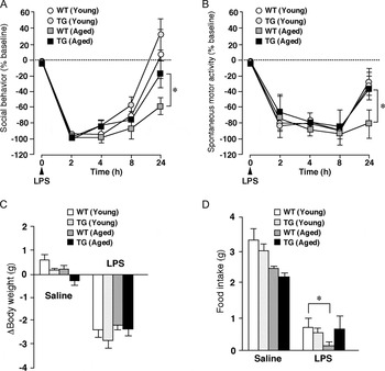
These observations prompted us to further elucidate the effect of human TFAM overexpression on the age-dependent prolongation of LPS-induced sickness behaviors. The motivation to engage in social behavior and locomotor activity were assessed in young and aged TG mice in comparison with their young and aged wild-type (WT) littermates at several time points after intraperitoneal injection of LPS. Changes in body weight and food intake were also determined for the 24 h period after LPS injection.
As shown in Fig. 1, LPS prominently reduced the mean score of the social behavior and locomotor activity between 2 and 8 h post-injection in all groups. The reduction of social behavior and the locomotor activity in the young mice of both genotypes were restored to the control level by 24 h after LPS injection. However, the aged WT mice still suffered from behavioral deficits in both motivation and locomotor activity even at 24 h after LPS injection (Fig. 1A,B). In contrast, the social behavior and locomotor activity of the aged TG mice were mostly restored to the control level at 24 h after LPS injection (Fig. 1A). Furthermore, there was a significant reduction in body weight (Fig. 1C) and food intake (Fig. 1D) 24 h after LPS injection. However, there were no significant differences in the mean reduction in body weight among the four groups (Fig. 1C). On the other hand, the mean food intake of the aged WT mice was significantly lower than those of remaining three groups (Fig. 1D). Therefore, TFAM overexpression can markedly improve the age-dependent prolonged LPS-induced sickness behaviors.
The effects of human TFAM overexpression on the age-dependent prolonged LPS-induced sickness behaviors. (A,B) The motivation to engage in social behavior (A) and locomotor activity (B) were assessed in both the young (2–4 months) and aged (19–22 months) mice of each genotype at several time points after the intraperitoneal injection of Escherichia coli LPS (0.33 mg kg−1, ~10 µg mouse−1; serotype 055:B5, Sigma). To assess the motivation to engage in social exploratory behavior, a novel juvenile nonspecific was introduced into the test subject's cage for a 5 min period. Social behavior was determined as the amount of time that the experimental subject spent investigating (e.g. ano-genital sniffing, trailing and grooming) the juvenile (Bluthé et al., Reference Bluthé, Dantzer and Kelley1991). Different juveniles were presented on each occasion to avoid habituation. On the other hand, to assess the locomotor activity, mice were placed in a novel home cage, which provided a floor area of 28 × 18 cm, and the locomotor activity of mice was scored for 3 min. Each symbol and vertical bar represents the mean ± SEM of six animals, respectively. Asterisks indicate significant differences between the values (*P < 0.05, two-way ANOVA with a post-hoc Tukey's test). (C,D) Changes in body weight (C) and food intake (D) were also determined for the 24 h period after LPS injection. Each column and vertical bar represents the mean ± SEM of six animals, respectively. An asterisk indicates a significant difference between the values (*P < 0.05, two-way ANOVA with a post-hoc Tukey's test).

IMPROVEMENT OF THE AGE-DEPENDENT LPS-INDUCED EXCESSIVE INFLAMMATORY RESPONSES AND OXIDATIVE STRESS IN MICROGLIA BY TFAM OVEREXPRESSION
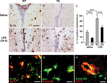
Sickness behaviors are well known to be due to the central actions of proinflammatory cytokines (Dantzer, Reference Dantzer2004), and microglia are considered to be the principal source of proinflammatory cytokines in the brain. Therefore, after the evaluation of behaviors, mice were subjected to the immunohistochemical analyses for IL-1β, because IL-1β is a master regulator of neuroinflammation (Basu et al., Reference Basu, Krady and Levison2004) and a potential key player during systemic inflammation. In the saline-treated groups, the immunoreactivity of IL-1β was barely detectable in the brain parenchyma of both the young TG mice and their WT littermates (data not shown). On the other hand, relative strong immunoreactivity for IL-1β was observed especially in the periventricular area of the striatum of the aged WT mice (Fig. 2A). In contrast, only modest immunoreactivity for IL-1β was observed in the periventricular regions of the aged TG mice (Fig. 2B). At 24 h after LPS injection, the immunoreactivity for IL-1β in the young WT mice had markedly declined (data not shown). The immunoreactivity for IL-1β in the LPS-treated aged WT mice remained elevated (Fig. 2C,E), which corresponded closely with a delayed recovery from LPS-induced sickness behaviors. In good agreement with the recovery from LPS-induced sickness behaviors of the aged TG mice, the immunoreactivity for IL-1β in the brain was markedly decreased (Fig. 2D,E). Double immunostaining experiments showed that IL-1β-positive cells in the periventricular regions corresponded well with Iba1-positive microglia in the aged mice (Fig. 2F). The immunoreactivity for 8-oxo-deoxyguanosine (8-oxo-dG), a major DNA peroxidation product, was mainly localized in Iba1-positive microglia (Fig. 2G). It is considered that the immunoreactivity for 8-oxo-dG indicated the presence of oxidative damages of the mtDNA in microglia, because the immunoreactivity for 8-oxo-dG corresponded closely to that for cytochrome b, a marker of the mitochondria (Fig. 2H). Therefore, it is considered that the age-dependent oxidative damage of the mtDNA mainly in microglia produces excessive ROS, which subsequently induce the increased production and secretion of IL-1β by microglia in the periventricular area of the striatum.
The effects of human TFAM overexpression on the age-dependent exaggerated LPS-induced IL-1β production in microglia. (A–D) After the evaluation of sickness behaviors, animals were subjected to the immunohistochemical analyses for IL-1β. The immunoreactivity for IL-1β in the periventricular area of the striatum of the saline- (A,B) or LPS-treated (C,D) aged TG mice and their WT littermates. (A,C), the aged WT mice; (B,D), the aged TG mice (B). Scale bar = 50 µm. (E) The mean IL-1β-positive cells in the periventricular area of the striatum of the saline- or LPS-treated aged TG mice and their WT littermates. Each column and vertical bar represent the mean ± SEM of five experiments, respectively. Asterisks indicate significant differences between the values (**P < 0.01, two-way ANOVA with a post-hoc Tukey's test). (F,G) Immunofluorescent confocal laser scanning microscopic images of IL-1β (F) and 8-oxo-dG (G) in Iba1-positive microglia. Scale bars = 30 µm in (F), 15 µm in (G). (H) The immunoreactivity for 8-oxo-dG in microglia corresponded closely with that for cytochrome b (Cyt b), a marker for the mitochondria. Scale bar = 4 µm. (F)–(H) are modified versions of Fig. 3B that appeared in Behavioral Brain Research, 2009.

TFAM overexpression can reduce the age-dependent oxidative damage of the mtDNA in microglia, thus improving the age-dependent exaggerated cytokine responses and the prolonged sickness behaviors. IL-1β-expressing microglia were mainly found in the hippocampus and the periventricular area of the striatum, including the subventricular zone (SVZ), after LPS injection. Besides the subgranular zone in the hippocampal dentate gyrus, the SVZ is also known to be an important source of neural stem cells. The proliferation of neural stem cells in the SVZ is also essential for the maintenance of mental health, because antidepressants can reverse the decline of proliferation of neural stem cells in the SVZ induced by chronic stress or corticosteroid use (Hitoshi et al., Reference Hitoshi, Maruta, Higashi, Kumar, Kato and Ikenaka2007; Lau et al., Reference Lau, Qui, Helmester, Lee, Tang and So2007). Therefore, it is reasonable to consider that TFAM overexpression can also improve the age-dependent impairment of neurogenesis in the SVZ, because IL-1β is known to suppress the proliferation of neural stem cells that are necessary for improving depressive behavior (Kaneko et al., Reference Kaneko, Kudo, Mabuchi, Takemoto, Fujimaki and Wati2006).
POTENTIAL MECHANISM UNDERLYING THE IMPROVEMENT OF THE AGE-DEPENDENT PROLONGED LPS-INDUCED SICKNESS BEHAVIORS BY TFAM OVEREXPRESSION
It is commonly accepted that LPS-induced inflammatory signal transduction is mediated by NF-κB in microglia/macrophages. Therefore, NF-κB activation is considered to be crucial for the generation of sickness behaviors triggered by microglia-mediated inflammation (Laflamme et al., 2001). NF-κB is released from the inhibitor of NF-κB (IκB)/NF-κB dimmer, and translocates from the cytoplasm into the nucleus, inducing the expression of proinflammatory genes and the subsequent inflammatory response (Hartlage-Rübsamen et al., Reference Hartlage-Rübsamen, Lemke and Schliebs1999; Takeuchi et al., Reference Takeuchi, Jin, Wang, Zhang, Kawanokuchi and Kuno2006; Park et al., Reference Park, Woo, Kim, Hyun, Kim and Lee2007). Increasing evidence suggests that LPS-induced NF-κB activation through TLR4 is dependent on the production of ROS (Baeuerle and Henkel Reference Baeuerle and Henkel1994; Janssen-Heininger et al., Reference Janssen-Heininger, Poynter and Baeuerle2000). Several lines of evidence indicate that the nuclear translocation of NF-κB can be triggered by ROS (Flohé et al., Reference Flohé, Brigelius-Flohe, Saliou, Traber and Packer1997; Janssen-Heininger et al., Reference Janssen-Heininger, Poynter and Baeuerle2000), and broad range of antioxidants abolishes NF-κB activation (Zhang et al., Reference Zhang, Spapen, Nguyen, Benlabed, Buurman and Vincent1994; Blackwell et al., Reference Blackwell, Blackwell, Holden, Christmen and Christman1996; Devaraj et al., Reference Devaraj, Li and Jialal1996; Fox et al., Reference Fox, Brower, Bellezzo and Leingang1997). These observations indicate that NF-κB activation is facilitated by conditions associated with an increased intracellular redox state (Toledano and Leonard, Reference Toledano and Leonard1991). Besides the ROS-dependent NF-κB activation pathway, LPS can directly cause the robust transcriptional activity of an inhibitor of NF-κBα (IkBα) following the signaling of IL-1β in microglia (Quan et al., Reference Quan, Whiteside, Kim and Herkenham1997; Laflamme and Rivest, Reference Laflamme and Rivest1999). The existence of an ROS-independent NF-κB activation pathway explains why TFAM overexpression was not sufficient to completely restore the number of IL-1β-positive microglia in the LPS-treated aged mice compared to the saline-treated control level (Fig. 2E).
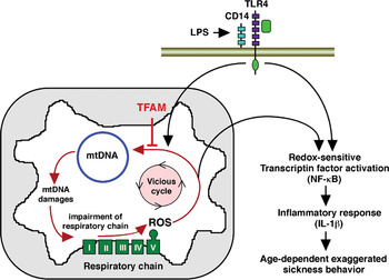
In conclusion, LPS induces the generation of ROS from the actively respirating mitochondria (Dikalov et al., Reference Dikalov, Dikalova and Mason2002) as well as NADPH oxidase, and overproduction of ROS, in turn, triggers NF-κB-dependent inflammatory responses in the aging microglia. The overexpression of human TFAM improves the age-dependent prolonged LPS-induced sickness behaviors by ameliorating the mtDNA damage and the resulting redox-regulated inflammatory response (Fig. 3). It is therefore of interest that resveratrol, a polyphenol present in red wines, improves the mitochondrial functions, and increases the amount of TFAM expressed in the mitochondria (Lagouge et al., Reference Lagouge, Argmann, Gerhart-Hines, Meziane, Lerin and Daussin2006).
A possible mechanism underlying the effect of TFAM overexpression on the age-dependent prolonged LPS-induced sickness behaviors. The ROS damages the mtDNA and in turn, the damaged mtDNA impairs the respiratory chain, forming a vicious cycle. Increased intracellular ROS activate the redox-sensitive NF-κB to provoke excessive inflammation in the aged brain. Systemic administration of LPS further activates NF-κB through two different pathways, the mitochondria-derived ROS-dependent and the direct ROS-independent pathways, resulting in prolonged sickness behaviors. TFAM could inhibit ROS-induced mtDNA damage and the resulting redox-regulated inflammatory responses to improve the age-dependent prolonged LPS-induced sickness behaviors. This figure is a modified version of Fig. 3A that appeared in Behavioral Brain Research, 2009.

ACKNOWLEDGEMENTS
The authors express thanks to Dr. T. Ide (Department of Cardiovascular Medicine, Graduate School of Medical Sciences, Kyushu University, Japan) and Dr. D. Kang (Department of Clinical Chemistry and Laboratory Medicine, Kyushu University, Fukuoka, Japan) for providing us with human TFAM transgenic mice. The authors also express thanks to Dr. M. Ochi-Shindou (Neurobiology Research Unit, Okinawa Institute of Science and Technology, Okinawa Japan) for conducting a part of behavioral studies. This study was supported by Granti-in-Aid for Scientific Research from the Ministry of Education, Science and Culture, Japan.
Statement of interest
None.