I. INTRODUCTION
Tamsulosin hydrochloride (brand name Flomax®) is an active drug used in the treatment of Benign Prostatic Hyperplasia and Chronic Prostatitis. The IUPAC name (CAS Registry number 106463-17-6) is 5-[(2R)-2-[2-(2-ethoxyphenoxy)ethylamino]propyl]-2-methoxybenzenesulfonamide hydrochloride. This molecule works as a selective alpha 1 adrenoreceptor blocker in the human prostate and bladder neck, relaxing prostatic muscles, and reducing the pain of urination. Tamsulosin hydrochloride also decreases maximum urethral closure pressure and improves several cytometry parameters related to bladder storage and emptying. A two-dimensional (2D) molecular diagram of the tamsulosin cation is shown in Figure 1.
2D molecular diagram of the tamsulosin cation.

A star quality powder pattern based on this synchrotron data set is included in the Powder Diffraction File as entry 00-065-1415. Powder patterns for Forms I and II of (R)-tamsulosin are reported in Gangakhedkar (Reference Gangakhedkar2006; Ranbaxy). Powder data for both amorphous and crystalline (R)-tamsulosin hydrochloride are reported in Ham (Reference Ham2009; LEK Pharmaceuticals). Single-crystal structures of three forms of (R)-tamsulosin free base and (R)-tamsulosin hydrochloride (Nanubolu et al., Reference Nanubolu, Sridhar and Ravikumar2014; JOWCIJ) (R)-tamsulosin have been published during the course of this work.
This work was carried out as part of a project (Kaduk et al., Reference Kaduk, Crowder, Zhong, Fawcett and Suchomel2014) to determine the crystal structures of large-volume commercial pharmaceuticals and include high-quality powder diffraction data for these pharmaceuticals in the Powder Diffraction File (Gates-Rector et al., Reference Gates-Rector and Blanton2017).
II. EXPERIMENTAL
Tamsulosin hydrochloride was a commercial reagent, purchased from Sigma-Aldrich (Lot #051M4734V), and was used as-received. The white powder was packed into a 1.5-mm diameter Kapton capillary and rotated during the measurement at ~50 Hz. The powder pattern was measured at 295 K at beamline 11-BM (Lee et al., Reference Lee, Shu, Ramanathan, Preissner, Wang, Beno, Von Dreele, Ribaud, Kurtz, Antao, Jiao and Toby2008; Wang et al., Reference Wang, Toby, Lee, Ribaud, Antao, Kurtz, Ramanathan, Von Dreele and Beno2008) of the Advanced Photon Source at Argonne National Laboratory using a wavelength of 0.413676 Å from 0.5° to 50° 2θ with a step size of 0.001° and a counting time of 0.1 s step−1.
The pattern was indexed on a primitive monoclinic unit cell with a = 7.62978, b = 9.27653, c = 32.29631 Å, β = 92.491°, V = 2297.5 Å3, and Z = 4 using N-TREOR (Altomare et al., Reference Altomare, Cuocci, Giacovazzo, Moliterni, Rizzi, Corriero and Falcicchio2013). Analysis of the systematic absences using EXPO2014 (Altomare et al., Reference Altomare, Cuocci, Giacovazzo, Moliterni, Rizzi, Corriero and Falcicchio2013) suggested the space group P21, which was confirmed by successful solution and refinement of the structure. A reduced cell search in the Cambridge Structural Database (Groom et al., Reference Groom, Bruno, Lightfoot and Ward2016) yielded 11 hits, among which was (R)-tamsulosin hydrochloride (Nanubolu et al., Reference Nanubolu, Sridhar and Ravikumar2014; JOWCIJ), which was published after this structure was solved.
The structure was solved by charge flipping as implemented in JANA2006 (Petricek et al., Reference Petricek, Dusek and Palatinus2014). All of the non-H atoms, apart from two ring carbon atoms were located, and the missing atoms were added using Materials Studio (Dassault, 2019).
Rietveld refinement was carried out using GSAS-II (Toby and Von Dreele, Reference Toby and Von Dreele2013). Only the 0.7–22.0° portion of the pattern was included in the refinement (d min = 1.084 Å). All non-H bond distances and angles were subjected to restraints, based on a Mercury/Mogul Geometry Check (Bruno et al., Reference Bruno, Cole, Kessler, Luo, Motherwell, Purkis, Smith, Taylor, Cooper, Harris and Orpen2004; Sykes et al., Reference Sykes, McCabe, Allen, Battle, Bruno and Wood2011) of the molecule. The results were exported to a .csv file. The Mogul average and standard deviation for each quantity were used as the restraint parameters and were incorporated using the new GSAS feature Restraints/Edit Restraints/Add MOGUL Restraints, which reads the bond distance and angle restraints from the .csv file. The restraints contributed 2.0% to the final χ 2. The hydrogen atoms were included in calculated positions, which were recalculated during the refinement using Materials Studio (Dassault, 2019). The U iso of the two Cl anions and the Uiso of chemically equivalent atoms in the two cations were constrained to be identical. A common U iso was refined for the O and N atoms of the sulfonamide groups, another U iso for the C atoms of the phenyl rings, and another U iso for the non-H atoms of the ethoxy groups. The U iso for each hydrogen atom was constrained to be 1.3× that of the heavy atom to which it is attached. The background was modeled using a 12-term shifted Chebyshev polynomial. The final refinement was begun from the results of the density functional theory (DFT) calculation.
The final refinement of 214 variables using 21 303 observations and 136 restraints yielded the residuals R wp = 0.0915 and GOF = 1.67. The largest peak (0.58 Å from O26) and hole (1.88 Å from C23) in the difference Fourier map were 0.43 and −0.30(8) eÅ−3, respectively. The Rietveld plot is included in Figure 2. The largest errors in the fit are in the shapes of some of the strong low-angle peaks.
The Rietveld plot for the refinement of tamsulosin hydrochloride. The blue crosses represent the observed data points, and the green line is the calculated pattern. The cyan curve is the normalized error plot. The vertical scale has been multiplied by a factor of 10× for 2 > 9.5°.

A density functional geometry optimization (fixed experimental unit cell) was carried out using CRYSTAL14 (Dovesi et al., Reference Dovesi, Orlando, Erba, Zicovich-Wilson, Civalleri, Casassa, Maschio, Ferrabone, De La Pierre, D-Arco, Noël, Causà and Kirtman2014). The basis sets for the H, C, N, and O atoms were those of Gatti et al. (Reference Gatti, Saunders and Roetti1994), and the basis sets for S and Cl were that of Peintinger et al. (Reference Peintinger, Vilela Oliveira and Bredow2013). The calculation was run on eight 2.1 GHz Xeon cores (each with 6 GB RAM) of a 304-core Dell Linux cluster at IIT, using 8 k-points and the B3LYP functional, and took ~24 days.
III. RESULTS AND DISCUSSION
This synchrotron power pattern of tamsulosin hydrochloride is in excellent agreement with that calculated from JOWCIJ (Nanubolu et al., Reference Nanubolu, Sridhar and Ravikumar2014), and in good agreement with that reported by Ham (Reference Ham2009) (Figure 3). The agreement is good enough to conclude that the patterns represent the same material.
Comparison of the synchrotron powder pattern (blue) of tamsulosin hydrochloride to the pattern calculated from JOWCIJ (red) (Nanubolu et al., Reference Nanubolu, Sridhar and Ravikumar2014) and that reported by Ham (green) (Reference Ham2009).

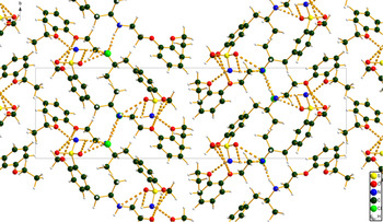
The refined atom coordinates of tamsulosin hydrochloride and the coordinates from the DFT optimization are reported in the CIFs deposited with ICDD. The root-mean-square (rms) Cartesian displacement of the non-hydrogen atoms in the Rietveld-refined and DFT-optimized structures are 0.196 and 0.125 Å for molecules 1 and 2 (Figure 4). The maximum displacements are 0.527 and 0.234 Å, at methyl groups C28 and C43. The good agreement between the refined and optimized structures provides evidence that the experimental structure is correct (van de Streek and Neumann, Reference van de Streek and Neumann2014). The agreement between the DFT-optimized structure from this study and that of JOWCIJ is excellent, except for the disordered groups in JOWCIJ (Figure 5). This discussion concentrates on the CRYSTAL-optimized structure. The asymmetric unit (with atom numbering) is illustrated in Figure 6, and the crystal structure is presented in Figure 7.
(a) Comparison of the Rietveld-refined (red) and VASP-optimized (blue) structures of cation 1 in tamsulosin hydrochloride. The rms cartesian displacement is 0.196 Å. (b) Comparison of the Rietveld-refined (red) and VASP-optimized (blue) structures of cation 2 in tamsulosin hydrochloride. The rms cartesian displacement is 0.125 Å.

Comparison of the DFT-optimized structure of tamsulosin hydrochloride of this study (blue) and the disordered structure of JOWCIG (green).

The asymmetric unit of tamsulosin hydrochloride, with the atom numbering. The atoms are represented by 50% probability spheroids/ellipsoids.

The crystal structure of tamsulosin hydrochloride, viewed down the a-axis.

The crystal structure is characterized by two slabs of tamsulosin hydrochloride molecules perpendicular to the c-axis. These slabs exist in the (0-13) and (031) planes. Tamsulosin hydrochloride is also layered in the bc-plane and line up looking down the a, b, and c-axes. In between, the slabs exist van der Waals intermolecular bonding and there are hydrogen bonds in the middle of each slab. The edges of the slabs contain methoxy phenol rings which interact with others through van der Waals interactions. In the single-crystal structure JOWCIJ (Nanubolu et al., Reference Nanubolu, Sridhar and Ravikumar2014), the two ethoxide groups O26–C27–C28 and O54–C55–C56 (in our nomenclature) are disordered in the ratio of ~60/40. In the difference Fourier map, there were small peaks (~0.4 eÅ−3) near each of these oxygen atoms, but no significant features in the regions of the ethyl groups. Either our sample really is different from that of Nanubolu et al. (Reference Nanubolu, Sridhar and Ravikumar2014), or the powder data are not sensitive enough to detect such disorder.
Almost all the bond distances, bond angles, and torsion angles fall within the normal ranges indicated by a Mercury/Mogul Geometry check (Macrae et al., Reference Macrae, Sovago, Cottrell, Galek, McCabe, Pidcock, Platings, Shields, Stevens, Towler and Wood2020). The S1–O2, S1–O3, S29–O30, and S29–O31 bonds are flagged as unusual (optimized = 1.492, 1.502, 1.493, 1.500 Å; average = 1.433(19) Å; Z-score = 3.1, 3.6, 3.1, 3.5). The S1–N4 and S29–N32 bonds are also flagged as unusual (1.673 and 1.681 Å; average = 1.600(20) Å; Z-score = 3.6 and 4.0). In the Rietveld-refined structure, these sulfonamide bond distances fall within the normal ranges, indicating perhaps an inaccuracy in the calculation. The O19–C18–C17–N16 torsion angle lies in a minor trans population of a mainly gauche distribution. The C21–O26–C27–C28 torsion lies in a minor gauche population of a mainly trans distribution. The C36–C37–C41–C42 and C38–C37–C41–C42 torsions lie on the tails of broad distributions. The C41–C42–N44–C45 torsion lies in a minor population of a broad distribution. The C43–C42–N44–C45 torsion lies on the tail of a broad distribution. The O47–C46–C45–N44 torsion lies in a minor trans population of a mainly gauche distribution.
The two-independent tamsulosin cations have different conformations (Figure 8). The rms Cartesian displacement is 1.859 Å. Inverting one of the molecules decreases the displacement to 0.509 Å. Quantum chemical geometry optimizations of the isolated tamsulosin cations (DFT/B3LYP/6-31G*/water) using Spartan ‘18 (Wavefunction, 2018) indicated that the observed solid-state conformation of molecule 1 is 0.8 kcal mol−1 lower in energy than the local minimum. The difference is within the expected error limits of such calculations, so the two conformations should be considered equivalent in energy. Molecular mechanics conformational analysis indicated that the minimum-energy conformation folds the cation on itself, to make the two six-membered rings approximately parallel. This geometry shows that intermolecular interactions are important in determining the solid-state conformations.
Comparison of the two-independent tamsulosin cations in tamsulosin hydrochloride. Cation 1 is in purple, and cation 2 is in orange.

Analysis of the contributions to the total crystal energy using the Forcite module of Materials Studio (Dassault, 2019) suggests that angle distortion terms are dominant in the intramolecular deformation energy. Since the Mogul analysis shows that all angles fall within the normal ranges, it is not clear in which angles the distortion arises. The intermolecular energy is dominated by van der Waals and (especially) electrostatic attractions, which in this force-field-based analysis include hydrogen bonds. The hydrogen bonds are better analyzed using the results of the DFT calculation.
As expected, each of the hydrogens on the protonated nitrogen atoms makes a strong hydrogen bond to one of the chloride anions (Table I). The result is to link the cations and anions into columns along the b-axis. One hydrogen of each sulfonamide group also makes a hydrogen bond to a chloride anion. The other hydrogen of each sulfonamide group forms bifurcated hydrogen bonds to two ether oxygens. The energies of these N–H⋯O hydrogen bonds were calculated using the correlation from Wheatley and Kaduk (Reference Wheatley and Kaduk2019). Several C–H⋯O hydrogen bonds to the oxygens of the sulfonamide groups and to an ether oxygen also contribute to the crystal energy.
Hydrogen bonds (CRYSTAL14) in tamsulosin hydrochloride.

The volume enclosed by the Hirshfeld surface (Figure 9; Hirshfeld, Reference Hirshfeld1977; Turner et al., Reference Turner, McKinnon, Wolff, Grimwood, Spackman, Jayatilaka and Spackman2017) is 1114.22 Å3, 99.00% of half the unit cell volume. The molecules are, thus, not tightly packed. All the significant-close contacts (red in Figure 9) involve the hydrogen bonds. The volume/non-hydrogen atom is relatively large, at 19.4 Å3.
The Hirshfeld surface of tamsulosin hydrochloride. Intermolecular contacts longer than the sums of the van der Waals radii are colored blue, and contacts shorter than the sums of the radii are colored red. Contacts equal to the sums of radii are white.

The Bravais–Friedel–Donnay–Harker (Bravais, Reference Bravais1866; Friedel, Reference Friedel1907; Donnay and Harker, Reference Donnay and Harker1937) model suggests that we might expect platy morphology for tamsulosin hydrochloride, with {001} as the principal faces. A fourth-order spherical harmonic model was included in the refinement. The texture index was 1.058, indicating that preferred orientation was present in this rotated capillary specimen. The powder pattern of tamsulosin hydrochloride from this synchrotron data set is included in the Powder Diffraction File 00-065-1415.
IV. DEPOSITED DATA
The Crystallographic Information Framework (CIF) files containing the results of the Rietveld refinement (including the raw data) and the DFT geometry optimization were deposited with the ICDD. The data can be requested at info@icdd.com.
ACKNOWLEDGEMENTS
We thank Lynn Ribaud and Saul Lapidus for their assistance in the data collection, and Andrey Rogachev for the use of computing resources at IIT.
FUNDING
Use of the Advanced Photon Source at Argonne National Laboratory was supported by the U.S. Department of Energy, Office of Science, Office of Basic Energy Sciences, under Contract No. DE-AC02-06CH11357. This work was partially supported by the International Centre for Diffraction Data. This work was also supported by the Illinois Math and Science Academy's SIR department.
CONFLICTS OF INTEREST
The authors have no conflicts of interest to declare.