Biofilms are an attached growth state of bacteria
Relevance of biofilms
Biofilms are surface-associated communities of bacteria encased in an extracellular matrix. Biofilms are encountered in almost every imaginable environment. It has been estimated that many bacteria in the environment adopt the biofilm lifestyle (opposed to the free-swimming or planktonic life style). Geesey et al.Reference Geesey, Mutch, Costerton and Green1 demonstrated that in the water column of streams in Montana, most bacteria were found associated with surfaces. In industry, biofilms cause many problems, including fouling of ship hulls, promoting corrosion in pipes, and contaminating food processing equipment. They can also be beneficial in industry. For example, they are a key feature of wastewater treatment plants. In the clinic, it has been estimated that biofilms cause up to 60% of all bacterial infections in developed countries.
There are several reasons why the biofilm lifestyle is advantageous. One of the primary reasons is that biofilms provide protection from a range of stressors, from antibiotics to host immune response and protozoan grazing. They can also facilitate acquisition of nutrients in cases where the surface is a nutrient source (e.g., a chicken in a poultry processing plant). Biofilms also promote genetic exchange, providing a high local cell density and a stable structured environment for genetic exchange events, such as conjugation and transformation.Reference Molin and Tolker-Nielsen2
Because of their widespread importance, there has been an explosion of biofilm-related research in the past 10 years. Scientists and engineers have been probing the molecular mechanisms underpinning biofilm formation and antimicrobial tolerance, while engineers and material scientists have struggled to design surfaces that prevent microbial attachment. This work has usually centered on a few key species for which we know the most about biofilm formation. Recent research has revealed that biofilm development can be at the confluence of many other types of social behavior for bacteria. One such bacterial species for which this is the case is the Gram-negative bacterium, Pseudomonas aeruginosa.
Pseudomonas aeruginosa: A model organism for studying sociomicrobiology
P. aeruginosa is an environmentally ubiquitous bacterium that routinely grows attached to surfaces (e.g., water pipes, or soil and sand particles) as a biofilm.Reference Emtiazi, Schwartz, Marten, Krolla-Sidenstein and Obst3 It is also a metabolically versatile organism, able to use a variety of compounds as sole carbon and energy sources. Its genome is rather large (6.3 million base pairs Mb),Reference Stover, Pham, Erwin, Mizoguchi, Warrener, Hickey, Brinkman, Hufnagle, Kowalik, Lagrou, Garber, Goltry, Tolentino, Westbrock-Wadman, Yuan, Brody, Coulter, Folger, Kas, Larbig, Lim, Smith, Spencer, Wong, Wu, Paulsen, Reizer, Saier, Hancock, Lory and Olson4 probably attesting to its ability to thrive in a variety of environmental conditions. P. aeruginosa is also capable of engaging in a variety of social behaviors. Besides biofilm formation, it can chemically communicate within a group in a process called quorum sensing (see the Renner and Weibel article in this issue), as well as participate in a type of social surface motility called swarming (see the Wilking et al. article).
This bacterium is also an opportunistic human pathogen, causing both chronic and acute infections in susceptible individuals, including cystic fibrosis patients, burn victims, contact lens wearers, and the immunocompromised.Reference Lyczak, Cannon and Pier5 A P. aeruginosa infection typically stems from environmental sources, as this ubiquitous organism transitions from environment to host and readily forms biofilms in vivo during chronic infections that show increased resistance to antibiotics and components of the immune system. Because of its importance in both environmental and clinical settings, it is a paradigm for studying social behaviors in bacteria in the emerging field of sociomicrobiology. Many other problematic biofilm bacteria are difficult to culture in the laboratory; research with P. aeruginosa is often useful to gain insight into parallel mechanisms used by these other bacteria.
Factors influencing P. aeruginosa biofilm formation
Several genetic and environmental determinants have been shown to influence biofilm development in P. aeruginosa. The nutritional environment is a key factor impacting biofilm structure. P. aeruginosa is capable of forming undifferentiated “flat” biofilms as well as highly structured biofilms containing void spaces and large cell aggregates (Figure 1). The carbon source upon which P. aeruginosa is grown dictates the architecture of the biofilm it forms. Other key nutrients such as iron have been shown to influence biofilm formation. Under conditions of iron starvation, biofilm formation is inhibited.Reference Banin, Vasil and Greenberg6 Several gene encoded functions contribute to biofilm development, including surface structures such as pili and flagella, as well as secreted polysaccharides. This review will focus on two important social behaviors and their contribution to biofilm formation: quorum sensing and surface motility.
P. aeruginosa flow-cell biofilms grown under differing media conditions: (a) defined phosphate buffer medium with succinate and (b) 1% tryptic soy broth. Scale bars ~50 μm. Images courtesy of Joshua Shrout and Dao Nguyen.

Quorum sensing
P. aeruginosa can engage in intercellular cell-cell signaling, called quorum sensing, to coordinate gene expression. All living organisms express genes to enable specific functions; most genes are not expressed constantly. Rather, genes are expressed conditionally in response to some specific cue or environmental signal. P. aeruginosa regulates expression of a subset of its genes in response to a critical concentration of self-produced extracellular communication molecules. This phenomenon is called quorum sensing because induction or repression of quorum sensing-controlled gene expression has been shown to require a critical local cell density or “quorum” of bacteria. In liquid batch culture (e.g., the well-mixed, small-volume conditions in test tubes), this is dependent upon the bacterial cell density. In a growing culture, the bacteria reach a critical cell density at which the concentration of extracellular signal initiates a quorum sensing response from the bacteria.Reference Fuqua, Parsek and Greenberg7–Reference Taga and Bassler10P. aeruginosa and several other bacterial species are capable of monitoring their own population density and use this form of communication to coordinate expression of particular genes.
Quorum sensing in P. aeruginosa
There are several different types of quorum sensing signaling mechanisms that use a range of different signal types and detection mechanisms.Reference Fuqua, Parsek and Greenberg7 One of the most common for the Proteobacteria (of which P. aeruginosa is a member) involves acyl-homoserine lactone (AHL) signal molecules. The AHL structure consists of a fatty acid chain with an amide bond linkage to a lactonized homoserine.Reference Fuqua, Parsek and Greenberg7, Reference Fuqua and Greenberg8, Reference Fuqua and Greenberg11, Reference Whitehead, Barnard, Slater, Simpson and Salmond12 AHL-based quorum sensing appears to be highly conserved among the Proteobacteria—greater than 50 species have been identified to produce AHLs. AHL molecules are described as amphipathic because they contain both polar and non-polar regions, since the homoserine lactone ring is hydrophilic and the fatty acid side chain is hydrophobic. This amphipathicity appears to facilitate free diffusion of AHLs within the aqueous environment inside and outside the cell, as well as across the phospholipid bilayer of cell membranes.
These AHLs are produced by proteins called acyl-homoserine lactone synthases. Originally described for the regulation of luciferase enzyme to generate bioluminescence in the marine bacterium Vibrio fischeri, the genes and proteins important for luciferase production all bear the designation “lux”. The LuxI protein, named “I” for inducer, is the AHL synthase for V. fischeri. Subsequently, proteins homologous to LuxI have been identified in many other bacteria, including P. aeruginosa, and are responsible for production of AHL signal molecules.Reference Fuqua, Parsek and Greenberg7–Reference Miller and Bassler9
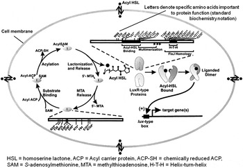
The second key component to AHL-mediated quorum sensing is the sensing of the AHL signal and initiating a coordinated response by individual bacteria when a threshold AHL concentration is reached. This sensing occurs through proteins in the LuxR family. Again, “lux” refers to production of luciferase by V. fischeri and “R” stands for regulator. These regulatory proteins initiate transcription of select genes when cued by a threshold concentration of AHL signal (Figure 2).
A representative quorum sensing circuit in Gram-negative bacteria.Reference Fuqua, Parsek and Greenberg7

There are two AHL signaling systems in P. aeruginosa. The LasI/LasR (named originally for control of elastase proteins, known virulence proteins that damage lung cells) and RhlI/RhlR (named for control of rhamnolipid production, another known virulence factor that is discussed later in the text) systems are reasonably well characterized and are known to have interdependence. For instance, transcription of rhlI and rhlR are induced by the las system.Reference de Kievit, Kakai, Register, Pesci and Iglewski13 A third LuxR homolog, QscR (named for quorum-sensing-control repressor), has been identified in P. aeruginosa; however, its function is poorly understood at present. No “QscI” has been identified, nor is such a LuxI homolog predicted based upon an analysis of the sequenced P. aeruginosa genome. It has been estimated that as many as 4–6% of the roughly 6000 P. aeruginosa genes are controlled by AHL quorum sensing.Reference Whiteley, Lee and Greenberg14–Reference Schuster, Lostroh, Ogi and Greenberg17 Expression of genes (called transcription) occurs when an RNA polymerase begins synthesis of a specific region of DNA. There are multiple variables that control such expression for different genes in different organisms, but, in general, gene expression requires RNA polymerase (and an accessory component called a “sigma factor”) to recognize DNA just before the actual gene; this upstream DNA is called the “promoter.” Variations in DNA binding target sequences may function to modulate RhlR/LasR/QscR binding to promoter regions, requiring different amounts of signal to affect expression of a particular gene or series of genes (called an operon). Additionally, non-AHL signaling has also been described for P. aeruginosa. The Pseudomonas quinolone signal, 2-heptyl-3-hydroxy-4-quilinone, plays a significant role in controlling expression of some virulence genes in P. aeruginosa and is integrated into the AHL quorum sensing regulatory circuit.Reference McGrath, Wade and Pesci18
Why is quorum sensing an important consideration for the biofilm mode of growth?
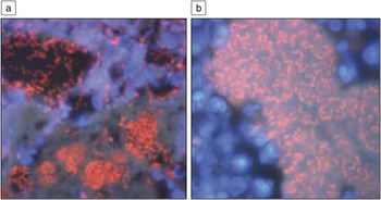
Although most of what we know about the molecular mechanisms underpinning quorum sensing is derived from the study of shaken liquid cultures, it is fairly obvious that dense, suspended growth in liquid is probably a rare occurrence for P. aeruginosa outside the laboratory. Thus the question is where in the environment might you encounter a “quorum” of P. aeruginosa? The probable answer is in situations where the environment is conducive to a somewhat robust period of growth that produces a high local cell density. A biofilm aggregate of P. aeruginosa cells (as observed in the lungs of cystic fibrosis patientsReference Bjarnsholt, Jensen, Fiandaca, Pedersen, Hansen, Andersen, Pressler, Givskov and Hoiby19 and chronic wounds;Reference Kirketerp-Moller, Jensen, Fazli, Madsen, Pedersen, Moser, Tolker-Nielsen, Hoiby, Givskov and Bjarnsholt20 see Figure 3) would be one such environment. The extracellular matrix enforces a high local cell density that would promote signaling, even under situations where the biofilm is cohabited by other species.
Intraluminal P. aeruginosa biofilms surrounded by polymorphonuclear-leukocytes visualized by (a) fluorescent in situ hybridization method using peptide nucleic acid and (b) 4’,6-diamidino-2-phenylindole.Reference Bjarnsholt, Jensen, Fiandaca, Pedersen, Hansen, Andersen, Pressler, Givskov and Hoiby19 Individual P. aeruginosa cells appear as red rods and are approximately 2 μm in length.

Unfortunately, studying signaling in a structured system such as a biofilm is difficult. What constitutes a quorum when you have a growing cell aggregate subject to the mass transfer effects of fluid flow? Quorum sensing certainly provides the organism with the opportunity to coordinate gene expression events that could coordinate building a biofilm by its members. Most of the work done to date has focused on P. aeruginosa, and the results have been interesting and complicated, as one might imagine.
What role does QS plays in P. aeruginosa biofilms?
Several researchers have investigated the role of quorum sensing in biofilm formation. In 1998, Davies et al. reported that a mutation in the lasI gene, rendering the bacteria unable to make the signal 3-oxododecanoyl-AHL, had a tremendous impact on biofilm structure.Reference Davies, Parsek, Pearson, Iglewski, Costerton and Greenberg21 Normal “wild-type” (i.e., all genes present) biofilms consisted of large cell aggregates separated by large channels and void spaces empty of cells. The mutant biofilms (missing the lasI gene) were flat and homogenous and were easily washed away by the detergent sodium dodecyl sulfate (SDS), while the wild-type biofilm was resistant to SDS treatment. This finding generated a huge degree of interest; if signaling controls biofilms, then disrupting signaling could be the silver bullet for eradicating biofilms. However, subsequent work showed that the relationship between quorum sensing and biofilm formation was more complicated than first thought. Some researchers reported that quorum sensing did not influence biofilm formation at all,Reference Heydorn, Ersboll, Kato, Hentzer, Parsek, Tolker-Nielsen, Givskov and Molin22 while others reported a significant effect. Reference Davies, Parsek, Pearson, Iglewski, Costerton and Greenberg21, Reference de Kievit, Gillis, Marx, Brown and Iglewski23 The literature at the time created confusion in the community, with researchers at a loss as to why some labs saw an effect, while others using the same strains did not. In retrospect, it is not a surprise. Biofilm culturing conditions have a profound effect on when quorum sensing is important.Reference Kirisits, Margolis, Purevdorj-Gage, Vaughan, Chopp, Stoodley and Parsek24–Reference Stoodley, Jacobsen, Dunsmore, Purevdorj, Wilson, Lappin-Scott and Costerton26 Additionally, several different quorum sensing-regulated factors can influence biofilm structure in different ways.Reference de Kievit, Gillis, Marx, Brown and Iglewski23, Reference Folsom, Richards, Pitts, Roe, Ehrlich, Parker, Mazurie and Stewart27, Reference Whiteley, Bangera, Bumgarner, Parsek, Teitzel, Lory and Greenberg28 Finally, the quorum sensing mechanism is subject to regulation by environmental conditions, which can modulate the expression of different key quorum sensing regulators.Reference Shrout, Chopp, Just, Hentzer, Givskov and Parsek29, Reference Bollinger, Hasset, Iglewski, Costerton and McDermott30
Rhamnolipids are surfactants whose production is quorum sensing-controlled. The rhlAB operon encodes two genes required for rhamnolipid synthesis, and expression of this operon requires the rhl quorum sensing system. P. aeruginosa is defective for rhamnolipid production and forms flat, homogenous biofilms. Davey et al. showed that rhamnolipids are important for maintaining the spaces between cell aggregates relatively free of cells.Reference Davey, Caiazza and O’Toole31 This may provide several important functions that include providing better access to nutrients to cells deep within these channels. Pamp and Tolker-Nielsen provided evidence that rhamnolipids are important for facilitating bacterial migration in the later stages of biofilm formation where mature microcolonies are formed.Reference Pamp and Tolker-Nielsen32 Furthermore, Jensen et al. and Alhede et al. showed that rhamnolipids form a protective shield against attaching phagocytic cells from the innate immune system.Reference Alhede, Bjarnsholt, Jensen, Phipps, Moser, Christophersen, Christensen, van Gennip, Parsek, Hoiby, Rasmussen and Givskov33, Reference Jensen, Bjarnsholt, Phipps, Rasmussen, Calum, Christoffersen, Moser, Williams, Pressler, Givskov and Hoiby34 The galactophilic lectin, LecA, is another important quorum sensing-regulated factor that can impact biofilm structure.Reference Winzer, Falconer, Garber, Diggle, Camara and Williams35 Lectins are small secreted proteins that bind to carbohydrates. Mutations in the lecA gene reduced biofilm formation on steel coupons.
Other quorum sensing-regulated functions include the operon encoding the extracellular polysaccharide Psl. Psl is a primary scaffolding EPS that aids in the attachment of the bacterium to a surface and forms a robust extracellular matrix holding biofilm cells together. The secreted iron siderophore, pyoverdine, is also controlled by quorum sensing, and Banin et al. demonstrated that mutant strains unable to produce it formed flat biofilms with reduced biomass.Reference Banin, Vasil and Greenberg36 These siderophores are proteins that have high affinity for iron and act to sequester iron for the bacterium even when dissolved concentrations are very low.
The final quorum sensing-regulated function to be discussed is a type of surface motility called swarming. Swarming motility’s dependence on quorum sensing is nutritionally conditional and will be discussed in more detail later in this review.
With so many different biofilm-relevant functions regulated by quorum sensing, perhaps the biggest surprise is that there are any biofilm culturing conditions for which a wild-type strain and a quorum sensing mutant form identical biofilms.Reference Heydorn, Ersboll, Kato, Hentzer, Parsek, Tolker-Nielsen, Givskov and Molin22, Reference de Kievit, Gillis, Marx, Brown and Iglewski23, Reference Shrout, Chopp, Just, Hentzer, Givskov and Parsek29 This point also highlights the importance of environmental conditions. As one might expect, P. aeruginosa can fine tune its quorum sensing response in accordance with the environment. Under certain environmental conditions, a quorum sensing response might induce only a subset of potential quorum sensing-regulated genes. Thus, growth conditions can modulate quorum sensing and its subsequent effects on biofilm formation.
Environmental parameters known to influence quorum sensing in P. aeruginosa biofilms
Different labs have demonstrated that nutritional, chemical, and physical environments can all have a significant effect on quorum sensing. The hydrodynamic environment is one such factor. Aquatic biofilms subjected to different flow regimes will have mass transfer effects on signal accumulation. Kirisits et al. demonstrated that the flow rate can impact the onset of quorum sensing in a developing biofilm.Reference Kirisits, Margolis, Purevdorj-Gage, Vaughan, Chopp, Stoodley and Parsek24 Using fluorescent quorum sensing reporters, they found that at higher flow rates, a larger amount of biofilm biomass was needed to initiate quorum sensing. Perhaps the most surprising result was that at very high flow rates, the biofilm community never fully induced quorum sensing, even after several days of incubation. The chemical environment can also affect quorum sensing. Obviously anything in the biofilm environment that might impede the diffusion of the acyl-HSL signal may dampen the onset of quorum sensing. Biomolecules in the extracellular matrix, such as EPS, could serve as a sink for signals. Additionally, high pH environments can chemically cleave the AHL ring into multiple parts, destroying the signal. The half-life of signals in such an environment would be on the order of minutes.
Shrout et al. also showed that the nutritional environment influences the importance quorum sensing has for biofilm formation.Reference Shrout, Chopp, Just, Hentzer, Givskov and Parsek29 Depending upon the carbon source used for growth, quorum sensing was seen to either have no effect or a major influence on biofilm development. When grown on glucose or the amino acid glutamate, wild-type and quorum sensing mutant strains formed biofilms with identical structures. The glucose-grown biofilms were characterized by the presence of large aggregates, while glutamate-grown biofilms were flat and homogenous. However, on succinate, the wild-type strain formed flat, homogenous biofilms, while the quorum sensing mutant produced biofilms with large aggregates. The explanation for these different phenotypes was linked to swarming motility. A quorum sensing mutant was defective for swarming motility when grown on succinate. Thus, unable to move on the surface, P. aeruginosa grew as clonal aggregates. The wild-type strain, able to move, spread evenly on the surface and multiplied, producing a flat, homogenous biofilm. When grown on glucose, neither strain was unable to move on a surface, and both produced biofilms characterized by cell aggregates. Correspondingly, when grown on glutamate, both strains were observed to move freely on the surface, producing flat, homogenous bioflms.
The points listed should be a warning to interested researchers. Experimental design can have a huge effect on the nature of quorum sensing and its role in biofilm development. Assumptions and transposition of quorum sensing and biofilm behavior should be applied sparingly when examining a new experimental system. Conducting the appropriate background experiments is the only way to be certain how these social behaviors affect the biology in previously untested systems.
Surface motility
Three surface motility modes have been observed for P. aeruginosa: swarming, twitching, and sliding.Reference Mattick37, Reference Murray and Kazmierczak38
Swarming motility is observed as groups of cells spread over surfaces. In the laboratory, swarming is routinely studied by discerning motility on hydrated semisolid surfaces (e.g., agar). P. aeruginosa swarms as groups of cells use their polar flagella to propel through a thin liquid film that forms on semisolid surfaces. The earliest reports suggested that P. aeruginosa swarmed when the bacterium produces a surfactant called rhamnolipid.Reference Caiazza, Shanks and O’Toole39–Reference Köhler, Curty, Barja, van Delden and Pechére41 Rhamnolipid production is linked with quorum sensing because rhamnolipid production gene expression is under quorum sensing control. The quorum sensing regulator RhlR activates expression of the rhlA and rhlB genes only when sufficient RhlI-produced signal (butyryl homoserine lactone) is present.Reference Ochsner, Fiechter and Reiser42 These genes rhlA and rhlB encode enzymes required to produce halo-alkanoic acid and mono-rhamnolipid precursor molecules needed for rhamnolipid production. P. aeruginosa swarming is influenced via the production of rhamnolipid because rhamnolipid lowers the local surface tension within the thin liquid layer, which eases cell motility.
Some additional details of P. aeruginosa swarming control are now known. Several regulatory genes have been identified that show specificity to swarming (e.g., pvdQ Reference Overhage, Lewenza, Marr and Hancock43, Reference Nadal Jimenez, Koch, Papaioannou, Wahjudi, Krzeslak, Coenye, Cool and Quax44 and metR Reference Yeung, Torfs, Jamshidi, Bains, Wiegand, Hancock and Overhage45).
Swarming is also influenced by local concentrations of bis(3’–5’)-cyclic-diguanidine-monophosphate (c-di-GMP). This molecule, c-di-GMP, acts as a secondary messenger to relay and amplify information about specific environmental conditions within the cell, allowing the cell to regulate itself accordingly. When c-di-GMP levels are low, this promotes surface swarming; conversely, as c-di-GMP levels rise, this cues production of P. aeruginosa matrix polysaccharides and the initiation of a sessile biofilm.
Twitching is a surface motility that requires Type IV pili motion. The action of the many Type IV pili present on the cell works by extension and retraction of the pili to pull the cell forward. It is not currently clear if the population directly affects twitching motility. However, population effects upon twitching are observed when rhamnolipid is present, which requires a sufficient quorum population for production. Work by Pamp and Tolker-Nielsen and Glick et al. showed that Type IV pili motility is facilitated by rhamnolipid.Reference Pamp and Tolker-Nielsen32, Reference Glick, Gilmour, Tremblay, Satanower, Avidan, Deziel, Greenberg, Poole and Banin46
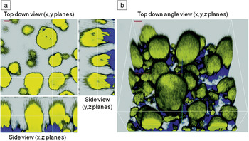
Both flagella and Type IV pili motility are important to biofilm development. These appendages serve to assist in attachment of cells to surfaces and in additional stages of biofilm development. For example, many P. aeruginosa biofilms show the development of “mushroom caps” (e.g., Figure 4b). These caps form under certain environmental conditions (namely sufficient iron and certain carbon sources) and require both flagella and Type IV pili. Interestingly, the Type IV pili appear capable of sensing extracellular DNA present from the initiating biofilm cells.Reference Barken, Pamp, Yang, Gjermansen, Bertrand, Klausen, Givskov, Whitchurch, Engel and Tolker-Nielsen47, Reference Klausen, Aaes-Jørgensen, Mølin and Tolker-Nielsen48
Mushroom-shaped multicellular biofilm structures with yellow caps and cyan or yellow stalks. Confocal laser scanning microscope images were acquired in a four-day-old biofilm that was initiated with a 1:1 mixture of yellow fluorescent P. aeruginosa PA01 wild type and cyan fluorescent P. aeruginosa pilA derivative and grown on glucose minimal medium. Red scale bars, 20 μm.Reference Klausen, Aaes-Jørgensen, Mølin and Tolker-Nielsen48

Sliding is the least understood of these surface motilities. P. aeruginosa has been shown to be surface motile even for a flagellum Type IV pili double-mutant (that is missing the genes necessary to synthesize both a flagellum and type IV pili). The requirement for the global regulatory gene, retS,Reference Murray and Kazmierczak38 for sliding motility may suggest a specific link to sensing of metabolic cues and biofilm formation, as retS has also been linked to such activity.
Why would bacteria want to move in structured communities, and what are the signals/cues they are responding to?
Work done in the last decade has transformed our view of biofilms as being communities of static cells, spatially constrained on the surface. We found that biofilm communities can consist of actively motile and immobile subpopulations. Klausen et al. demonstrated that growth on defined medium in glucose gave rise to an interesting phenomenon mentioned previously, where an immobile aggregate of cells could serve as a “stalk,” which is subsequently colonized by a motile population that crawls up the stalk, forming a “cap” (see Figure 4).Reference Klausen, Aaes-Jørgensen, Mølin and Tolker-Nielsen48 Additionally, motility has been shown to be a feature of the microbiology of other spatially structured systems. One example is the photosynthetic mats observed in regions of geothermal activity, such as those seen in Yellowstone National Park. In these photosynthetic mats (which exhibit many properties of biofilms), phototrophic bacteria move up and down vertically within the mat in response to changing light gradients during the day.
Of course, one of the primary benefits to being motile is the ability to move toward and away from favorable/unfavorable compounds, a process called “chemotaxis.” Thus, it makes sense that being able to move within a biofilm community, which is subject to numerous chemical gradients, would be a tremendous advantage. One particular scenario where motility may be important relates to the acquisition of nutrients. Nutrients can quickly become limiting within the interiors of the dense, large, cell aggregates present in biofilms. Bacteria might favorably reposition themselves in the biofilm community in response to changing nutritional gradients.Reference Nielsen, Tolker-Nielsen, Barken and Molin49 The movement of phototrophs (organisms that use light for energy) within photosynthetic mats may be an example of this, with the phototrophs repositioning themselves to optimally utilize their energy source, light. Therefore, from this standpoint, motile species might possess a distinct advantage over non-motile species in a mixed species biofilm community.
Another potential compound that could influence motility is intercellular signaling compounds, such as acyl-HSLs. Although signal taxis has not been shown to occur in P. aeruginosa, the quorum sensing control of motility has been shown to occur in a variety of bacterial species. As signal concentrations build in a biofilm community, one response to high cell densities may be the induction of surface motility. This might serve to regulate local population densities in a biofilm. To regulate local population densities, quorum sensing might allow biofilm bacteria to leave the local environment when population densities are high and, presumably, ambient nutrient conditions are not favorable for continuous growth.
Summary
Some key questions remain unanswered for the contribution of cell-cell signaling and motility to biofilm development: What cues attachment of cells to surfaces? When do motile communities transition to sessile biofilm cells? Which community signals regulate these processes?
In addition to improving upon the more “classical” microbiology and molecular techniques used to study bacteria, answering the many remaining questions of biofilm development will likely require an interdisciplinary approach that draws upon methods from several scientific disciplines.
Our understanding of biofilm development has already been advanced using mathematical in silico experiments. Predictive mathematical simulations can be utilized to assess the actions of single cells within groups. The impact of these mathematical experiments to discern biological events continues to be realized with better representation of molecular and microscale variables in descriptive equations and improvements in computational speed to simulate greater complexity.
There is a need to improve our understanding of the chemistry between the organic and inorganic constituents of attachment surfaces, with biochemical constituents presented by biofilm bacteria. Research in surface chemistry and biochemistry will continue to be useful to study cell attachment to surfaces and other cells.
Biofilms represent an intersection of biology with physical and chemical processes. Biophysics research will continue to be important for understanding the mechanics of cell movement and the coordination among these bacterial communities. Such analysis will also be useful to understand the many molecular events transpiring during biofilm development, including quorum sensing signal diffusion, matrix assembly of polysaccharides, interaction of cell surface molecules with surfaces, and extraction of nutrients by cells from surfaces in a dynamic environment.
Acknowledgments
This work was supported, in part, by MCB0822405 (NSF) and R01 A1077628 (NIH) to M.R.P. Joshua Shrout is supported by the Indiana Clinical and Translational Science Institute (NIH # UL1RR025761).