Introduction
Percutaneous balloon microcompression is one of the methods used to treat trigeminal neuralgia. This procedure, which was first introduced by Mullan and Lichtor in 1983, is performed by inserting a balloon catheter through the foramen ovale into the region of the Gasserian ganglion and inflating the balloon for 1–10 minutes.Reference Mullan and Lichtor1, Reference Lichtor and Mullan2 Percutaneous balloon microcompression improves the symptoms of trigeminal neuralgia by selectively injuring the myelin sheath of the large myelinated fibres that mediate light touch.Reference Strojnik and Smigoc3 It is indicated for patients with trigeminal neuralgia which proves intractable despite medical therapy, or when the side effects of medications exceed the patient's tolerability.Reference Strojnik and Smigoc3
This paper reports a case in which patulous eustachian tube was a complication of percutaneous balloon microcompression performed for trigeminal neuralgia.
Case report
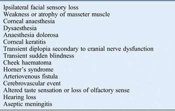
A 41-year-old man with highly active multiple sclerosis and left-sided trigeminal neuralgia was treated with percutaneous balloon microcompression of the left Gasserian ganglion. The treatment had good results in terms of pain relief. However, two weeks after the procedure, the patient experienced intermittent episodes of autophony (defined as an abnormal appreciation of one's own bodily sounds, notably voice, breathing and heartbeat) in the left ear that lasted from 30 minutes to 2 hours. He described hearing his own voice and breath sounds, which could be relieved by lying down. He also developed left-sided facial numbness and experienced difficulty in mastication; these are recognised complications of percutaneous balloon microcompression (Table I).
Percutaneous balloon microcompression complicationsReference Strojnik and Smigoc3, Reference Muñoz, Aedo and Der4

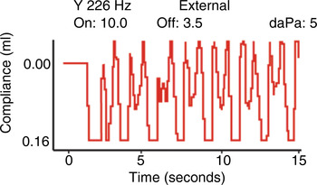
The patient was seen in the audiovestibular medicine out-patient clinic. Otoscopic examination revealed free movement of the left tympanic membrane during respiration. The tympanic membrane (on either side) appeared otherwise normal. Audiometry was within normal limits (Figure 1). Compliance of the left middle ear was recorded; this showed a respiration-synchronous pattern (Figure 2). Subsequent examination of the postnasal space and palate was normal. These findings were consistent with a diagnosis of patulous eustachian tube.
Pure tone audiogram (250–8000 Hz).

Middle-ear compliance recording (left ear) showing a respiration-synchronous pattern (maximum compliance = 0.40 ml).

The mechanism and benign nature of the symptoms were explained to the patient, and he was referred to the otolaryngology clinic. Here, at review six months after the percutaneous balloon microcompression, the patient reported that his symptoms had resolved.
Discussion
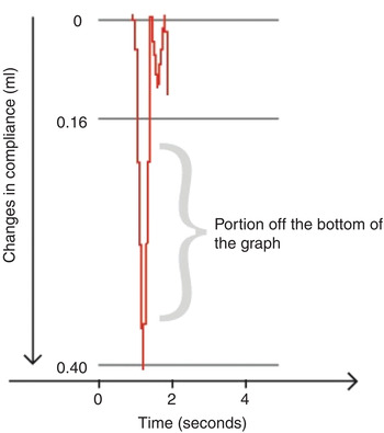
Patulous eustachian tube is defined as a permanently open eustachian tube. It was first described by Schwartze in 1864, following his observation of a fibrous and atrophic eardrum that moved synchronously with breathing.Reference Muñoz, Aedo and Der4, Reference Grimmer and Poe5 Typical symptoms are autophony, a sense of aural fullness and tinnitus, often pulsatile.Reference Muñoz, Aedo and Der4 The cyclical changes in middle-ear compliance during the breath cycle can be recorded in patulous eustachian tube patients using tympanometry with steady pressure (Figures 2 and 3).
Reconstruction of middle ear compliance during a complete breath cycle: inspiration (down slope) and expiration (up slope).

After percutaneous balloon microcompression, patients may experience sensory and motor deficit to areas innervated by the trigeminal nerve branches. The tensor veli palatini is innervated by the medial pterygoid nerve, a branch of the mandibular division of the trigeminal nerve, and is the only muscle of the palate not innervated by the vagus nerve. This muscle is attached to the lateral cartilaginous lamina of the eustachian tube and assists in its opening during swallowing or yawning to allow air pressure to equalise across the tympanic membrane.Reference Lopez, Hamlyn and Zakzewska6
Percutaneous balloon microcompression could cause dysfunction of the tensor veli palatini muscle secondary to injury of the medial pterygoid nerve. The exact pathophysiological mechanism in this case is unclear, but the eustachian tube orifice was clearly dysfunctional after percutaneous balloon microcompression. It is possible that the tensor veli palatini was in a (tonic) state of hyperfunction, rather than palsy, as a result of percutaneous balloon microcompression, thus giving rise to patulous eustachian tube.
• Percutaneous balloon microcompression is used for refractory trigeminal neuralgia
• The procedure is associated with transient sensory and motor deficits of trigeminal nerve branches
• This case report shows an association between percutaneous balloon microcompression and patulous eustachian tube
• A history of intermittent autophony and measurement of middle-ear compliance are key for patulous eustachian tube diagnosis
• In this case report, the patulous eustachian tube symptoms resolved within six months
We did not find any reports of similar cases. This suggests either that patulous eustachian tube is an extremely rare complication of percutaneous balloon microcompression or that it has not been reported because of the transient nature of the symptoms.