





Introduction
Idiopathic pulmonary fibrosis (IPF) is a chronic interstitial lung disease of unknown aetiology, characterized by progressive dyspnoea and irreversible decline in pulmonary function (Ref. Reference Raghu, Remy-Jardin, Myers, Richeldi, Ryerson, Lederer, Behr, Cottin, Danoff, Morell, Flaherty, Wells, Martinez, Azuma, Bice, Bouros, Brown, Collard, Duggal, Galvin, Inoue, Jenkins, Johkoh, Kazerooni, Kitaichi, Knight, Mansour, Nicholson, Pipavath, Buendía-Roldán, Selman, Travis, Walsh and Wilson1). Studies have demonstrated significant regional variations in the epidemiological data of IPF, with incidence rates ranging from 0.09 to 1.30 per 10,000 population and prevalence rates ranging from 0.33 to 4.51 per 10,000 population (Ref. Reference Maher, Bendstrup, Dron, Langley, Smith, Khalid, Patel and Kreuter2). Currently, the treatment of IPF is primarily based on pharmacotherapy. Specifically, the antifibrotic agents approved by the U.S. Food and Drug Administration (FDA) approach the disease through distinct mechanisms: pirfenidone attenuates fibroblast proliferation and collagen synthesis by inhibiting transforming growth factor-β (TGF-β) signalling (Ref. Reference Conte, Gili, Fagone, Fruciano, Iemmolo and Vancheri3), whereas nintedanib is a multitargeted tyrosine kinase inhibitor targeting the vascular endothelial growth factor (VEGF) receptor, fibroblast growth factor (FGF) receptor and platelet-derived growth factor (PDGF) receptor pathways (Ref. Reference Wollin, Wex, Pautsch, Schnapp, Hostettler, Stowasser and Kolb4). However, these established agents, along with emerging therapies like nerandomilast, can only slow the progression of IPF but are unable to reverse pulmonary function impairment, leading to a poor median survival (Refs Reference King, Bradford, Castro-Bernardini, Fagan, Glaspole, Glassberg, Gorina, Hopkins, Kardatzke, Lancaster, Lederer, Nathan, Pereira, Sahn, Sussman, Swigris and Noble5–Reference Richeldi, Azuma, Cottin, Kreuter, Maher, Martinez, Oldham, Valenzuela, Clerisme-Beaty, Gordat, Wachtlin, Liu, Schlecker, Stowasser, Zoz and Wijsenbeek7).
Crucially, IPF is an independent risk factor for LC. Patients with IPF have a 3.34-fold higher incidence than the general population and a markedly shorter median survival than those with LC alone (Ref. Reference Yoon, Nouraie, Chen, Zou, Sellares, Veraldi, Chiarchiaro, Lindell, Wilson, Kaminski, Burns, Trejo Bittar, Yousem, Gibson and Kass8). Large-scale epidemiological data suggest that the prevalence of LC in IPF patients is approximately 10%, and the all-cause mortality of patients with IPF associated with lung cancer (IPF-LC) is significantly higher than that of patients with IPF alone (Ref. Reference Karampitsakos, Spagnolo, Mogulkoc, Wuyts, Tomassetti, Bendstrup, Molina-Molina, Manali, Unat, Bonella, Kahn, Kolilekas, Rosi, Gori, Ravaglia, Poletti, Daniil, Prior, Papanikolaou, Aso, Tryfon, Papakosta, Tzilas, Balestro, Papiris, Antoniou, Bouros, Wells, Kreuter and Tzouvelekis9). IPF patients who develop LC are predominantly elderly males with a smoking history, typically presenting with non-small–cell lung cancer (NSCLC) that preferentially arises within or adjacent to fibrotic areas (Ref. Reference Yoo, Jeong, Chung, Lee, Kwon and Chung10). Notably, evidence indicates that antifibrotic therapy in IPF may reduce both the incidence of LC and cancer-related mortality (Ref. Reference Naoi, Suzuki, Mori, Aono, Kono, Hasegawa, Yokomura, Inoue, Hozumi, Karayama, Furuhashi, Enomoto, Fujisawa, Nakamura, Inui, Nakamura and Suda11). These findings suggest that IPF and LC may share common signalling pathways that foster a microenvironment conducive to both fibrogenesis and tumorigenesis. A recent systematic review elucidated the shared pathogenesis between IPF and LC, detailing their common genetic and epigenetic alterations, aberrant activation of signalling pathways such as TGF-β and PI3K–AKT–mTOR, and overlapping cellular mechanisms including fibroblast activation and epithelial–mesenchymal transition (EMT), thereby underscoring the link of their convergent pathobiology (Ref. Reference Zhang, Qi, Wei, Li and Tian12).
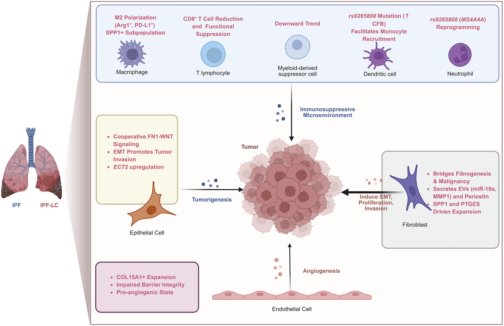
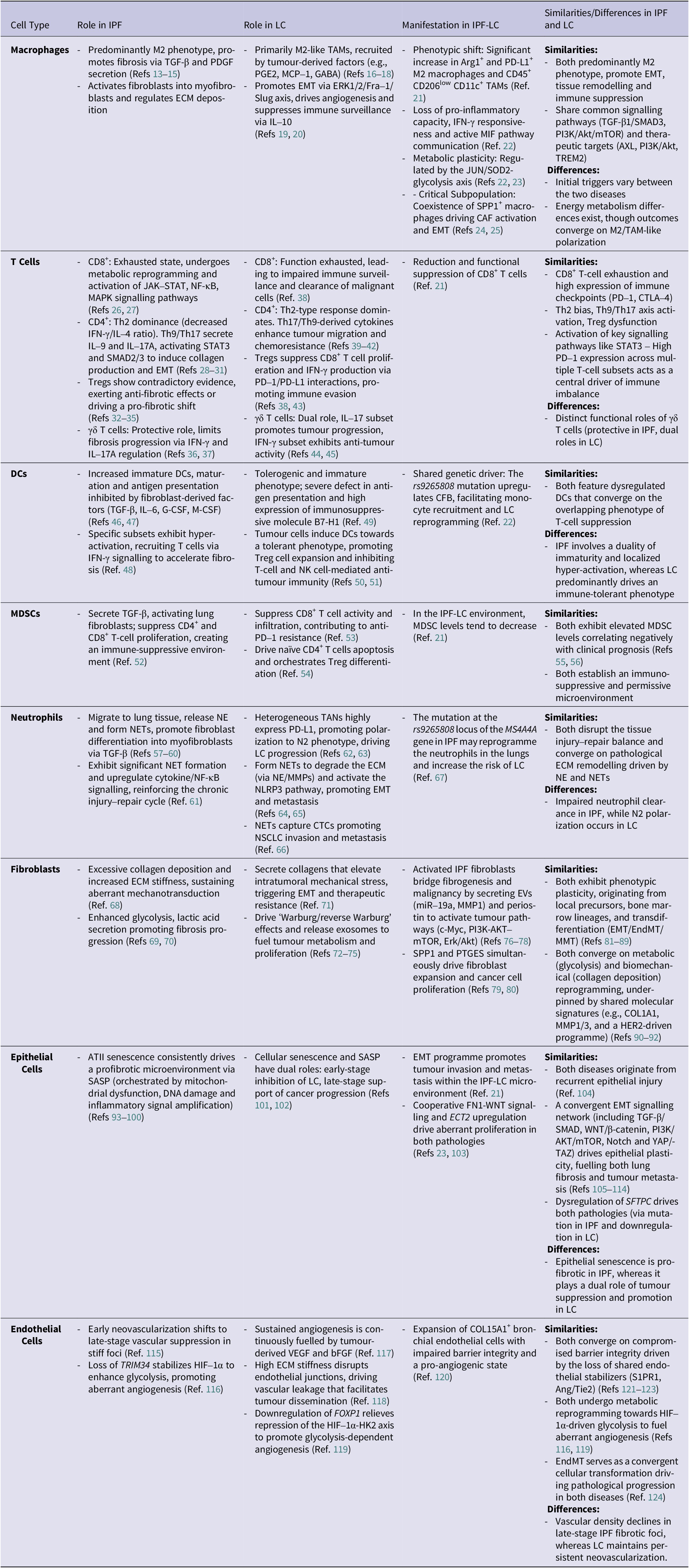
Despite these insights, the functional states and phenotypic transitions of specific cells within the shared IPF and LC microenvironments remain poorly defined. While current literature largely focuses on shared signalling pathways and generalized molecular mechanisms, this review distinctly explores their co-pathogenesis from a cell-type–specific perspective. By systematically comparing shared cellular components and mechanisms, we elucidate how their convergence drives disease onset and progression within the comorbid IPF-LC microenvironment (Figure 1). Given the limited research on the joint microenvironment of IPF-LC, part of this discussion is derived from comparative analyses of cellular behaviours and mechanisms in the respective microenvironments of IPF and LC, identifying overlapping features between the two (Table 1). Ultimately, we hope this work will provide new perspectives for the discovery of early diagnostic biomarkers and the development of therapeutic strategies targeting the shared regulatory pathways underlying fibrosis and tumour development.
The microenvironment of IPF-LC (created with BioRender.com). Notes: ↑indicates upregulation; CFB: complement factor B; ECT2: epithelial cell transforming sequence 2; EMT: epithelial–mesenchymal transition; EVs: extracellular vesicles; IPF: idiopathic pulmonary fibrosis; IPF-LC: idiopathic pulmonary fibrosis associated with lung cancer; MMP1: matrix metalloproteinase 1; PTGES: prostaglandin E synthase; SPP1: secreted phosphoprotein 1.

Figure 1. Long description
At the center of the diagram is a large, irregular pink mass labeled Tumor. Surrounding this central mass are four primary cellular zones that interact with it via arrows.
1. Top Zone: A blue box labeled Immunosuppressive Microenvironment contains five immune cells. From left to right: a Macrophage (M 2 Polarization, Arg 1 plus, P D dash L 1 plus, S P P 1 plus subpopulation); a T lymphocyte (C D 8 plus T Cell Reduction and Functional Suppression); a Myeloid-derived suppressor cell (Downward Trend); a Dendritic cell (r s 9 2 6 5 8 0 8 Mutation, up-arrow C F B, Facilitates Monocyte Recruitment); and a Neutrophil (r s 9 2 6 5 8 0 8 M S 4 A 4 A Reprogramming). An arrow points from this zone down toward the tumor.
2. Right Zone: A purple star-shaped Fibroblast is shown next to a gray box. Text indicates it Bridges Fibrogenesis and Malignancy, Secretes E V s (m i R dash 1 9 a, M M P 1) and Periostin, and has S P P 1 and P T G E S Driven Expansion. An arrow points from the fibroblast to the tumor labeled Induce E M T, Proliferation, Invasion.
3. Bottom Zone: A layer of flat pink Endothelial Cells is shown at the base. An arrow points upward to the tumor labeled Angiogenesis. A pink box to the left lists C O L 1 5 A 1 plus Expansion, Impaired Barrier Integrity, and Pro-angiogenic State.
4. Left Zone: An orange Epithelial Cell is shown next to a yellow box. The box lists Cooperative F N 1 dash W N T Signaling, E M T Promotes Tumor Invasion, and E C T 2 upregulation. An arrow points from this cell to the tumor labeled Tumorigenesis.
On the far left, a small anatomical illustration compares a healthy lung labeled I P F to a lung with a dark mass labeled I P F dash L C.
The microenvironment of IPF and LC

Table 1. Long description
The table details the roles of eight cell types in I P F and L C:
* Macrophages: In I P F, predominantly M2 phenotype promotes fibrosis via T G F-beta. In L C, M2-like T A Ms promote E M T and suppress immune surveillance. In I P F-L C, there is a phenotypic shift to Arg 1 super plus and P D-L 1 super plus M2 macrophages.
* T Cells: Both diseases show C D 8 super plus exhaustion and Th 2 dominance. I P F features protective gamma delta T cells, while L C shows a dual role for them.
* D Cs: Both feature immature phenotypes and T-cell suppression. I P F involves localized hyper-activation, while L C is predominantly immune-tolerant.
* M D S Cs: Both exhibit elevated levels that establish an immunosuppressive environment correlating with poor prognosis.
* Neutrophils: Both converge on pathological E C M remodeling via N E and N E Ts. I P F shows impaired clearance, while L C shows N 2 polarization.
* Fibroblasts: Both exhibit phenotypic plasticity and metabolic reprogramming toward glycolysis and excessive collagen deposition.
* Epithelial Cells: Both originate from recurrent injury and convergent E M T signaling. A T I I senescence is profibrotic in I P F but has a dual role in L C.
* Endothelial Cells: Both involve compromised barrier integrity and H I F-1 alpha-driven glycolysis. Vascular density declines in late I P F but persists in L C.
A comprehensive literature search was performed across three major databases: PubMed, Web of Science and Google Scholar, covering the period from January 2005 to December 2025. The search strategy included combinations of keywords such as ‘idiopathic pulmonary fibrosis’, ‘lung cancer’, ‘microenvironment’ and ‘cellular mechanisms’, alongside specific cell types such as macrophages, T cells, dendritic cells (DCs), myeloid-derived suppressor cells (MDSCs), neutrophils, fibroblasts, epithelial cells and endothelial cells. Studies were selected based on their relevance to immune and stromal cell behaviour, shared pathogenic pathways and translational therapeutic targets in both diseases.
Macrophages
Phenotypic convergence and shared signalling networks
Macrophages are key participants in innate immunity and in the maintenance of pulmonary homeostasis, exhibiting high plasticity across different pathological contexts (Ref. Reference Aegerter, Lambrecht and Jakubzick125). During the progression of IPF, macrophages act as the primary regulatory hub within the immune–cell interaction network by communicating intensely with specific populations, including T cells, natural killer T (NKT) cells, neutrophils and DCs (Ref. Reference Lv, Gao, Ma, Liu, Tian, Yang, Li, Zhao, Li, Zhang, Zhu, Zhang and Wu126). Furthermore, they play pivotal roles in the establishment of the tumour-associated microenvironment and in tumour immune evasion, invasion and metastasis (Ref. Reference De Zuani, Xue, Park, Dentro, Seferbekova, Tessier, Curras-Alonso, Hadjipanayis, Athanasiadis, Gerstung, Bayraktar and Cvejic127). In both IPF and LC, M2 polarization emerges not merely as a phenotypic shift but as a convergent molecular response driven by shared signalling networks under chronic tissue injury.
Under disease-associated signals, macrophages predominantly polarize into the M2 phenotype. Studies have shown that in IPF-LC mouse models, M2-type macrophages with high expression of Arg1 and programmed death-ligand 1 (PD-L1) are markedly increased, accompanied by a reduction and functional suppression of CD8+ T cells. A prevailing consensus in the literature indicates that these alterations collectively create a profoundly immunosuppressive microenvironment that accelerates tumour progression (Ref. Reference Barravecchia, Lee, Manassa, Magnuson, Ferris, Cavanaugh, Steele, Espinoza, Galban, Ramnath, Frankel, Pasca di Magliano and Galban21). However, it remains unclear whether this M2 polarization reflects identical epigenetic alterations in both diseases or independent processes that merely overlap in their downstream fibrotic and immunosuppressive phenotypes.
Individually, M2 macrophages in IPF drive fibrogenesis by secreting profibrotic mediators such as TGF-β and PDGF, which activate fibroblasts via shared gene regulatory networks, including TGF-β1/SMAD3 and Wnt/β–catenin pathways (Refs Reference Hou, Shi, Chen, Lv, Chen, Cao, Xiang and Han13–Reference Wu, Zhang, Jiang, He, Zhang, Chen, Tong, Nie and Pang15). Similarly, in LC, M2-like tumour-associated macrophages (TAMs) predominate, recruited by tumour-derived factors such as prostaglandin E2 (PGE2), monocyte chemoattractant protein-1 (MCP-1) and gamma-aminobutyric acid (GABA) (Refs Reference Dong, Wang, Nie, Xu, Bai, Lu, Jian, Wang and Zheng16–Reference Kawaguchi, Ohshio, Watanabe, Shiratori, Okamoto, Ueda, Kataoka, Suzuki and Hanaoka18). These TAMs remodel the tumour microenvironment (TME) by promoting EMT via the ERK1/2/Fra-1/Slug axis, stimulating angiogenesis and suppressing immune surveillance through IL-10 secretion (Refs Reference Guo, Song, Hao, Zhao, du, Li, Kuang, Yang, Wang, Deng and Wang19, Reference Zhao, Zhang, Liu, Xue, Chen, Zou and Jiang20). Despite varying initial triggers, IPF and LC converge on similar macrophage-driven cascades to maintain a fibrotic and tumorigenic microenvironment.
Metabolic reprogramming and immune suppression
In the coexisting IPF-LC microenvironment, macrophages not only undergo phenotypic changes but also exhibit features of functional suppression and altered metabolic states. During IPF-to-LC progression, single-cell RNA sequencing (scRNA-seq) and genetic analyses reveal that macrophages lose their pro-inflammatory capacity and IFN-γ responsiveness, accompanied by widespread T- and B-cell suppression. Moreover, fibroblasts can communicate with macrophages via the highly active macrophage migration inhibitory factor (MIF) pathway, ultimately orchestrating an immune-evasive microenvironment (Ref. Reference Gu, Xu, Jiao, Cai, Gu, Wu, Cheng, Chen, Chen and Liu22). Notably, in microenvironments where IPF and LC coexist, macrophages exhibit pronounced metabolic plasticity. Genetically, this metabolic reprogramming is tightly regulated by shared transcriptional networks, such as the JUN/SOD2-glycolysis axis, while simultaneously polarizing towards a profibrotic phenotype driven by elevated lipid metabolism and oxidative phosphorylation. Despite the differences in energy metabolism, the outcome converges on the polarization of macrophages towards M2/TAM-like phenotypes. These reprogrammed macrophages secrete multiple immunoregulatory and profibrotic factors, including TGF-β, IL-10 and CCL18, thereby promoting EMT and the formation of an immunosuppressive microenvironment, ultimately driving the comorbidity of IPF and LC (Refs Reference Gu, Xu, Jiao, Cai, Gu, Wu, Cheng, Chen, Chen and Liu22, Reference Ren, Zi, Zhang, Liao and Chen23).
Characterization of shared cellular subpopulations
Macrophages play a key role in both fibrosis and tumour progression, and based on this, researchers have gradually identified specific macrophage subpopulations in the IPF-LC microenvironment. For instance, Barravecchia et al. found a distinct subpopulation of TAMs (CD45+, CD206low, CD11c+) significantly increased in the IPF-LC mouse model, suggesting the presence of polarized macrophage populations in the fibrosis–cancer coexisting microenvironment (Ref. Reference Barravecchia, Lee, Manassa, Magnuson, Ferris, Cavanaugh, Steele, Espinoza, Galban, Ramnath, Frankel, Pasca di Magliano and Galban21). Among the various macrophage subpopulations, secreted phosphoprotein 1 (SPP1)+ macrophages have garnered significant attention from researchers. Analysis based on the Human Lung Cell Atlas (HLCA) revealed the coexistence of SPP1+ profibrotic monocyte-derived macrophages in both pulmonary fibrosis and LC (Ref. Reference Sikkema, Ramírez-Suástegui, Strobl, Gillett, Zappia, Madissoon, Markov, Zaragosi, Ji, Ansari, Arguel, Apperloo, Banchero, Bécavin, Berg, Chichelnitskiy, Chung, Collin, Gay, Gote-Schniering, Hooshiar Kashani, Inecik, Jain, Kapellos, Kole, Leroy, Mayr, Oliver, von Papen, Peter, Taylor, Walzthoeni, Xu, Bui, de Donno, Dony, Faiz, Guo, Gutierrez, Heumos, Huang, Ibarra, Jackson, Kadur Lakshminarasimha Murthy, Lotfollahi, Tabib, Talavera-López, Travaglini, Wilbrey-Clark, Worlock, Yoshida, Chen, Hagood, Agami, Horvath, Lundeberg, Marquette, Pryhuber, Samakovlis, Sun, Ware, Zhang, van den Berge, Bossé, Desai, Eickelberg, Kaminski, Krasnow, Lafyatis, Nikolic, Powell, Rajagopal, Rojas, Rozenblatt-Rosen, Seibold, Sheppard, Shepherd, Sin, Timens, Tsankov, Whitsett, Xu, Banovich, Barbry, Duong, Falk, Meyer, Kropski, Pe’er, Schiller, Tata, Schultze, Teichmann, Misharin, Nawijn, Luecken and Theis24). Further, Chen et al. utilized scRNA-seq to identify that SPP1+ macrophages, located at the terminal end of the developmental trajectory, may play a key role in driving the progression from IPF to LC. The underlying mechanism may involve not only the induction of fibroblast transformation into cancer-associated fibroblasts (CAFs), but also interactions with these inflammation-associated CAFs and stimulation of EMT in alveolar epithelial cells (Ref. Reference Chen, Guo, Ai, Wang, Wang, Chen, Liu, Zhuang, Liu, Zhao, Liang and Gu25). These findings establish SPP1+ macrophages as a mechanistic link bridging fibrogenesis and tumorigenesis, rather than representing a phenotypic overlap.
Clinical implications and targeted therapies
Consequently, targeting these shared populations or their regulatory pathways, such as AXL, PI3K/Akt or triggering receptor expressed on myeloid cells-2 (TREM2), represents a promising therapeutic strategy (Refs Reference Kim, Kim, Im, Baek, Choi, Lee, Kim, Lee, Jeong, Ban, Kim, Ji, Lee, Kim, Lee, Yang, Yun, Choi and Rho128–Reference Li, Yuan, Li, He, Shen, Luo, Tai, Li, Deng and Luo131). Given the critical role of macrophages in the IPF and LC microenvironments, expanding efforts are directed towards macrophage-targeted therapies. Liu et al. developed a novel delivery system for treating LC combined with pulmonary fibrosis: macrophage membrane-coated rapamycin micelles (MM@TPGS/F127@RAPA) administered via intratracheal instillation. The design leverages the high similarity in macrophage biological behaviours between IPF and LC, including the recruitment of macrophages mediated by similar chemokines and the immune microenvironment characterized by M2 polarization. The encapsulated rapamycin targets shared signalling pathways, such as TGF-β1/SMAD3 and PI3K/AKT/mTOR, thereby enabling precise drug delivery and synergistic therapy (Ref. Reference Liu, Cai, Sun, Zhao, xu, Gu, Peng, di, Zhou, Gu, Ai and Yang132). Notably, with the continuous identification of specialized macrophage subsets, such as SPP1+ macrophages, targeting these cells that exhibit both profibrotic and pro-tumour characteristics holds promise as a new treatment approach to intervene in fibrosis and reduce the risk of LC.
T cells
In addition to macrophages, T cells constitute another essential component of the immune system and play pivotal roles in the pathogenesis and immune responses of both IPF and LC. Based on surface markers, they are broadly classified into CD8+ and CD4+ T cells, both of which undergo significant functional dysregulation in the disease microenvironment.
CD8+ T cells
As classical cytotoxic effector cells, CD8+ T cells primarily function by eliminating infected or malignant cells. However, in IPF, their function becomes markedly dysregulated. Although CD8+ T cells are abundantly infiltrated in the lung tissue of patients with IPF, they do not exhibit effective cytotoxic activity. Instead, subsets of these cells display features of exhaustion, undergoing metabolic reprogramming and activation of signalling pathways such as JAK–STAT, NF–κB and MAPK, thereby promoting fibroblast activation and driving the progression of fibrosis (Ref. Reference Wei, Jin, Li, Wang, Zheng, Feng, shi, Kong, Ma and Wang26). In both diseases, exhausted CD8+ T cells highly express immune checkpoint molecules such as programmed cell death protein 1 (PD-1) and cytotoxic T lymphocyte–associated protein 4 (CTLA4), which impair immune surveillance and the clearance of senescent or malignant cells (Refs Reference Yadav, Anbalagan, Khatun, Prabhakaran, Matsunaga, Manges, McLachlan, Lasky, Kolls and Thannickal27, Reference Guo, Zhang, Zheng, Zheng, Song, Zhang, Kang, Liu, Jin, Xing, Gao, Zhang, Dong, Hu, Ren, Kirchhoff, Roider, Yan and Zhang38). This progressive loss of anti-fibrotic and anti-tumour capacity represents a crucial mechanism for chronic disease progression and a primary target for immunotherapy.
CD4+ T cells
CD4+ T cells possess a variety of immune regulatory functions, primarily categorized into T helper (Th) cells and regulatory T cells (Tregs). A consistent consensus in the literature indicates that both IPF and LC are characterized by a systemic shift towards Th2 dominance, representing a shared immune–metabolic adaptation. In bleomycin (BLM)-induced pulmonary fibrosis, this is evidenced by a significant reduction in Th1 cytokine IFN-γ levels and a notable increase in Th2 cytokine IL-4 levels, leading to a decreased IFN-γ/IL-4 ratio (Refs Reference Xing, Qiang, Bao, Yang, Hou, Tao, Meng, Zhang, Zhang and Sun28, Reference Mojiri-Forushani, Hemmati, Khodadadi and Rashno29). Similarly, in the TME of NSCLC, a distinct Th2 skewing is observed, with Th2 and Treg cells predominating in both the tumour epithelium and stroma, while Th1 cells are significantly reduced (Ref. Reference Frafjord, Buer, Hammarström, Aamodt, Woldbæk, Brustugun, Helland, Øynebråten and Corthay39).
Other CD4+ subsets, including Th9 and Th17 cells, act as pathogenic drivers by activating STAT3 and SMAD2/3 signalling. Specifically, IL-9 and IL-17A secreted by these cells induce collagen production in fibroblasts, promote EMT in epithelial cells and enhance tumour cell migration and chemoresistance (Refs Reference Deng, Yang, Luo, She, Yu and Tang30, Reference Celada, Kropski, Herazo-Maya, Luo, Creecy, Abad, Chioma, Lee, Hassell, Shaginurova, Wang, Johnson, Kerrigan, Mason, Baughman, Ayers, Bernard, Culver, Montgomery, Maher, Molyneaux, Noth, Mutsaers, Prele, Stokes Peebles, Newcomb, Kaminski, Blackwell, van Kaer and Drake31, Reference Wang, Yang, Zhang, Wang, Zhang, He, Chen, Zhang, Qin, Wang and Zhang40–Reference Heim, Yang, Tausche, Hohenberger, Chiriac, Koelle, Geppert, Kachler, Miksch, Graser, Friedrich, Kharwadkar, Rieker, Trufa, Sirbu, Neurath, Kaplan and Finotto42).
Tregs play a central role in maintaining immune system homeostasis, but their function is disrupted in both IPF and LC. However, the literature presents contradictory evidence regarding their precise role in fibrosis. While some studies suggest Tregs exert anti-fibrotic effects, others indicate they undergo phenotypic instability and drive a pro-fibrotic shift from Th1 to Th2 cells (Refs Reference Kamio, Azuma, Matsuda, Usuki, Inomata, Morinaga, Kashiwada, Nishijima, Itakura, Kokuho, Atsumi, Hayashi, Yamaguchi, Fujita, Saito, Abe, Kubota and Gemma32–Reference Birjandi, Palchevskiy, Xue, Nunez, Kern, Weigt, Lynch, Chatila and Belperio35). Furthermore, in the LC microenvironment, activated Tregs directly inhibit CD8+ T-cell proliferation and IFN-γ production via PD-1 and PD-L1 interactions, thereby promoting immune evasion (Refs Reference Guo, Zhang, Zheng, Zheng, Song, Zhang, Kang, Liu, Jin, Xing, Gao, Zhang, Dong, Hu, Ren, Kirchhoff, Roider, Yan and Zhang38, Reference Kim, Park, Son, Lee, Chung, Cho, Shim, Park, Kim, in Yoon, Kim, Jung, Cho, Park, Rha and Ha43).
γδ T cells
Additionally, non-conventional T cells, such as γδ T cells, are activated in both diseases and participate in early immune responses by secreting cytokines such as IL-17 and IFN-γ, which influence chronic inflammation, fibrosis and TME remodelling. However, their functional roles differ between the two diseases. In the BLM-induced pulmonary fibrosis model, γδ T cells are considered key regulatory cells, playing a protective role in lung fibrosis by producing IFN-γ and suppressing Th17 immune responses (Refs Reference Segawa, Goto, Iizuka, Kaneko, Yokosawa, Kondo, Matsumoto and Sumida36, Reference Russo, Savino, Mirolo, Buracchi, Germano, Anselmo, Zammataro, Pasqualini, Mantovani, Locati and Teixeira37). In contrast, the role of γδ T cells in NSCLC are dualistic: γδ T cells producing IFN-γ exhibit anti-tumour activity and are associated with favourable long-term overall survival, while γδ T17 cells producing IL-17 may promote tumour progression by mediating pro-tumour inflammatory responses (Refs Reference Xu, Ke, Shang, Liu, Fu, Wang and Jin44, Reference Song, Ma, Chen, Miao, Tao and Liu45). It remains unclear whether these contrasting roles reflect disease-specific mechanisms or dynamic phenotypic shifts dictated by the evolving tissue microenvironment.
IPF and LC exhibit several key similarities in T cell-mediated immunity, including CD8+ exhaustion, Th2 skewing, activation of the Th9/Th17 axis and dysfunction of Tregs. Notably, high expression of PD-1 across multiple T-cell subsets acts as a central driver of immune tolerance in both microenvironments. However, there is currently a lack of reports on the exact spatial distribution and epigenetic state of T cells in the coexisting IPF-LC microenvironment. Further multi-omics investigation is needed to elucidate their immune characteristics and provide a foundation for the development of targeted therapeutic strategies.
Dendritic cells
DCs, as classic antigen-presenting cells, play a crucial role in initiating, regulating and maintaining immune responses and microenvironment homeostasis, and thus critically impact pulmonary diseases (Ref. Reference Upham and Xi133). While traditional descriptive studies depict DCs as merely ‘immature’ in IPF and ‘tolerogenic’ in LC, recent genomic evidence reveals a deeper mechanistic link.
A key shared genetic driver is the rs9265808 mutation in DCs, which upregulates complement factor B (CFB). This specific genetic variant represents a shared mechanism that facilitates monocyte recruitment and tumour reprogramming, thereby increasing the risk of LC in patients with preexisting pulmonary fibrosis (Ref. Reference Gu, Xu, Jiao, Cai, Gu, Wu, Cheng, Chen, Chen and Liu22).
Beyond genetics, DCs are heavily remodelled by distinct pathological microenvironments. In IPF, the number of immature DCs is significantly increased, particularly in areas of epithelial hyperplasia and fibrosis (Ref. Reference Marchal-Sommé, Uzunhan, Marchand-Adam, Kambouchner, Valeyre, Crestani and Soler46). Fibroblast-derived factors in IPF, such as TGF-β, IL-6, granulocyte colony-stimulating factor (G-CSF) and macrophage colony-stimulating factor (M-CSF), inhibit DC maturation and antigen presentation, thereby fostering a local immunosuppressive and chronic inflammatory environment that drives the fibrotic process (Ref. Reference Freynet, Marchal-Sommé, Jean-Louis, Mailleux, Crestani, Soler and Michel47). Conversely, some DC subsets in IPF exhibit hyper-activation, recruiting T cells via CCL22, CD40 and CD58 under the dominance of IFN-γ signalling to accelerate fibrosis (Ref. Reference Serezani, Pascoalino, Bazzano, Vowell, Tanjore, Taylor, Calvi, McCall, Bacchetta, Shaver, Ware, Salisbury, Banovich, Kendall, Kropski and Blackwell48). In contrast, in NSCLC, DCs within the TME predominantly shift towards a tolerogenic and immature phenotype, characterized by a severe defect in antigen presentation and high expression of the immunosuppressive molecule B7-H1 (PD-L1) to promote tumour immune escape (Ref. Reference Perrot, Blanchard, Freymond, Isaac, Guibert, Pachéco and Lebecque49). Tumour cells drive this tolerogenic shift by downregulating costimulatory molecules (CD80/CD86) and pro-inflammatory cytokines (IL-12, IL-23), while increasing the secretion of anti-inflammatory cytokines (IL-10, IL-27). Consequently, these reprogrammed DCs facilitate immune evasion by expanding Tregs and suppressing effector T and NK cells (Ref. Reference Lu, Xu, Gu, Chang, Wei, Rong, Qin, Chen and Zhou50). Moreover, CCR8+ Tregs in the TME interact with CCL5+ DCs to inhibit the ability of DCs to produce IL-12 via the JAK–STAT pathway, weakening CD8+ T-cell activation and anti-tumour immunity (Ref. Reference Chen, Wang, Tang, Shi, Cheng, Zhao, Li and Zhou51).
Conceptually, DC dysregulation in IPF and LC likely represents independent processes that ultimately converge on the overlapping phenotype of T-cell suppression. Specifically, this involves both immaturity and hyper-activation in IPF and systemic immune tolerance in cancer. Targeting these pathways offers promising therapeutic avenues. For instance, antigen-targeted DC vaccines have demonstrated feasibility in NSCLC, and modulating the complement pathway may intervene in the transition from IPF to LC (Refs Reference Gu, Xu, Jiao, Cai, Gu, Wu, Cheng, Chen, Chen and Liu22, Reference Ingels, De Cock and Stevens134). Furthermore, as a complementary clinical strategy, managing CFB-related systemic depletion through targeted nutritional and physical support might be beneficial, though this approach requires further validation (Ref. Reference Gu, Xu, Jiao, Cai, Gu, Wu, Cheng, Chen, Chen and Liu22).
Myeloid-derived suppressor cells
MDSCs are a subset of myeloid cells with immunosuppressive functions, and they are primarily classified into two subgroups based on phenotype and morphological characteristics: polymorphonuclear myeloid-derived suppressor cells (PMN-MDSCs) and monocytic myeloid-derived suppressor cells (M-MDSCs) (Ref. Reference Hegde, Leader and Merad135). Their levels are significantly elevated in the peripheral blood and lung tissues of patients with both IPF and LC, where they correlate negatively with pulmonary function and clinical prognosis (Refs Reference Yamauchi, Safi, Blattner, Rathinasamy, Umansky, Juenger, Warth, Eichhorn, Muley, Herth, Dienemann, Platten, Beckhove, Utikal, Hoffmann and Umansky55, Reference Fernandez, Greiffo, Frankenberger, Bandres, Heinzelmann, Neurohr, Hatz, Hartl, Behr and Eickelberg56). In both diseases, MDSCs share common molecular signalling networks that drive disease progression. In IPF, B7H3 secreted by lung fibroblasts, alveolar epithelial cells and macrophages can activate MDSCs. Subsequently, these activated MDSCs induce TGF-β secretion, myofibroblast differentiation and T-cell suppression, thereby promoting both fibrosis and immune evasion (Ref. Reference Liu, Gonzalez De Los Santos, Rinke, Fang, Flaherty and Phan52). Similarly, in NSCLC, local pathogenic signals actively recruit MDSCs. Specifically, intrinsic expression of tripartite motif 28 (TRIM28) in tumour cells activates the NF–κB signalling pathway to upregulate CXCL1, driving MDSC accumulation. These recruited MDSCs subsequently suppress the activity and infiltration of CD8+ T cells, ultimately leading to resistance to anti-PD-1 therapy (Ref. Reference Liang, Sun, Chen, Wang, Wang, Qin, Zhao and Geng53). Furthermore, studies have found that M-MDSCs derived from LC can promote the apoptosis of naïve CD4+ T cells and induce their differentiation into Tregs (Ref. Reference Wan, Mu, Zhao, Li, Xu and Zhang54).
Collectively, MDSCs establish a permissive environment for both fibrogenesis and tumorigenesis by dampening effector immune responses and stimulating pro-fibrotic signalling.
However, this field contains a notable paradox: despite strong consensus on their individual pro-tumorigenic and pro-fibrotic roles, recent in vivo studies reveal a downward trend in MDSC levels within the coexisting IPF-LC microenvironment (Ref. Reference Barravecchia, Lee, Manassa, Magnuson, Ferris, Cavanaugh, Steele, Espinoza, Galban, Ramnath, Frankel, Pasca di Magliano and Galban21). This contradictory finding suggests that immune cell recruitment patterns may shift significantly when fibrotic and TMEs coexist. For instance, it remains to be seen whether other populations take on a more dominant immunosuppressive role in this context. Further studies are needed to clarify these dynamics.
Neutrophils
Neutrophils, traditionally recognized as early responders in tissue injury, can drive chronic inflammation and tissue remodelling when their accumulation persists (Ref. Reference Metzemaekers, Gouwy and Proost136). Crucially, recent genomic evidence reveals that this dysregulation is not merely a secondary inflammatory response, but is associated with a shared genetic susceptibility. Specifically, mutations at the rs9265808 locus in MS4A4A act as a critical genetic link that reprogrammes pulmonary neutrophils, increasing the risk of malignant transformation in patients with preexisting IPF (Ref. Reference Xie, Tan, Chen, Yao, Luo, Ma, Feng and Jiang67).
Beyond their shared genetic drivers, neutrophil dysfunction in IPF and LC exhibits disease-specific patterns that converge on pathological extracellular matrix (ECM) remodelling. Neutrophils are among the first immune cells to accumulate during the early stages of wound healing, and their effective recruitment to the site of injury is essential for initiating the repair and fibrotic processes (Ref. Reference Leslie, Millar, Del Carpio Pons, Burgoyne, Frost, Barksby, Luli, Scott, Simpson, Gauldie, Murray, Finch, Carruthers, Ferguson, Sleeman, Rider, Howarth, Fox, Oakley, Fisher, Mann and Borthwick137). However, when this process is dysregulated, persistent neutrophil accumulation can intensify fibrosis. In BLM-induced pulmonary fibrosis mouse models, neutrophils migrate from the bone marrow to the lung tissue but fail to reverse-migrate, leading to their prolonged survival and persistent activation (Ref. Reference Warheit-Niemi, Huizinga, Edwards, Wang, Murray, O’Dwyer and Moore57). These neutrophils release neutrophil elastase (NE) and neutrophil extracellular traps (NETs), which activate TGF-β and promote the differentiation of pulmonary fibroblasts into myofibroblasts (Refs Reference Takemasa, Ishii and Fukuda58–Reference Gregory, Kliment, Metz, Kim, Kargl, Agostini, Crum, Oczypok, Oury and Houghton60). Clinical evidence from IPF biopsies confirms significant NET formation and upregulation of cytokine/NF-κB signalling pathways, reinforcing the chronic injury–repair cycle (Ref. Reference Divolis, Synolaki, Tringidou, Tzouvelekis, Boumpas, Skendros and Galani61).
Conversely, in NSCLC, neutrophil dysregulation is characterized by phenotypic plasticity rather than mere clearance failure. Regulated by microenvironmental signals like TGF-β, tumour-associated neutrophils (TANs) polarize into either an N1 phenotype with anti-tumour properties or an N2 phenotype with pro-tumour effects (Ref. Reference Fridlender, Sun, Kim, Kapoor, Cheng, Ling, Worthen and Albelda62). N2-type TANs drive LC progression via a positive feedback loop involving IL-10/STAT3 signalling and PD-L1 upregulation (Ref. Reference Zhang, Sun, Zuo and Feng63). While fibrosis is characterized by impaired clearance and cancer by N2 polarization, these distinct processes ultimately converge on the formation of NETs. Within the TME, these NETs promote EMT and metastasis by degrading the ECM through NE and matrix metalloproteinases (MMPs), or by activating the NF-κB/NOD-like receptor protein 3 (NLRP3) pathway (Refs Reference Wang, Liu, Chen, Fang, Li, Yuan, Qian, Yin, Yu, Fu, Zhang and Li64, Reference Lee, Lee, Lawler and Kim65). Furthermore, NETs can capture circulating tumour cells (CTCs) in the vasculature, enhancing their retention and facilitating metastatic site formation (Ref. Reference Cools-Lartigue, Spicer, McDonald, Gowing, Chow, Giannias, Bourdeau, Kubes and Ferri66).
Through the secretion of pro-inflammatory cytokines, NE and the formation of NETs, neutrophils disrupt the balance between tissue injury and repair, thereby driving the pathological progression of both IPF and LC. Consequently, targeting the neutrophil axis through approaches such as inhibiting NE, degrading NETs, promoting neutrophil clearance or modulating the MS4A4A locus represents a promising therapeutic strategy (Ref. Reference Xie, Tan, Chen, Yao, Luo, Ma, Feng and Jiang67). Regarding neutrophil plasticity, it remains unclear how the N1/N2 polarization spectrum adapts when a heavily fibrotic ECM directly intersects with an emerging tumour niche, highlighting the need for further spatial transcriptomic investigation.
Fibroblasts
While immune cells exhibit marked similarities between IPF and LC, non-immune cells within their respective microenvironments also display a series of parallel pathological alterations. In particular, fibroblasts, epithelial cells and endothelial cells interact closely with immune cells, collectively shaping microenvironmental features that drive both fibrotic progression and tumour development.
Cellular origins and phenotypic plasticity
Firstly, from the perspective of cellular origins, fibroblasts in both IPF and LC exhibit pronounced heterogeneity and derive from multiple sources. While resident pulmonary fibroblasts serve as the primary reservoir for expansion, the fibrotic and tumour stroma are significantly augmented by alternative lineages through shared epigenetic reprogramming and transdifferentiation programmes (Refs Reference Cao, Li, Zheng, Zhang, Qian, Ren, Wang, Wang, Wu, Zhang and Xu81, Reference Walsh, Worrell, Fabre, Hinz, Kane and Keane82). Specifically, both microenvironments recruit bone-marrow–derived fibrocytes or mesenchymal stem cells and harness the plasticity of non-mesenchymal cells via EMT and endothelial–mesenchymal transition (EndMT) (Refs Reference Walsh, Worrell, Fabre, Hinz, Kane and Keane82–Reference Wong, Cheung, Chen, Chan, Yu, Lo, Kang and To85). This cellular repertoire is further diversified by the transdifferentiation of pericytes, adipocytes and pleural mesothelial cells undergoing mesothelial–mesenchymal transition (MMT) (Refs Reference Wong, Cheung, Chen, Chan, Yu, Lo, Kang and To85–Reference Liu, Yu, Lu, Cheng, Liang, Wang, Chen, Huang, Song, He, Xiong, Xin, Ma and Ye88). Notably, recent studies have identified SMAD3-driven macrophage-to-myofibroblast transition as a novel mechanism contributing to CAF generation, revealing immune cells as an unexpected progenitor pool (Ref. Reference Tang, Chung, Xue, Xiao, Meng, Huang, Zhou, Chan, Tsang, Cheng, Lee, Leung, Lam, To, Tang and Lan89). These diverse origins reflect a profound phenotypic plasticity, where distinct cellular lineages converge into a unified pathogenic stroma under chronic injury.
Biomechanical and metabolic convergence
Excessive fibroblast activation constitutes a central pathological engine that drives the aberrant ECM remodelling shared by IPF and LC. In both diseases, this activation represents a convergent cellular response to continuous mechanical stress and biochemical stimuli. In IPF, hyperactivated fibroblasts secrete excessive collagens, leading to increased ECM stiffness, which acts as an abnormal mechanotransduction platform that sustains a vicious cycle of fibroblast activation (Ref. Reference Yan, Wang, Su, Zou, Zhou, Shou and Huo68). Similarly, in the TME, myofibroblast-derived collagens such as COL5A1 elevate intratumoral mechanical stress, mechanically triggering EMT and enhancing therapeutic resistance (Ref. Reference Zhu, Wang, Wang, Huang, Li, Chen, Chen, Zhang, Li, Liu and Chen71).
Genetically and metabolically, this structural remodelling is underpinned by shared transcriptional networks. Fibroblasts in both diseases display enhanced glycolysis and lactate secretion. In IPF, the glycolysis driven by 6-phosphofructo-2-kinase/fructose-2,6-biphosphatase 3 (PFKFB3) stabilizes HIF-1α via succinate, promoting fibroblast-to-myofibroblast differentiation (Ref. Reference Xie, Tan, Banerjee, Cui, Ge, Liu, Bernard, Thannickal and Liu69). Additionally, epigenetic modifications play a crucial role: glycolysis-derived lactate functions as a paracrine signal, to induce histone lactylation in macrophages and promote the expression of pro-fibrotic genes (Ref. Reference Cui, Xie, Banerjee, Ge, Jiang, Dey, Matthews, Liu and Liu70). In LC, this metabolic shift manifests as the ‘Warburg’ and ‘reverse Warburg’ effects, where CAFs secrete lactate and energy substrates to support tumour growth, while cancer cell-derived lactate promotes M2 macrophage polarization via CAFs (Refs Reference Cruz-Bermúdez, Laza-Briviesca, Vicente-Blanco, García-Grande, Coronado, Laine-Menéndez, Alfaro, Sanchez, Franco, Calvo, Romero, Martin-Acosta, Salas, Garcia and Provencio72–Reference Duda, Janczara, McCubrey, Gizak and Rakus74). Beyond direct metabolite secretion, CAFs can also modulate tumour metabolism through extracellular vesicle communication. Specifically, CAF-derived exosomes are enriched in long noncoding RNA (lncRNA) ANRIL, which acts as a competing endogenous RNA to sponge miR-186-5p. This relieves the inhibition of hypoxia-inducible factor 1α (HIF-1α), thereby enhancing glycolytic activity and proliferation of LC cells (Ref. Reference Zhang, Huang, Tang and Hu75). This metabolic and structural convergence is underpinned by a shared molecular program. Bioinformatics analyses consistently confirm this molecular convergence, identifying shared hub genes, such as COL1A1, COL3A1, COL4A1, POSTN, TIMP3 and MMP1/3, which are enriched in both IPF fibroblast foci and LC stroma (Refs Reference Kreus, Lehtonen, Skarp and Kaarteenaho90, Reference Yao, Li and Gao91). Furthermore, research has found that a HER2-driven regulatory programme promotes cell invasion and matrix remodelling across both fibrotic foci and lung adenocarcinoma stroma (Ref. Reference Liu, Geng, Liang, Coelho, Yao, Deng, Wang, Dai, Huang, Xie, Liu, Rowan, Taghavifar, Kulur, Liu, Stripp, Hogaboam, Jiang and Noble92).
Intercellular communication in the coexisting microenvironment
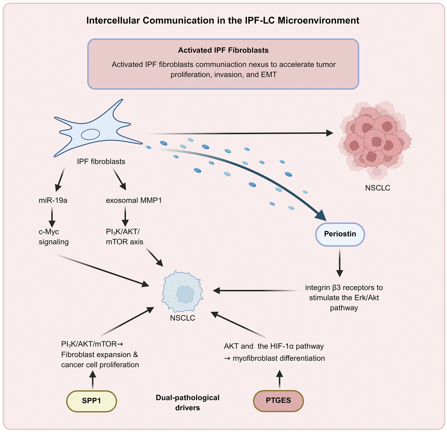
Within the coexisting IPF-LC microenvironment, activated IPF fibroblasts function as a pivotal communication nexus bridging the pathological gap between fibrogenesis and malignancy (Figure 2). This crosstalk is primarily driven by extracellular vesicles (EVs), exosomes and secreted soluble factors. For instance, senescent IPF fibroblasts release EVs enriched with miR-19a to activate c-Myc signalling, while exosomal MMP1 triggers the PI3K–AKT–mTOR axis in NSCLC cells. Collectively, these dual pathways accelerate tumour proliferation, invasion and EMT (Refs Reference Lei, Zhong, Zhang, Zheng, Xu, Li, Huang and Ren76, Reference Fujita, Fujimoto, Miyamoto, Kaneko, Kadota, Watanabe, Kizawa, Kawamoto, Watanabe, Utsumi, Wakui, Minagawa, Araya, Ohtsuka, Ochiya and Kuwano77). Beyond vesicular transport, IPF fibroblasts secrete Periostin, which engages integrin β3 receptors to stimulate the Erk/Akt pathway and fuel tumour progression (Ref. Reference Yamato, Kimura, Fukui, Kanou, Ose, Funaki, Minami and Shintani78). Notably, molecules such as SPP1 and prostaglandin E synthase (PTGES) serve as dual-pathological drivers. While SPP1 activates the PI3K/Akt/mTOR pathway to promote both fibroblast expansion and cancer cell proliferation, PTGES facilitates myofibroblast differentiation by modulating AKT and the HIF-1α pathway (Refs Reference Lin, Lee, Liao, Chou and Yang79, Reference Yue, Xiong, Chen, Yang, Zhao, Shao, Wei, Gao, Huang and Chen80). However, it is still unclear whether IPF-specific myofibroblasts can directly transdifferentiate into tumour-promoting CAFs within this shared niche, or if they represent independent parallel lineages. Elucidating the spatiotemporal transcriptomic trajectories of these specific stromal cells is urgently needed.
Intercellular communication of fibroblasts in the IPF-LC microenvironment (created with BioRender.com). Notes: EMT: epithelial–mesenchymal transition; HIF-1α: Hypoxia-inducible factor 1α; IPF: idiopathic pulmonary fibrosis; IPF-LC: idiopathic pulmonary fibrosis associated with lung cancer; MMP1: matrix metalloproteinase 1; NSCLC: non-small–cell lung cancer; PTGES: prostaglandin E synthase; SPP1: secreted phosphoprotein 1.

Figure 2. Long description
The diagram is titled Intercellular Communication in the I P F-L C Microenvironment. At the top, a header box labeled Activated I P F Fibroblasts states they act as a communication nexus to accelerate tumor proliferation, invasion, and E M T.
Below this, a blue star-shaped I P F fibroblast is the primary source. It has three main output paths:
* A horizontal black arrow points directly to a cluster of pink N S C L C cells.
* A downward-curving blue arrow represents secreted factors leading to a Periostin node, which then points to integrin beta 3 receptors to stimulate the E r k forward slash A k t pathway, targeting a central N S C L C cell.
* Two vertical black arrows lead to signaling cascades: one through m i R-19a to c-Myc signaling, and another through exosomal M M P 1 to the P I 3 K forward slash A K T forward slash m T O R axis. Both cascades point toward the central N S C L C cell.
At the bottom, under the heading Dual-pathological drivers, are two oval nodes:
* S P P 1 on the left points upward to P I 3 K forward slash A K T forward slash m T O R leading to fibroblast expansion and cancer cell proliferation, which then points to the central N S C L C cell.
* P T G E S on the right points upward to A K T and the H I F-1 alpha pathway leading to myofibroblast differentiation, which also points to the central N S C L C cell.
Epithelial cells
Pulmonary epithelial cells, particularly alveolar type II (ATII) cells, are essential for maintaining alveolar homeostasis, epithelial repair and immune defence. Both IPF and LC originate from recurrent or persistent injury to the epithelial barrier, yet they ultimately diverge into fundamentally distinct pathological outcomes (Ref. Reference Ruaro, Salton, Braga, Wade, Confalonieri, Volpe, Baratella, Maiocchi and Confalonieri104). Genomic evidence suggests that these cells act as a mechanistic bridge. Specifically, basal cells (BCs) and ATII cells cooperatively promote EMT via the FN1–WNT axis, while upregulation of oncogenes like epithelial cell transforming sequence 2 (ECT2) in ATII cells drives aberrant proliferation in both pathologies (Refs Reference Ren, Zi, Zhang, Liao and Chen23, Reference Ulke, Mutze, Lehmann, Wagner, Heinzelmann, Günther, Eickelberg and Königshoff103). These findings further highlight the shared epithelial injury responses and aberrant repair mechanisms in IPF and LC, characterized by convergent cellular behaviours such as EMT and senescence.
Epithelial plasticity and the EMT signalling axis
Phenotypic evidence of this epithelial plasticity has been well-validated in the IPF-LC mouse model, where the significant loss of CK19+ and CK7+ epithelial cells confirms an active EMT programme that promotes tumour invasion and metastasis (Ref. Reference Barravecchia, Lee, Manassa, Magnuson, Ferris, Cavanaugh, Steele, Espinoza, Galban, Ramnath, Frankel, Pasca di Magliano and Galban21). This process is orchestrated by several distinct but converging signalling axes. At the core of this transition is the TGF-β/SMAD axis, acting as the master regulator in both diseases. It downregulates E-cadherin while upregulating mesenchymal markers such as vimentin and α-SMA, thereby conferring migratory capacities to alveolar epithelial cells in IPF and metastatic potential to cancer cells in NSCLC (Refs Reference Zhang, Van Der Zon, Ma, Mei, Cabukusta, Agaser, Madunić, Wuhrer, Zhang and Ten Dijke105–Reference Ding, Shen, Yu, Zheng, Liu, Wang, Liu, Zhao, Nian and Liu107). Working in tandem with TGF-β, the WNT/β–catenin pathway represents a critical convergent node. Specifically, differentiated embryo-chondrocyte expressed gene 1 (DEC1) facilitates fibrotic progression in IPF by modulating the integrated PI3K/AKT/GSK-3β/β-catenin cascade (Ref. Reference Hu, Zou, Ni, Zhang, Zheng, Liu and Cheng108). Furthermore, specific genetic mutations within epithelial cells link fibrotic remodelling to malignant transformation. For instance, the ATII marker surfactant protein C gene (SFTPC) serves as a dual driver: its mutation triggers epithelial dysfunction and inflammation underlying IPF, while its downregulation in NSCLC relieves SOX7-mediated inhibition of WNT signalling to fuel EMT (Refs Reference Zhang, An, Liu, Zhu, Pan, Gu, Zhao, Pu and Zhu109, Reference Nureki, Tomer, Venosa, Katzen, Russo, Jamil, Barrett, Nguyen, Kopp, Mulugeta and Beers138). Concurrently, the reactivation of developmental and mechanosensitive pathways further entrenches this plasticity. Aberrant Notch1 signalling impairs surfactant processing in IPF while driving tumour invasion via downstream effectors like SIX1 in LC (Refs Reference Huang, Lin, Wang, Gao, Yuan, Zhang, Chen and Chu110, Reference Wasnick, Korfei, Piskulak, Henneke, Wilhelm, Mahavadi, Dartsch, von der Beck, Koch, Shalashova, Weiss, Klymenko, Askevold, Fink, Witt, Hackstein, el Agha, Bellusci, Klepetko, Königshoff, Eickelberg, Schermuly, Braun, Seeger, Ruppert and Guenther111). Parallel to this, the PI3K/AKT/mTOR and MAPK/ERK1/2 pathways synergistically drive EMT, cytoskeletal remodelling, migration and invasion in NSCLC (Ref. Reference Zhou, Luo, Gan, Zhi, Zhou, Lv, Huang and Liang112). Furthermore, YAP/TAZ signalling acts as a mechanotransductive bridge, exacerbating ECM stiffening in IPF while maintaining the metastatic phenotype in LC (Refs Reference Ishikawa, Okubo, Matsushita, Tashiro, Warita and Naito113, Reference Zeyada, Eraky and El-Shishtawy114). These converging pathways illustrate that epithelial plasticity is not merely a phenotypic shift, but a shared pathological vulnerability driven by continuous microenvironmental stress.
Cellular senescence and the SASP microenvironment
ATII cell senescence, primarily triggered by p53 pathway activation, constitutes a cornerstone of IPF pathogenesis. Consequently, the resulting senescence-associated secretory phenotype (SASP) establishes a persistent profibrotic microenvironment (Ref. Reference Yao, Guan, Carraro, Parimon, Liu, Huang, Mulay, Soukiasian, David, Weigt, Belperio, Chen, Jiang, Noble and Stripp93). This senescent landscape is mechanistically orchestrated by three convergent clusters: mitochondrial dysfunction driven by the loss of regulators such as PTEN-induced putative kinase 1 (PINK1), adenine nucleotide translocase 1 (ANT1) and yes-associated protein 1 (YAP1) (Refs Reference Su, Guo, Wang, Ma, Zhang, Zhang, Geng, Jin, Guo, Yang, Niu, Ren, Wang, Ning, Li, He, Sun, Li, Li, Shan and Liang94–Reference Sui, Boatz, Shi, Hu, Li, Zhang, Königshoff and Kliment96); DNA damage-induced cell-cycle arrest, in which deficiencies in insulin-like growth factor binding protein 2 (IGFBP2), nuclear receptor subfamily 2 group F member 2 (NR2F2) and leucine-rich repeat kinase 2 (LRRK2) activate the p21 pathway by impairing autophagy (Refs Reference Chin, Ravichandran, Sanborn, Fleming, Wheatcroft, Kearney, Tokman, Walia, Smith, Flint, Mohanakumar, Bremner and Sureshbabu97–Reference Tian, Lv, Su, Wu, Li, Hu, Zhang and Wu99); and amplified inflammatory signalling, particularly the activation of the NF-κB axis following phosphatase and tensin homolog (PTEN) loss (Ref. Reference Tian, Li, Qiu, Dai, Zhang, Chen and Cai100).
However, during LC progression, cellular senescence and SASP play a dual, context-dependent role. In the early stages, epithelial cell senescence acts as a crucial barrier that suppresses tumour initiation (Ref. Reference Liu, Li, Wang, Liu, Feng, Wang, Huang, Huang, Wu, Cai, Cai, Xu, Cai and Li101). Yet, SASP derived from senescent bronchial epithelial cells paradoxically creates a microenvironment that enhances tumour cell adhesion, angiogenesis and migration, collectively supporting malignant progression (Ref. Reference Kuźnar-Kamińska, Mikuła-Pietrasik, Witucka, Romaniuk, Konieczna, Rubiś, Książek, Tykarski and Batura-Gabryel102). Thus, senescent epithelial cells bridge the gap between fibrogenesis and carcinogenesis, providing a unified molecular framework for the elevated LC risk observed in IPF lungs. This paradox reveals that while early senescence protects against tumorigenesis, chronic SASP eventually establishes a tumour-promoting microenvironment. Future studies must define the precise mechanisms driving this pro-oncogenic shift in fibrotic lungs.
Endothelial cells
Traditional research on both IPF and LC has largely focused on epithelial injury and fibroblast activation. In recent years, however, endothelial cells have increasingly attracted attention as a shared pathological component in these diseases, owing to their critical roles in angiogenesis, barrier maintenance and immune regulation.
Dysregulated angiogenesis and vascular remodelling
Regarding vascular dynamics, both diseases exhibit aberrant angiogenesis and compromised barrier integrity. For example, Engelbrecht et al. demonstrated that expanded COL15A1+ bronchial endothelial cells in both IPF and lung adenocarcinoma display impaired vascular barrier function and a pro-angiogenic phenotype (Ref. Reference Engelbrecht, Kooistra, Burg, Hariri, Nguyen, Brazee, Hla, Gochuico and Knipe120). In IPF, endothelial angiogenic capacity exhibits a strict stage-dependent divergence. In early, softer tissue environments, pro-angiogenic signals like VEGF promote neovascularization; conversely, in stiff fibrotic foci, angiogenesis is suppressed and vascular density declines, though PDGF and FGF may partially compensate by promoting pericyte-mediated vessel maturation (Ref. Reference Leonard-Duke, Bruce, Peirce and Taite115). In parallel, LC exhibits a sustained angiogenic profile, where tumour cells secrete VEGF and bFGF to activate endothelial cells and stimulate angiogenesis to support tumour growth and metastasis (Ref. Reference Du, Shi, Chen, Zhou and Che117). Despite these divergent trajectories, both diseases converge on compromised vascular integrity. In the TME, high ECM stiffness disrupts endothelial junctions, leading to abnormal vessel architecture and substantial leakage, thereby facilitating invasion and hematogenous dissemination (Ref. Reference Cappelli, Kanugula, Adapala, Amin, Sharma, Midha, Paruchuri and Thodeti118). Mechanistically, the loss of endothelial stabilizers like sphingosine-1-phosphate receptor 1 (S1PR1) or the Ang/Tie2 axis further disrupts junctions in both diseases, leading to vascular leakage that fuels inflammation and fibrosis (Refs Reference Hou, Jia, Cao, Zhang, Zhang, Wei, Xie, Dong and Wang121–Reference Wu, Zhang, Wang, Xu and Zheng123).
Endothelial metabolic reprogramming
Underpinning this dysfunction is a metabolic switch to glycolysis, primarily driven by the stabilization of HIF-1α. In both diseases, the loss of specific regulatory proteins precipitates this metabolic reprogramming. Specifically, reduced TRIM34 expression in IPF enhances endothelial glycolysis via stabilization of HIF-1α, thereby promoting endothelial proliferation, migration and aberrant angiogenesis (Ref. Reference Yang, Liu, Chen, Jing, Wang, Chen, Song and Fu116). Similarly, in LC, the downregulation of Foxp1 promotes glycolysis-dependent angiogenesis by relieving repression of the HIF1α–Hk2 metabolic axis (Ref. Reference Pi, Liu, Chang, Chen, Pan, Zhang, Zhuang, Liu, Wang, Tomlinson, Chan, Cheng, Yu, Zhang, Zhao, Liu, Liu and Zhang119). This shared metabolic reprogramming drives aberrant vascular architecture and local hypoxia, which synergistically accelerate disease progression.
EndMT as a convergent cellular transformation
EndMT represents a commonly validated mechanism in both IPF and LC. In pulmonary fibrosis, EndMT serves as a crucial source of myofibroblasts, driving collagen deposition; whereas in LC, it promotes tumour fibrosis and remodels local cytokine profiles. Recent work indicates that manipulating genetic regulators, such as enhancing endothelial heat shock protein family B (small) member 1 (HSPB1) function or inhibiting the JAK–STAT pathway, can effectively suppress EndMT (Ref. Reference Choi, Nam, Kim, Jang, Jin, Lee, Park, Ji, Cho and Lee124).
In summary, endothelial cells drive the progression of IPF and LC through shared pathological mechanisms involving dysregulated angiogenesis, mechanical and metabolic perturbations and EndMT. Endothelial cells have thus emerged from their traditional role as structural vascular components to become key pathological drivers in both diseases, highlighting their potential as shared therapeutic targets. A critical gap in current knowledge is how endothelial cells behave precisely at the spatial interface where avascular fibrotic tissue meets highly vascularized tumour tissue. Understanding this dynamic requires advanced spatial transcriptomics.
Therapy
The management of comorbid IPF and LC remains a formidable clinical conundrum. Current treatment modalities, including surgical resection, chemoradiotherapy and immunotherapy, frequently precipitate life-threatening acute exacerbations (AEs) or severe pneumonitis. Notably, the incidence of immune-related pneumonitis in patients with underlying IPF is approximately threefold higher than in the general population (Ref. Reference Czyżak and Majewski139). In the absence of unified guidelines, current strategies emphasize a stratified approach tailored to disease stage (Ref. Reference Tzouvelekis, Antoniou, Kreuter, Evison, Blum, Poletti, Grigoriu, Vancheri, Spagnolo, Karampitsakos, Bonella, Wells, Raghu, Molina-Molina, Culver, Bendstrup, Mogulkoc, Elia, Cadranel and Bouros140).
For patients with IPF and otherwise technically operable early-stage LC, surgical resection can still confer a significant survival benefit despite a higher incidence of postoperative acute exacerbation compared with the general population (Ref. Reference Karampitsakos, Spagnolo, Mogulkoc, Wuyts, Tomassetti, Bendstrup, Molina-Molina, Manali, Unat, Bonella, Kahn, Kolilekas, Rosi, Gori, Ravaglia, Poletti, Daniil, Prior, Papanikolaou, Aso, Tryfon, Papakosta, Tzilas, Balestro, Papiris, Antoniou, Bouros, Wells, Kreuter and Tzouvelekis9). Evidence indicates that perioperative administration of antifibrotic agents markedly decreases the risk of postoperative AEs in IPF patients undergoing LC surgery (Ref. Reference Bongiolatti, Salvicchi, Rosi, Bargagli, Mugnaini, Gonfiotti, Lavorini, Spagnolo, Dell’Amore, Rea and Voltolini141). Crucially, in addition to preventing AEs, these antifibrotic agents exhibit emerging roles in modulating tumour progression within the fibrotic niche. Nintedanib can disrupt the tumour vascular supply and inhibit the pro-tumorigenic effects of CAFs by suppressing their activation and reducing the secretion of pro-tumour cytokines such as IL-6 and IL-8 (Refs Reference Wollin, Wex, Pautsch, Schnapp, Hostettler, Stowasser and Kolb4, Reference Yamanaka, Harimoto, Yokobori, Muranushi, Hoshino, Hagiwara, Gantumur, Handa, Ishii, Tsukagoshi, Igarashi, Tanaka, Watanabe, Kubo, Araki and Shirabe142). Similarly, pirfenidone downregulates TGF-β signalling, which suppresses EMT and inhibits tumour cell proliferation. Additionally, it reprogrammes the immunosuppressive TME by reducing tumour-infiltrating MDSCs (Ref. Reference Wang, Zhou, Guo, Huang, Li, Lv, Han, Zheng, Xu, Chai, Li, Li and Zheng143).
In advanced cases, the focus has shifted from palliative monotherapy to synergistic combination regimens. Traditional chemotherapy offers modest benefits but is often constrained by a high risk of treatment-induced interstitial lung disease (Ref. Reference Watanabe, Taniguchi, Kondoh, Kimura, Kataoka, Nishiyama, Kondo and Hasegawa144). Recent studies suggest that combining pirfenidone or nintedanib with platinum-based chemotherapy or immune checkpoint inhibitors (ICIs) maintains an acceptable safety profile and may exert dual anti-tumour and antifibrotic effects (Refs Reference Otsubo, Kishimoto, Ando, Kenmotsu, Minegishi, Horinouchi, Kato, Ichihara, Kondo, Atagi, Tamiya, Ikeda, Harada, Takemoto, Hayashi, Nakatomi, Kimura, Kondoh, Kusumoto, Ichikado, Yamamoto, Nakagawa, Nakanishi and Okamoto145, Reference Yamamoto, Yano, Kuge, Okabe, Ishijima, Uenami, Kanazu, Akazawa, Yamaguchi and Mori146).
While conventional and repurposed antifibrotics remain foundational, the next frontier in IPF-LC management involves novel therapies designed to attack their shared fibro-tumorigenic niche. For instance, shared therapeutic targets such as Rho-associated protein kinase (ROCK), focal adhesion kinase (FAK) and the YAP/TAZ axis represent critical convergent nodes. Targeting these nodes reverses fibrosis and attenuates CAF activation, suppressing both fibrogenesis and tumour progression. Furthermore, novel microenvironment-targeted strategies are emerging, such as inhibition of lysyl oxidase-like 2 (LOXL2), integrin-mediated TGF-β activation or hyaluronan synthesis, all of which are under active preclinical or clinical investigation (Ref. Reference Tschumperlin and Lagares147). Ultimately, the future of IPF-LC therapy lies in targeting convergent molecular nodes within the shared pathogenic microenvironment, notably dysregulated immune checkpoints and profibrotic signalling, to transform a traditionally restrictive treatment landscape into one of precision, multimodal intervention.
Summary and perspectives
IPF and LC share pronounced microenvironmental similarities, deeply rooted in shared genetic susceptibilities, such as mutations at the rs9265808 locus, and common signalling pathways. These convergent features include immune dysregulation characterized by aberrant macrophage polarization and T-cell exhaustion, persistent activation of fibroblasts and associated remodelling mechanisms and abnormal interaction networks among epithelial, endothelial and stromal cells. These microenvironmental features not only drive the progression of fibrosis but also create favourable conditions for tumour initiation, invasion and metastasis. Critically, it is essential to distinguish whether these commonalities reflect fundamental shared mechanisms or independent disease-specific processes that simply overlap. Therefore, conceptualizing IPF-LC as a unified pathological entity rather than a mere coexistence of two diseases may facilitate a deeper understanding of their biology and inform more integrated clinical management.
Clinically, the treatment of patients with IPF-LC requires a comprehensive evaluation of both fibrotic progression and oncologic therapeutic needs. It is essential to carefully balance treatment risks and benefits, aiming to improve anti-tumour efficacy while minimizing the likelihood of triggering acute IPF AEs. Currently, antifibrotic agents such as nintedanib and pirfenidone, by targeting shared pathways involving fibroblasts and vasculature, have shown dual potential in stabilizing lung function and attenuating tumour progression within the fibrotic niche. However, robust clinical evidence supporting their anti-tumour efficacy remains limited, and no standardized treatment strategy has yet been established. Thus, balancing treatment risks and benefits continues to pose a substantial clinical challenge.
The shared microenvironmental features of IPF and LC offer opportunities for therapeutic innovation. Future research should focus on elucidating the precise spatiotemporal and interactive mechanisms between the two microenvironments, particularly the dynamic alterations in immune regulation, fibroblast activation, EMT and angiogenesis. Advanced multi-omics approaches, such as scRNA-seq and spatial transcriptomics, will be essential to uncover how cellular behaviours shift at the interface where fibrotic tissues and malignant niches physically and functionally intersect. Microenvironment-based combination strategies, for example, integrating antifibrotic agents or stroma-targeted modulators with immunotherapy or targeted therapy, hold promise for simultaneously controlling tumour progression and mitigating IPF deterioration. Moreover, microenvironmental monitoring and individualized therapeutic approaches may enable precision treatment for patients with IPF-LC. Overall, translating insights into the shared genetic, molecular and cellular microenvironment of these two diseases into clinical application will be pivotal for improving outcomes in this challenging patient population.
Abbreviations
- AEs
-
Acute exacerbations
- ANT1
-
Adenine nucleotide translocase 1
- ATII
-
Alveolar type II
- BCs
-
Basal cells
- BLM
-
Bleomycin
- CAFs
-
Cancer-associated fibroblasts
- CFB
-
Complement factor B
- CTCs
-
Circulating tumour cells
- CTLA4
-
Cytotoxic T lymphocyte–associated protein 4
- DCs
-
Dendritic cells
- DEC1
-
Differentiated embryonic chondrocyte expressed gene 1
- ECM
-
Extracellular matrix
- ECT2
-
Epithelial cell transforming sequence 2
- EMT
-
Epithelial–mesenchymal transition
- EndMT
-
Endothelial–mesenchymal transition
- EVs
-
Extracellular vesicles
- FAK
-
Focal adhesion kinase
- FDA
-
Food and Drug Administration
- FGF
-
Fibroblast growth factor
- GABA
-
Gamma-aminobutyric acid
- G-CSF
-
Granulocyte colony-stimulating factor
- HIF-1α
-
Hypoxia-inducible factor 1α
- HLCA
-
Human Lung Cell Atlas
- HSPB1
-
Heat shock protein family B (small) member 1
- ICIs
-
Immune checkpoint inhibitors
- IGFBP2
-
Insulin-like growth factor binding protein 2
- IPF
-
Idiopathic pulmonary fibrosis
- IPF-LC
-
Idiopathic pulmonary fibrosis associated with lung cancer
- LC
-
Lung cancer
- lncRNA
-
Long noncoding RNA
- LRRK2
-
Leucine-rich repeat kinase 2
- LOXL2
-
Lysyl oxidase-like 2
- MCP-1
-
Monocyte chemoattractant protein-1
- M-CSF
-
Macrophage colony-stimulating factor
- MDSCs
-
Myeloid-derived suppressor cells
- MIF
-
Migration inhibitory factor
- M-MDSCs
-
Monocytic myeloid-derived suppressor cells
- MMPs
-
Matrix metalloproteinases
- MMT
-
Mesothelial–mesenchymal transition
- NE
-
Neutrophil elastase
- NETs
-
Neutrophil extracellular traps
- NKT
-
Natural killer T cells
- NLRP3
-
NOD-like receptor protein 3
- NR2F2
-
Nuclear receptor subfamily 2 group F member 2
- NSCLC
-
Non-small–cell lung cancer
- PD-1
-
Programmed cell death protein 1
- PDGF
-
Platelet-derived growth factor
- PD-L1
-
Programmed death-ligand 1
- PFKFB3
-
6-phosphofructo-2-kinase/fructose-2,6-biphosphatase 3
- PGE2
-
Prostaglandin E2
- PINK1
-
PTEN-induced putative kinase 1
- PMN-MDSCs
-
Polymorphonuclear myeloid–derived suppressor cells
- PTEN
-
Phosphatase and tensin homolog
- PTGES
-
Prostaglandin E synthase
- ROCK
-
Rho-associated protein kinase
- S1PR1
-
Sphingosine-1-Phosphate Receptor 1
- SASP
-
Senescence-associated secretory phenotype
- scRNA-seq
-
Single-cell RNA sequencing
- SFTPC
-
Surfactant Protein C gene
- SIX1
-
Sine oculis homeobox homolog 1
- SPP1
-
Secreted phosphoprotein 1
- TAMs
-
Tumour-associated macrophages
- TANs
-
Tumour-associated neutrophils
- TGF-β
-
Transforming growth factor-β
- Th
-
T helper
- TME
-
Tumour microenvironment
- Tregs
-
Regulatory T cells
- TREM2
-
Triggering receptor expressed on myeloid cells-2
- TRIM28
-
Tripartite motif 28
- VEGF
-
Vascular endothelial growth factor
- YAP1
-
Yes-associated protein 1
Data availability statement
Not applicable.
Author contribution
M.C.: Writing – review and editing, Writing – original draft. Y.Z.: Writing – review and editing, Writing – original draft. B.W.: Writing – original draft. L.Z.: Writing – original draft. B.C.: Writing – original draft. X.S.: Conceptualization, Writing – review and editing. E.C.: Conceptualization, Writing – review and editing.
Acknowledgments
We apologize to all researchers whose relevant contributions were not cited due to space limitations.
Funding statement
This work was supported by grants from the Project of Zhejiang Basic Public Benefit Research of Zhejiang Province (No. WKJ-ZJ-2556 to B.W.), the National Natural Science Foundation of Zhejiang (No. LY22H160038 to X.S.), the Project of Huzhou Basic Public Benefit Research (No. 2022GY02 to L.Z.), and the Key Research and Development Project of the Science and Technology Department of Zhejiang Province (No. 2019C03041 to E.C.).
Competing interests
The authors declare that they have no competing interests.
Declaration of generative AI use
Generative AI and AI-assisted technologies were not used in the preparation of this work.
Consent for publication
Not applicable.
Clinical trial number
Not applicable.

 Open access

