Introduction
Aortic regurgitation is rarely seen as an isolated lesion in the neonatal period. The common causes of congenital aortic regurgitation include bi- and uni-cuspid valves, a prolapsed aortic cusp secondary to septal defects, and abnormal truncal valve associated with truncus arteriosus.Reference Donofrio, Engle and O’Loughlin1 Aortic regurgitation is also seen in association with genetic conditions such as Marfan’s and Turner’s syndromes. Another common cause of aortic regurgitation is iatrogenic, secondary to surgical and interventional procedures for aortic stenosis.Reference Brown, Ruzmetov, Vijay, Rodefeld and Turrentine2 In one single-centre study, investigators reviewed CHD patients from 1950 to 1973 and found an incidence of congenital aortic regurgitation to be 0.3% among all CHD.Reference Keith and Rowe3 Anderson stated that the most common cause of congenital isolated aortic regurgitation is an aortic-left ventricular tunnel, not involving defects in the valve itself.Reference Anderson4 Although there have been case reports of prolapsed aortic valve cusp as well as ruptured and absent aortic valve cusp leading to aortic regurgitation and dysplastic aortic valve in young children to our knowledge, there are no reported cases of this particular aortic valve morphology in neonates causing both a dysplastic coronary cusp and coronary artery origin obstruction. However, there have been some reports of dysplastic aortic valve causing myocardial ischaemia in adults. These reports include varying degrees of abnormal aortic valve morphology such as a hypoplastic left coronary cusp and abnormal cusp attachments the aortic wall causing myocardial ischaemia.Reference Ahmari, Schaff, Click and Chandrasekaran5,Reference Siu and Jim6
Case presentation
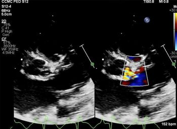
A 21-year-old female was referred at 30 weeks gestation for fetal echocardiography for suspicion of cardiomegaly on routine obstetric ultrasound screening. The fetal echocardiogram confirmed cardiomegaly with left ventricle dilation and evidence suggestive of endocardial fibroelastosis. Aortic valve morphology was not clearly characterised but an eccentric diastolic jet was noted, with diastolic flow reversal in the descending aorta and mild biventricular systolic dysfunction. The main differential diagnoses at that time included an aortic-left ventricular tunnel, ruptured aortic sinus, coronary artery fistula, and eccentric aortic regurgitation. There was no family history of CHD or connective tissue disorders. The neonate was delivered at term. Examination of the infant revealed no dysmorphic facial features or other physical anomalies. Genetic testing revealed normal karyotype, and fluorescence in situ hybridisation interpretation was negative for DiGeorge syndrome. A post-natal echocardiogram on day 0 of life, demonstrated that the aortic valve was tri-commissural but thickened and dysplastic. There was no evidence of aortic stenosis. The left coronary cusp appeared hypoplastic, tethered, and retracted, resulting in lack of coaptation. There was an eccentric diastolic jet that appeared to initiate high in the sinus in the region of left coronary cusp, resulting in moderate aortic regurgitation (Fig 1). The right coronary artery appeared larger than the left; however, antegrade flow was seen in both. Systolic function was preserved. No intervention was deemed necessary. Pre-natal evaluation by ultrasounds and limited post-natal evaluation by abdominal and head imaging revealed no associated non-cardiac anomalies.
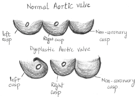
Pictorial representation of the dysplastic aortic valve from the perspective of the surgeon.

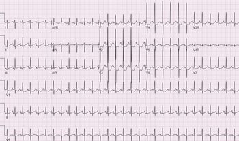
The neonate was discharged but presented on day 15 of life with cardiogenic shock and evidence of severely depressed left ventricular systolic function on repeat echocardiography. An electrocardiogram showed evidence of ischaemic changes with ST segment and T wave abnormalities (Fig 2). There was concern that the left coronary ostium was being obstructed by the abnormal valve leaflet. Both a cardiac MRI and cardiac catheterisation study were performed in an attempt to better delineate the anatomy of the aortic valve and coronary arteries. The MRI did not add any additional information to the echocardiographic findings. The aortic root angiogram confirmed a dysplastic aortic valve with severe aortic insufficiency. Both coronary arteries exhibited normal origins and distributions. A large right coronary artery was noted with faint opacification of the left coronary artery associated with small anterior descending and circumflex arteries. Of note, a non-specific filling defect was noted within the left aortic cusp. In order to better delineate coronary anatomy, selective coronary angiography was attempted. However, this was technically challenging due to patient size and development of arrhythmias, haemodynamically instability, and adverse ST segment changes. As a result, this was abandoned. Based on the available information, the infant was scheduled for surgical repair of the valve, without a clear aetiology for the cardiac ischaemia.
Parasternal short view on echocardiogram showing the dysplastic aortic valve and distorted left coronary cusp.

While the initial thought was that a Ross procedure might be required, direct surgical inspection showed that the left coronary leaflet was inserted abnormally on the aortic wall. In particular, the portion of the leaflet in the left-right commissure was rolled up and over back towards the left coronary ostium and not attached to the commissure itself. This splayed open the left-right commissure and was the mechanism of the insufficiency, and also obstructed view of and flow into the left coronary ostium, which itself was normal (Fig 3). It seemed to be relatively normal valve tissue; however, there was a sliver of extraneous tissue on the non-coronary leaflet. The portion of the left coronary leaflet was sharply divided off the aortic wall, realigned so that its way from the left coronary ostium and reattached to the aortic wall with fine Prolene sutures. The commissure was resuspended for added stability with fine Prolene sutures tied on the outside of the aorta. Once the valve cusp was reattached, direct visualisation confirmed patency of both coronary ostia with normal origins and courses. In confirmation of the small size of the left coronary seen on the angiogram and echocardiogram, there was evidence of collateralisation from the right coronary artery to the left. Endocardial fibroelastosis was noticed to be very mild, and it was decided not to excise it. The patient’s intra-operative and post-operative course was uncomplicated. He is now 10 months old, and his most recent echocardiograms have shown normal LV function, trivial aortic insufficiency, and mild supra-valvar aortic stenosis most likely at the site of suture lines. Endocardial fibroelastosis has not progressed and continues to mild with no evidence of systolic or diastolic dysfunction by echocardiogram. The previous ischaemic electrocardiographic changes have resolved.
Electrocardiogram, showing ST segment and T wave abnormality.

Discussion
Embryologic development of cardiac valves begins with the formation of the endocardial cushions. The semilunar aortic and pulmonary valves derive from a common truncal ridge. The endocardial cushions are highly proliferative and form primordial valve swellings which grow and elongate into fibrous leaflets, eventually forming the cusps of the semilunar valve leaflets. The extracellular matrix deposition on the valve cusps is tightly regulated by various factors such as bone morphogenic protein, transforming growth factor, and fibroblast growth factor and other signalling molecules. Defects in the process of proliferation and degradation of the extracellular matrix can lead to valve defects.Reference Combs and Yutzey7 The authors hypothesise that any defect in the process of proliferation and degradation of the extracellular matrix on the primordial valve could lead to such an aortic valve morphology as described above. Endocardial fibroelastosis is associated with hypoplastic left heart syndrome, aortic stenosis, and atresia. In our patient, the abnormal aortic valve, coronary ostium obstruction, diminished development of the left coronary artery, and slow chronic ischaemia during the fetal development could be the possible explanation of endocardial fibroelastosis.
Non-invasive imaging remains vital for the diagnosis of aortic valve disease, both congenital and acquired. Transthoracic echocardiogram is typically the first line to diagnose aortic valve disease and to monitor progression. Transthoracic imaging alone has been debated to be inadequate for defining aortic valve morphological abnormalities in adults.Reference Ahmari, Schaff, Click and Chandrasekaran5 Cardiac MRI is an emerging imaging modality with excellent spatial resolution, functional resolution, and myocardial tissue characterisation. There is growing evidence of use of MRI in determining valvar morphology as it has the advantage to visualise the morphology and motion of the valve from any desired image orientation without limitation of optimal windows, especially when the echocardiographic assessment is inconclusive.Reference West and Kramer8 In this particular case, aortic valve morphology prior to surgical repair could not be accurately established despite performing non-invasive as well as invasive imaging. This is attributed to the fact that there was no prior knowledge of this exclusive aortic valve defect to perhaps enable more detailed visualisation. Cardiac catheterisation is the gold standard of establishing coronary artery anatomy, whether it is anomalous origin of the coronary artery, external compression, or internal obstruction. In our patient, the left coronary artery and branches appeared smaller. Since blood flow contributes to cardiac chamber as well as vessel dimensions, diminished flow into the left coronary artery during cardiac development can be viewed as the aetiology for this finding. Whether performance of selective coronary angiography would have better delineated the obstruction or not, is conjectural.
Conclusion
To our knowledge, this is the first case of this unique and isolated abnormal aortic valve morphology causing valve regurgitation, coronary obstruction, and ischaemia. Inability of pre-operative evaluations to identify this valve abnormality demonstrates some limitations of non-invasive as well as angiographic imaging especially in infants. The embryological basis of this aortic valve abnormality is not known but may indicate an unknown gene mutation. Direct visual examination at the time of surgery made the definite diagnosis and determined the aetiology of myocardial ischaemia. Appropriate surgical intervention corrected the problem.
Acknowledgements
None.
Financial support
This research received no specific grant from any funding agency, commercial, or not for profit sectors.
Conflicts of interest
None.