Introduction
Nasal airway obstruction in a neonate is an uncommon occurrence. It may be due to choanal atresia, neonatal rhinitis or, more rarely, pyriform aperture stenosis, choanal stenosis or a nasal mass. Neonates are obligate nasal breathers, and therefore any cause of nasal obstruction may present with acute respiratory distress.
Here, we present an unusual case of nasal obstruction in a neonate, and we describe its successful management.
Case report
An eight-day-old boy was transferred to our tertiary referral centre for further assessment of respiratory difficulty of unknown cause.
The boy had been born at term following an uneventful pregnancy. Delivery had been by Ventouse suction due to prolonged rupture of membranes (more than 48 hours). The infant's Apgar scores had been 8 at 1 minute and 9 at 5 minutes, but some respiratory distress had been noted at around 24 hours. This had been characterised by predominate mouth-breathing and difficulty with nasal respiration. Oxygen requirements had been minimal, but subcostal and suprasternal recession had been noted. Antibiotics had been commenced due to prolonged rupture of membranes, although there had been no other evidence of infection. A small rise in PaCO2 levels had required continuous positive airway pressure ventilation. However, over the next few days the infant's respiratory difficulties had resolved and he had been discharged.
The child was readmitted to his local hospital within 24 hours with increasing respiratory difficulties, most apparent during feeding. On admission, oxygen saturations were 70 per cent despite oxygen therapy. They improved when the child was nursed supine with head and neck extension. Inspection of the pharynx with a laryngoscope revealed a normal epiglottis, and catheters were successfully passed through both nasal cavities.
The child's respiratory difficulties continued, and two days later he was transferred to our paediatric otolaryngology unit.
An urgent rigid laryngotracheobronchoscopy and examination of the post-nasal space under anaesthesia were undertaken. This revealed a large, cystic lesion which appeared to be arising from the roof of the posterior nasal space. The remainder of the upper airway was found to be normal. The child was intubated, and computed tomography (CT) and magnetic resonance imaging (MRI) were arranged.
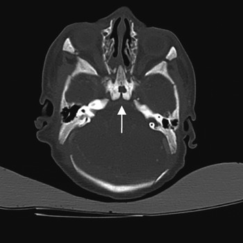
Imaging revealed a cephalocoele with an intracranial connection to the pituitary fossa. No neural tissue was seen within the cephalocoele, on either the CT or MRI scans (Figures 1 to 3).
Sagittal computed tomography scan showing a lesion of fluid density with intracranial extension (short arrow). No neural tissue is seen within the mass. Obstruction of the nasopharynx and oropharynx can be seen with endotracheal tube in situ. An area in the superior aspect of the posterior nasal cavity was also suggested as a possible abnormality (long arrow), but was thought to represent mucus lying within the nasal cavity.

Axial computed tomography scan showing the lesion's intracranial connection (arrow).

Sagittal, T2-weighted magnetic resonance imaging scan showing better characterisation of the lesion. The large nasopharyngeal mass can be seen to connect with the pituitary fossa. The anterior area identified on the computed tomography scan (see Figure 1) shows a small area of high signal change but no neural tissue; this may represents retained mucus in the nasal cavity.

A transpalatal excision of the nasopharyngeal cephalocele with closure of the intracranial connection and repair of the palate was undertaken. A lumbar drain was inserted at the commencement of the operation. A Dingman mouth gag retractor (more commonly used in cleft palate surgery) was used to achieve access to the oropharynx. The palate was then divided in the midline to visualise the nasopharynx (Figure 4). The cephalocoele was found to be adherent to the posterior septum and nasal roof in the posterior nasal space. The lesion was opened and the bony defect identified within the base of the lesion (Figure 5). The cephalocoele was excised and sent for histological analysis. Layered closure of the skull base defect was achieved using bone paté, periosteum, fascia (harvested from a post-auricular incision) and fibrin glue (Tissucol; Baxter Healthcare, Deerfield, USA). Firstly, the mucosa was removed from the bony opening and the defect was filled with bone paté. Fibrin glue was then applied to the bone paté. Finally, periosteum and temporalis fascia were laid over the repaired defect, each covered with a further layer of fibrin glue. A post-nasal pack was then inserted. Palatal repair was then undertaken with two myomucosal layers closed with 5/0 Monocryl sutures (Ethicon, Livingston, UK).
Surgical photograph showing the transpalatal incision, with the underlying cephalocoele visible (arrow).

Surgical photograph following cephalocoele removal, with the underlying bony defect visible (arrow).

Histological analysis revealed the cephalocoele to consist of fibrovascular tissue with respiratory-type epithelium present. The tissue contained multiple cystic spaces lined by choroid plexus epithelium. Glial tissue was present within the walls of the lesion. In view of the presence of glial tissue, appearances were thus consistent with an encephalocoele.
Post-operatively, the nasal pack was removed after two days, the child was extubated after three days, and the lumbar drain was left in situ for five days. The child made an uneventful recovery, with no evidence of cerebrospinal fluid (CSF) leakage or palatal dysfunction.
Follow-up endoscopic examinations and MRI scanning revealed no evidence of recurrence.
Discussion
Airway obstruction in a neonate may be caused by the abnormal development of normal airway structures (e.g. choanal atresia and pyriform aperture stenosis), or by a mass obstructing the airway. There are several causes for a nasal mass in a neonate. These include nasal dermoid cysts, gliomas, cephalocoeles, and rarer entities such as benign fibrous histiocytomas and hamartomas.Reference Shrier, Wang, Patel, Monajati, Chess and Numaguchi1–Reference Kim, Low, Billman, Wickersham and Kearns4 Due to the normal obligate nasal breathing pattern seen in neonates, any cause of nasal obstruction may present with features of respiratory distress.
When a nasal cavity mass is found to contain neural tissue, the three important differential diagnoses are cephalocoele, nasal glioma and teratoma. A cephalocoele is an abnormal protrusion of the brain or its coverings. It may contain brain tissue (encephalocoele) or solely meninges (meningocoele). Nasal gliomas are similar to cephalocoeles but have lost their intracranial connection. Teratomas may be differentiated by examining the entire specimen for germ cell layers.Reference Patterson, Kapur and Chandra5
Encephalocoeles have been reported as a cause of nasal mass in newborns, and are usually described with typical associated features such as cleft lip and hypertelorism.Reference Lewin6, Reference Kennedy, Gruber, Billmire and Crone7 They can be subdivided according to their origin. The majority (75 per cent) occur in the occipital region. Fifteen per cent are sincipital, occurring on the dorsum of the nose, the orbit or the forehead. The remaining 10 per cent are basal, and can be classified according to the site of the protrusion;Reference Mylanus, Marres, Vlietman, Kollee, Freihofer and Thijssen8 types include transethmoidal, sphenoethmoidal, trans-sphenoidal, sphenomaxillary and spheno-orbital.Reference Suwanwela and Suwanwela9 Basal encephalocoeles are rare congenital disorders, occurring in approximately one in every 40 000 live births.Reference Mylanus, Marres, Vlietman, Kollee, Freihofer and Thijssen8 The trans-sphenoidal subtype is the least common, occurring in only approximately one in 700 000 live births.Reference Kahyaoglu, Cavusoglu, Musluman, Kaya, Yilmaz and Sahin10 They may present with a variety of problems, including respiratory difficulty, cranial defects (e.g. cleft palate, hypertelorism and optic malformations), meningitis and endocrine abnormalities. Some cases may only become apparent in adulthood, presenting with persistent CSF rhinorrhoea.Reference Blaivie, Lequeux, Kampouridis, Louryan and Saussez11, Reference Schick, Brors and Prescher12
Our patient's trans-sphenoidal cephalocoele had an intracranial connection to the pituitary fossa (i.e. it was trans-sellar). This is only the 10th documented case of such a cephalocoele.Reference Kahyaoglu, Cavusoglu, Musluman, Kaya, Yilmaz and Sahin10, Reference Raman Sharma, Mahapatra, Pawar, Thomas and Al-Ismaily13, Reference Herman, Sauvaget, Guichard and Tran Ba Huy14
In order to understand how a cephalocoele may develop, it is important to consider the development of the skull base, and in particular that of the sphenoid bone. There are, in total, fourteen ossification centres in the sphenoid bone, which come together approximately between the third and seventh or eighth months of fetal life. The process is complete when the anterior and posterior components of the sphenoid bone become fused. If this complex process of ossification is not successfully completed, then there is the risk of localised areas of bone deficiency, and/or herniation of normally intracranial contents.Reference Blaivie, Lequeux, Kampouridis, Louryan and Saussez11, Reference Schick, Brors and Prescher12, Reference Gray15
Three different approaches have been described for the repair of basal cephalocoeles.
The transcranial route is the standard method of repair. However, it involves a craniotomy, with all that procedure's associated risks and possible complications. This approach has the advantage of good repositioning of herniated tissue, but some studies report morbidity of up to 70 per cent and mortality of up to 50 per cent.Reference Kennedy, Gruber, Billmire and Crone7 The main complications encountered include removal of functioning neural tissue, anosmia, intracerebral haemorrhage and frontal lobe dysfunction; there is also some evidence of increased recurrence rates.Reference Gursan, Aydin, Atlas and Ertas16
• A cephalocoele is a rare but important cause of airway obstruction in neonates
• Imaging is crucial for guiding management
• These lesions may have an intracranial connection to the pituitary fossa
• Surgery via a transpalatal approach gives good access and results, even in patients without a cleft palate
The second repair method described is an endoscopic approach via the nasal cavity. This is the preferred route for repair of lesions in an adult. It may be used in the paediatric population, but the documented cases have involved older children.Reference Castelnuovo, Bignami, Pistochini, Battaglia, Locatelli and Dallan17 There are no documented cases of endoscopic management in the neonatal population.
The third repair method, the transpalatal approach, was chosen in the presented case. This approach has also been used to correct other documented cases of neonatal cephalocoele.Reference Kennedy, Gruber, Billmire and Crone7 Its advantages include good access (particularly in cases with associated cleft palate), better cosmesis, decreased recurrence rates, and the opportunity to formally repair skull base defects at the time of primary surgery.
From our review of the literature, it can be surmised that the choice of approach must be based on each individual patient's case, influenced by the lesion's location, size and associated features. As highlighted in the presented case, imaging is crucial to this planning process, but it cannot completely exclude the presence of neural tissue within the lesion.
Conclusion
Although there are more common causes of neonatal nasal obstruction causing respiratory distress, the presented case illustrates that cephalocoele should be considered as a possible aetiology. This condition is an important differential diagnosis, due to the potential complications of misdiagnosis, including removal of functioning neural tissue, CSF leakage and meningitis.
The presented case also shows that respiratory difficulties may not be immediately apparent at birth.
In this case, radiographic imaging enabled identification of the location and extent of the lesion, aiding the appropriate choice of surgical approach. A transpalatal approach was used to excise the lesion, giving good results, with no subsequent CSF leakage, recurrence or palatal dysfunction.