

Introduction
Endolymphatic hydrops is characterised by the classic association of episodic vertigo, a sense of aural fullness, fluctuating progressive hearing loss and tinnitus. Loudness intolerance and diplacusis are also often present. In most cases, it is impossible to establish the aetiology, and a diagnosis of idiopathic endolymphatic hydrops or Ménière's disease is established.
Vertigo due to endolymphatic hydrops, whether idiopathic or secondary to a known aetiology, is amenable to either medical or surgical treatment. Treatment is usually symptomatic, with anti-emetics, sedatives, tranquilisers, diuretics or vasodilators administered on an empirical basis. Psychological support of these patients is also an important part of treatment. Surgical treatment is usually performed when conservative therapy has failed and intractable incapacitating vertigo cannot be controlled by other means. In this situation, it is desirable to recommend a procedure that offers a high likelihood of vertigo control with maximum hearing preservation.
Different surgical procedures for intractable vertigo have been described. These can be categorised as conservative procedures (endolymphatic sac surgical procedures, sacculotomy, cryotherapy, labyrinth fistulisation) and destructive procedures (vestibular neurectomy and labyrinthectomy).
The first surgical procedure for treating Ménière's disease was described by Portmann in 1927, who opened the endolymphatic sac as a means to diminish endolymphatic pressure.Reference Portmann 1 In 1938, Hallpike and Cairns demonstrated the pathological findings of endolymphatic hydrops in Ménière's disease patients,Reference Hallpike and Cairns 2 but by that time the surgical procedure described by Portmann was seldom used and reports by other otologists were often not enthusiastic about the procedure's value. In 1962, William House described good results after performing a subarachnoid shunt for drainage of endolymphatic hydrops.Reference House 3 This renewed the interest in the technique, but it was not until 1967, when Kimura reliably reproduced endolymphatic hydrops in guinea pigs by obliterating the endolymphatic duct and sac, that this type of surgery really caught the attention of a larger group of surgeons.Reference Kimura 4 Since then, various shunting techniques have emerged, including subarachnoid shunts, mastoid shunts and endolymphatic duct valves.
In1976, Paparella and Hanson described a technique in which they emphasised a wide exposition of dura avoiding the posterior semicircular canal and sac drainage via a T-tube that served as a valve.Reference Paparella and Hanson 5 The results of this procedure in 75 patients were excellent, with control of vertigo in 94 per cent of patients and even a significant improvement in cochlear function in 30 per cent. Later on, Paparella and colleagues referred to this surgical technique as endolymphatic sac enhancement.Reference Paparella and Goycoolea 6 Since then, others have described variations in the surgical technique for endolymphatic sac surgery, with variable results.Reference Brown 7 , Reference Brinson, Chen and Arriaga 8
A large number of authors consider endolymphatic sac decompression an effective surgical procedure. Success rates for vertigo control with this technique range between 60 and 90 per cent.Reference Brinson, Chen and Arriaga 8 However, some question its long-term efficacy, and others think that vertigo control is more the result of a placebo effect than a result of the procedure itself.Reference Thomsen, Bretlau, Tos and Johnsen 9 , Reference Bretlau, Thomsen, Tos and Johnsen 10
When endolymphatic homeostasis is disturbed and hydrops occurs, the sac appears to be an excellent site for decompression of the endolymphatic space, at least conceptually. The sac is relatively distant from the cochlear and vestibular organs, which suggests that this type of surgery might be a good alternative for hearing preservation.
In 2014, Sood et al. performed a systematic review and meta-analysis to analyse current endolymphatic surgery techniques and assess their efficacy in controlling vertigo and maintaining hearing.Reference Sood, Lambert, Nguyen and Meyer 11 This study demonstrated that endolymphatic sac decompression alone or with a mastoid shunt may be effective at controlling vertigo in the short term (between 12 and 24 months of follow up) and in the long term (over 24 months) in at least 75 per cent of Ménière's disease patients in whom medical therapy had failed. Although not statistically significant, there was a trend for sac decompression alone to provide better vertigo control and better hearing preservation than shunting procedures. In addition, there was a statistically significant difference in hearing preservation between shunting with and without Silastic®, in favour of not using Silastic.Reference Sood, Lambert, Nguyen and Meyer 11
A novel surgical technique for the treatment of Ménière's disease has recently been described: endolymphatic duct blockage. In this technique, the sac is not incised or dissected from the posterior fossa dura. The surgeons dissect all the bone found around the endolymphatic duct in order to identify as much of it as possible, then they block the dissected endolymphatic duct with two small titanium clips. Saliba et al. reported (in 2015) on a non-blinded, randomised controlled trial comparing this technique against endolymphatic sac decompression.Reference Saliba, Gabra, Alzahrani and Berbiche 12 They found that 96.5 per cent of the patients in the endolymphatic blockage group had achieved complete control of vertigo spells, compared with 37.5 per cent of the endolymphatic sac decompression group, with no significant difference between the pre-operative and the post-operative hearing levels in both groups. Their study suggested that this novel technique might be better than traditional endolymphatic sac decompression. In a later study, published in 2016, the same group reported a total absence of Ménièré's attacks in 89.9 per cent of their patients with this technique in a larger group than that of their previous study.Reference Gabra, Asmar, Berbiche and Saliba 13
The current study aimed to describe the results obtained with endolymphatic sac drainage surgery in a group of 95 patients with Ménière's disease, managed in a tertiary referral centre, who failed to obtain vertigo control after a course of medical management.
Materials and methods
A total of 95 patients with Ménière's disease were operated on by the senior author in a tertiary referral centre in São Paulo, Brazil, from January 1986 to December 2012. Patients in this series became candidates for surgery when their vertigo became incapacitating and could not be controlled following extensive medical and psychological management. Some of these patients were already candidates for labyrinthectomy as they had greater than 50 dB hearing loss and/or less than 50 per cent discrimination in the affected ear. Symptoms had been present in these patients from 1 to 10 years (average, 4.7 years). The average duration of significant, difficult-to-control vertigo prior to surgery was 18 months. Our patients experienced 10–15 Ménière's episodes per month in the 6 months prior to surgery (approximately 1 episode every 2–3 days), even with medical therapy.
There were 36 male patients (38 per cent) and 59 female patients (62 per cent), with an age range of 28 to 67 years (average, 47.5 years). The majority of patients had unilateral disease (88 patients; 92.63 per cent) and only seven cases were bilateral Ménière's (7.36 per cent). Of the patients with bilateral disease, five were operated on bilaterally and only two unilaterally (in the worst hearing ear in one patient, and on the side with worse vestibular function in the other).
The bone conduction hearing level for unilateral cases, calculated using 0.5, 1, 2 and 3 kHz thresholds, was 40 dB in 3 cases, 50 dB in 32 cases, 60 dB in 39 cases and 70 dB in 14 cases. For the 7 bilateral cases, the bone conduction in the 12 ears operated on was 40 dB in 2 ears, 50 dB in 4 ears, 60 dB in 5 ears and 70 dB in 1 ear.
The follow-up period for the 95 patients ranged from 3 to 15 years after surgery (average, 9 years), with 45 patients (13.3 per cent) followed for a period longer than 10 years.
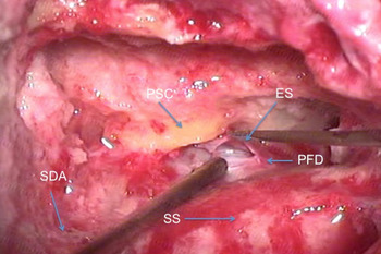
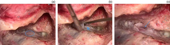
The method of endolymphatic sac decompression and drainage employed by the senior author was previously described by Paparella and colleagues.Reference Paparella and Hanson 5 , Reference Paparella and Goycoolea 6 , Reference Paparella and Sajjadi 14 It starts with wide exposition of the mastoid cortex, a simple mastoidectomy with complete removal of pre-sigmoid, infra-labyrinth and retro-labyrinth mastoid air cells, and identification of the horizontal and posterior semicircular canals. After that, the bone that covers the sigmoid sinus, the posterior cranial fossa and the middle cranial fossa (at the level of the tegmen antri) is thinned. The more medial portion of the lateral sinus, all accessible dura in Trautmann's triangle, and often an adjacent small portion of dura of the middle cranial fossa, are exposed by removal of the thin bone with curettes and elevators. Sometimes a small island of bone might be left covering the sigmoid sinus to prevent its damage, and because by pressing it the retro-labyrinthian area might be better visualised. The sac is then identified as a white, dense thickening in the dura, pointing towards the inferior portion of the posterior semicircular canal. The sac in most cases is clearly differentiated from adjacent dura by a lack of blood vessels coursing across the dura, and by the thickness of the white sac as compared to the thinness of the dura elsewhere. After identifying the endolymphatic sac, a small opening in the sac is performed with a joint knife, often beneath the bony edge (Figure 1). Retracting the dura with a suction tip or elevator allows further identification of the sac under the bony edge. The lumen of the sac is then identified by blunt probing, using right and left curved probes. A Silastic ‘T’ shape is cut from silicone rubber 0.127 mm (0.005 inch) sheeting. The T portion of the tube is then pushed into the small opening and it is allowed to coil upon it, thus forming a tubular drain (Figure 2). An absorbable gelatine sponge saturated in a steroid and antibiotic solution is then placed in the mastoid cavity. The wound is subsequently closed in an ordinary fashion.
Endolymphatic sac decompression and drainage in the right ear. PSC = posterior semicircular canal; ES = endolymphatic sac (opened); PFD = posterior fossa dura; SDA = sinodural angle; SS = sigmoid sinus
Endolymphatic sac decompression, drainage and enhancement in the left ear. (a) The endolymphatic sac is identified in the posterior fossa dura (circled area). (b) The sac is opened (arrowhead). (c) A Silastic sheet (arrow) is placed to keep the sac opened.
Our results are described according to both the American Academy of Otolaryngology–Head and Neck Surgery (AAO-HNS) Committee on Hearing and Equilibrium guidelines for quantifying efficacy in controlling vertigo and preserving hearing, 15 and the classification used by Paparella and colleagues, which for the authors is an easily understandable way of demonstrating results with this technique.Reference Paparella and Hanson 5 The classifications of treatment results proposed by Paparella and the AAO-HNS are described in Tables I and II respectively. Importantly, in the classification proposed by Paparella, groups A, B and C represent patients in whom a significant improvement in vertigo was observed after surgical treatment. As the grade of improvement is not clarified in this classification, we considered ‘controlled’ vertigo as a minimum 80 per cent reduction in vertigo spells when comparing one year from the pre-operative stage with one year after surgery. We considered improved hearing to be a greater than 20 dB improvement in bone conduction hearing thresholds or a better than 20 per cent improvement in discrimination scores.
Paparella's classification of treatment results in endolymphatic hydropsReference Paparella and Hanson 5

Summary of AAO-HNS committee on hearing and equilibrium reporting guidelines 15

Numerical value = (X/Y) × 100 rounded to the nearest whole number, where ‘X’ is the average number of definitive spells per month for 6 months in the 18 to 24 months after therapy and ‘Y’ is the average number of definitive spells per month for the 6 months before therapy. AAO-HNS = American Academy of Otolaryngology–Head and Neck Surgery
Results
Our primary objective when performing these surgical procedures was the elimination of incapacitating vertigo, and of the total dependence on labyrinth suppressants, central nervous system suppressants and diuretics presented by our patients. Total absence of vertiginous symptoms was accomplished in 34 patients (35.78 per cent of patients in class A according to the AAO-HNS classification); most of them had previously been incapacitated because of the intensity and regularity of their vertigo. Another 51 patients greatly improved after surgery compared to their previous 6 months (53.68 per cent of patients in AAO-HNS class B). Vertigo control was obtained in 94.3 per cent of patients with unilateral idiopathic endolymphatic hydrops in this series (83 patients in the Paparella and the AAO-HNS groups A, B and C), leaving only five patients who did not respond well to endolymphatic sac surgery. All of these patients, who were previously totally dependent on labyrinth suppressants, central nervous system suppressants and diuretics, were able to stop taking these medications, which is another indication of great improvement. Regarding the AAO-HNS classification, four patients showed no improvement and one patient showed worsening in the number of Ménière's episodes.
It is important to note that around 14 per cent of the patients also had a significant improvement in hearing following the procedure. Hearing was undisturbed in almost 74 per cent of our patients, with a total of 88 per cent of patients in Paparella groups A and B (77 patients with undisturbed or improved hearing). Hearing deteriorated in five of the unilateral patients (12 per cent), mostly to a severe hearing loss. None of the patients developed anacusis.
Considering only the bilateral idiopathic endolymphatic hydrops group, vertigo control was obtained in 85.7 per cent of cases (six patients in the Paparella and the AAO-HNS groups A, B and C). Hearing was undisturbed in 43 per cent of the operated ears and improved in 28 per cent, with a total of 71 per cent of patients in Paparella groups A and B together. As mentioned earlier, two of the bilateral Ménière's disease patients underwent surgery to one ear only (the worst hearing ear in one case and in the ear with worse vestibular function in the other case); after surgery, one of the patients was classified as being in Paparella group A and the other in Paparella group B based on their hearing in the operated ear, even though nothing was done for the contralateral ear.
The unilateral and bilateral disease patients’ post-operative results, after long-term follow up, according to the Classification of Treatment Results in Endolymphatic Hydrops proposed by Paparella,Reference Paparella and Hanson 5 are summarised in Table III. The results for the number of definitive Ménièré's spells in the six months before surgery compared with the six months after surgery, as suggested by the AAO-HNS, are summarised in Table IV. 15
Post-operative results according to paparella's classification*

*Of treatment results in endolymphatic hydrops, for unilateral and bilateral disease patients after long-term follow up.Reference Paparella and Hanson 5
Post-operative results according to AAO-HNS committee on hearing and equilibrium reporting guidelines*

*For quantifying efficacy in controlling vertigo and preserving hearing, in unilateral and bilateral disease patients. 15 AAO-HNS = American Academy of Otolaryngology–Head and Neck Surgery
The follow-up period for the 95 patients ranged from 3 to 15 years post-surgery (average, 9 years). No major complications occurred during the surgical procedures and there were no surgery-related problems during the follow-up period for the 95 patients. If cerebrospinal fluid leak was suspected during surgery, the site was packed with a small piece of muscle or abdominal fat and fibrin glue. During the immediate post-operative period, patients were managed with complete bed rest for 3 to 4 days. No patients required further management.
Hearing deteriorated significantly in only 6 of the 45 patients (13.3 per cent) who were followed for a period longer than 10 years, which may suggest a mechanism of endolymphatic hydrops stabilisation following sac surgery.
Discussion
The theory behind decompression is that removal of mastoid bone provides pressure relief and allows for expansion of the endolymphatic sac, thereby decreasing episodic vertigo attacks. Placement of a Silastic sheet in the form of a tube in the sac lumen may further augment its inner diameter, enhancing its function and serving as a means of drainage.Reference Paparella and Sajjadi 14
We attempted to evaluate the subjective symptoms such as vertigo intensity and level of incapacity as objectively as possible. When evaluating hearing results in Ménière's patients, it is important to consider the fluctuating nature of the disease and the possibility of spontaneous vertigo control associated with the natural history of the disease.
In our experience, endolymphatic sac drainage has been safe, and has not led to any major complications. Some authors have repeatedly suggested that endolymphatic sac surgery is no more effective than a placebo.Reference Thomsen, Bretlau, Tos and Johnsen 9 , Reference Bretlau, Thomsen, Tos and Johnsen 10 Regardless of all uncertainties surrounding Ménière's disease treatment, endolymphatic sac surgery is both effective in controlling vertigo and safe for preserving hearing. It is the authors’ opinion that sac surgery is still an excellent treatment option for medically refractory Ménière's disease with serviceable hearing. As mentioned earlier, a new technique called endolymphatic duct blockage has been recently described.Reference Saliba, Gabra, Alzahrani and Berbiche 12 , Reference Gabra, Asmar, Berbiche and Saliba 13 Excellent results have been obtained by directly blocking the endolymphatic duct with clamps; this is physiologically different to what surgeons have been doing for years, which is enhancing or promoting endolymphatic drainage. Attempts to drain the endolymphatic sac with traditional surgical procedures may ultimately promote fibrosis or scarring in the endolymphatic sac and duct, acting in the same way as the endolymphatic duct blockage technique; however, this is just theoretical.
In some of our patients with bilateral disease, improvement was noted even though only the more severely affected ear was operated on. The rationale for the good results in these cases could be based on either psychological or physiological grounds.
Our results with this procedure have been gratifying, and 90 per cent of our incapacitated endolymphatic hydrops patients have been able to return to a functional life free of vertigo (94.3 per cent in the unilateral group and 85.7 per cent in the bilateral group). Furthermore, a significant improvement of cochlear function was observed in 14 of these patients (13.6 per cent of the unilateral patients and 28.5 per cent of the bilateral patients were in Paparella group A). In addition, hearing preservation (Paparella groups A and B together) was obtained in 86.6 per cent of patients (87.4 per cent in the unilateral group and 71.4 per cent in the bilateral group). This demonstrates the safety of the surgical technique.
A meta-analysis by Sood et al. showed vertigo control in the long term, after approximately three years of follow up, for 81.6 per cent of patients who underwent sac decompression alone compared to 75.7 per cent for those who underwent current mastoid shunt techniques (categories A and B from the AAO-HNS classification, representing total resolution and great improvement of vertigo respectively).Reference Sood, Lambert, Nguyen and Meyer 11 In our series, 89.46 per cent of patients were in these categories.
Most authors who speak against endolymphatic sac surgery for the treatment of endolymphatic hydrops promote chemical labyrinthectomy with aminoglycosides, especially gentamicin. In 2004, Chia et al. performed a meta-analysis with 980 patients from 27 studies, comparing 5 different delivery methods for gentamicin to the inner ear.Reference Chia, Gamst, Anderson and Harris 16 When the five groups were considered together, complete control of vertigo was obtained in 73.6 per cent of patients, with a significantly greater vertigo control rate achieved by the titration method (81.7 per cent). Overall improvement in vertigo was observed in 90.2 per cent of patients. However, hearing loss was observed in 25.1 per cent of patients, suggesting that gentamicin is not that safe for hearing preservation given that one in every four patients will experience hearing loss. The meta-analysis by Sood et al. showed that with endolymphatic sac decompression alone, post-operative hearing was stable or improved in 72.8 per cent of patients, and when using mastoid shunts (with and without Silastic) post-operative hearing was stable or improved in 71.4 per cent.Reference Sood, Lambert, Nguyen and Meyer 11 In our series, hearing preservation was accomplished in 87.4 per cent of our unilateral patients and in 71.4 per cent of the bilateral patients (86.3 per cent total). Our results for hearing preservation are better than those reported in the international literature for endolymphatic sac surgery and the results obtained with gentamicin, especially for our unilateral group.
The natural history and progression of endolymphatic hydrops complicates the analysis of long-term surgical results given that a large number of patients will ultimately stop having vertigo episodes by the time their posterior labyrinth is completely destroyed and their hearing is lost.Reference Silverstein, Smouha and Jones 17 The large follow-up period for our patients shows good long-term control for vertigo and good hearing preservation, as hearing deteriorated in only 13.3 per cent of the 45 patients that were followed for a period longer than 10 years.
Kitahara et al. suggested another interesting treatment modality,Reference Kitahara, Kubo, Okumura and Kitahara 18 which may be a useful adjunct for that which we performed. These authors performed endolymphatic sac decompression with intra-endolymphatic sac application of steroids, as a new therapeutic strategy for intractable Ménière's disease. They divided their patients in three groups: endolymphatic sac decompression only, endolymphatic sac decompression accompanied by intra-endolymphatic sac application of steroids, and a control group (which received no such treatment). The intra-endolymphatic sac application of large doses of steroids had additional effects to those of endolymphatic sac expanding surgery, especially on hearing. The results for the treated groups were, of course, superior to those of the group who received no surgical treatment for intractable Ménière's disease, both in terms of vertigo control and hearing improvement, for at least seven years.
-
• Vertigo due to endolymphatic hydrops, whether idiopathic or secondary to a known aetiology, is amenable to medical or surgical treatment
-
• Treatment includes anti-emetics, sedatives, tranquilisers, diuretics or vasodilators, and psychological support is important
-
• Surgical treatment, whether conservative or destructive, is usually performed when medical therapy has failed
-
• Many authors consider endolymphatic sac decompression an effective surgical procedure, but others still doubt its benefits
-
• Ninety per cent of incapacitated endolymphatic hydrops patients returned to a vertigo-free life, with hearing preservation in 86 per cent
In our series, revision endolymphatic sac surgery was not needed. Nevertheless, a large number of papers report good results when patients who showed improvement after surgery and a subsequent relapse of vertiginous symptoms years later underwent revision surgery.Reference Huang 19 , Reference Paparella 20
Conclusion
Endolymphatic sac drainage is still an excellent non-destructive surgical option for patients with incapacitating endolymphatic hydrops, providing a high percentage of vertigo control and hearing preservation. Together with medical and psychological therapy, it is an important adjunct to help patients suffering with severe Ménière's disease. In our opinion, this treatment has important advantages over other forms of clinical and surgical treatment for this disease.



